Animals
Mice were housed in the Animal Center of the Affiliated Drum Tower Hospital of Nanjing University Medical School under controlled air pressure and temperature conditions with free access to food and water. Mouse use and experimental procedures were approved by the Institutional Animal Care and Use Committees of Nanjing Drum Tower Hospital (2021AE01035).
Generation of Dnajb7 knockout mice via CRISPR/Cas9 technology
Dnajb7-deficient mice in the C57BL/6 genetic background were generated by the CRISPR/Cas9-mediated genome editing system (Cyagen Biosciences, Suzhou, China). The Dnajb7 exon containing the J-domain was targeted by two sgRNAs (5’-ACTGTTTAAAAGGCCCTCGA-3’ and 5’-TGCCACACTATTTACAAGAA-3’). To obtain knockout mice, Cas9 mRNA and sgRNAs were coinjected into the cytoplasm of fertilized eggs. Genotypes of pups were determined by extracting genomic DNA. After genotyping, the F0 mice underwent serial mating to generate homozygous mutants.
Genotyping
Tail DNA from offspring was extracted and genotyped using PCR amplification (Tab. S1). The results of Sanger sequencing were analyzed using SnapGene (GSL Biotech, Chicago, IL, USA).
Fertility test
To investigate fertility in knockout mice, 10-week-old Dnajb7+/+ and Dnajb7−/− male mice were caged with two 10-week-old Dnajb7+/+ female mice for at least 8 weeks. The average litter size for each mouse line was calculated and recorded.
Histological analyses
Testes and epididymides from 10-week-old Dnajb7+/+ and Dnajb7−/− males were removed and fixed in Bouin’s solution overnight. Subsequently, tissues were dehydrated in increasing concentrations of ethanol (70%, 80%, 90%, 100%), cleared with xylene, embedded in paraffin and cut into 5-μm-thick sections, followed by hematoxylin and eosin (H&E; Sigma–Aldrich, USA) staining. For sperm staining, cauda epididymal sperm from 10-week-old male mice were isolated, fixed in 4% PFA, spread on clean glass slides and stained with H&E. Sections were analyzed under a microscope (LEICA DM2500, Germany).
Sperm counts and motility analyses
For the sperm count test, cauda epididymal sperm from 10-week-old Dnajb7+/+ and Dnajb7−/− males were released in PBS, fixed in 4% PFA and counted using a hemocytometer. For the sperm motility test, cauda epididymis sperm were resuspended in HTF (human tubal fluid) culture medium and analyzed using a computer-aided sperm analysis (CASA) system (Hamilton Thorne Biosciences, USA).
Quantitative RT–PCR
Total RNA was extracted from tissues and cells by TRIzol reagent (15596018, Thermo Fisher Scientific, MA, USA). The concentration and purity of RNA were determined by absorbance at 260/280 nm using a NanoDrop 2000 (Thermo Fisher Scientific, MA, USA). RNA was reverse transcribed using 5 × All-In-One RT MasterMix (G492, ABM, Canada). The cDNA was diluted and used for quantitative RT–PCR (qRT–PCR) with SYBR Green Master Mix (Q321, Vazyme, China). 18S rRNA was used to normalize gene expression. The primer sequences are listed in Table S1.
Cytoplasmic and nuclear extract preparation
Nuclear and cytoplasmic proteins were extracted from adult testes using a nuclear and cytoplasmic protein extraction kit according to the manufacturer’s instructions (P0027, Beyotime, China). GAPDH and Lamin B1 were used as loading controls for cytoplasmic and nuclear extracts, respectively.
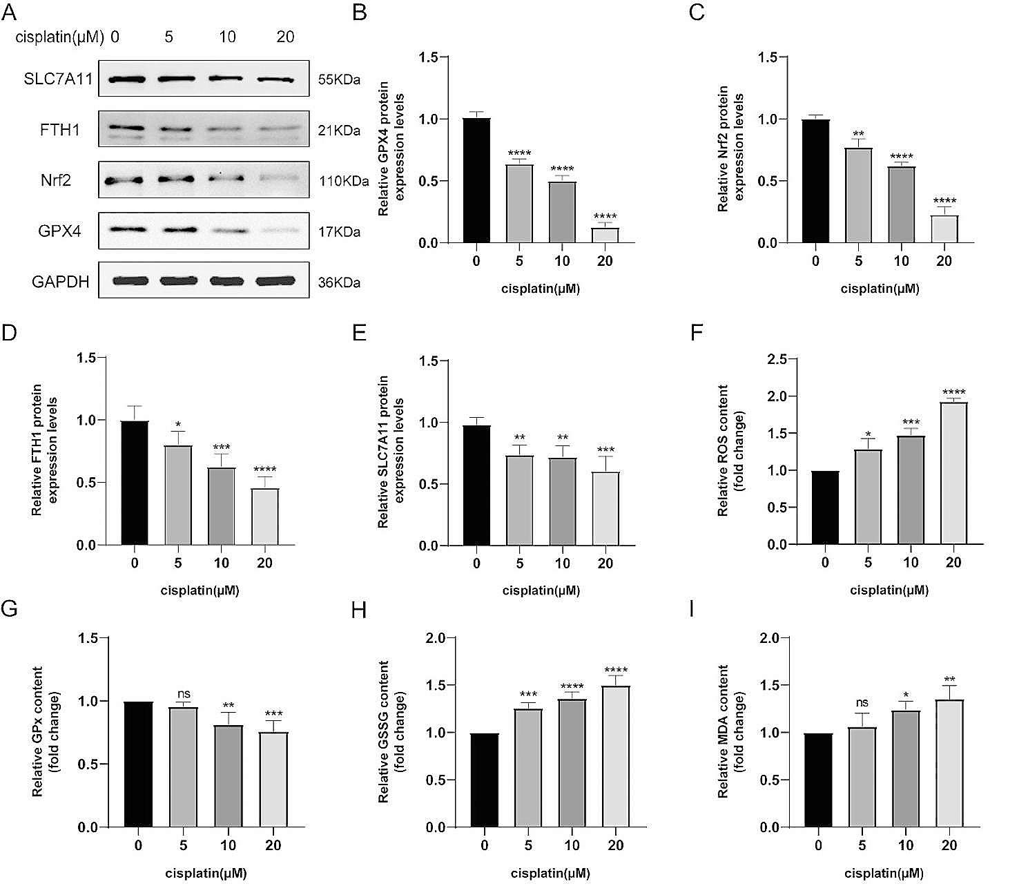
Western blotting
Tissues were collected from 10-week-old Dnajb7+/+ and Dnajb7−/− males, lysed in RIPA buffer containing protease inhibitor cocktail for 30 min on ice and centrifuged at 13000 rpm for 15 min at 4 °C. The concentration of proteins was determined by the bicinchoninic acid (BCA) protein assay (E11201, Vazyme, China). A total of 20 µg of protein was loaded and separated on 10% SDS–PAGE gels. The primary antibodies used were as follows: anti-DNAJB7 (diluted 1:1000 in TBST, 18540–1-AP, Proteintech, China), anti-DNAJB13 (diluted 1:1000 in TBST, 25118–1-AP, Proteintech, China), anti-Lamin B1 (diluted 1:10000 in TBST, 12987–1-AP, Proteintech, China), anti-GAPDH (diluted 1:10000 in TBST, 60004–1-Ig, Proteintech, China), anti-β-ACTIN (diluted 1:10000 in TBST, P30002M, Abmart, China), and anti-α-TUBULIN (diluted 1:5000 in TBST, 11224–1-AP, Proteintech, China).
Cell culture and transfection
The mouse DNAJB7 full-length coding sequence was fused to enhanced green fluorescent protein (EGFP). Human embryonic kidney 293 T cells (HEK293T) were obtained from ATCC (CRL-3216) and cultured in Dulbecco’s modified Eagle medium (DMEM, VivaCell, C3113-0500) supplemented with 10% fetal bovine serum (FBS, Gibco, 16000–044) and 1% penicillin–streptomycin (Gibco, 15140). HEK293T cells were transfected with plasmids harboring EGFP or EGFP-DNAJB7 using a Lipofectamine 3000 transfection kit (Invitrogen, L3000015). Expression was detected 28 h post-transfection under a fluorescence microscope (Nikon ECLIPSE 80i).
Immunofluorescence
Samples were fixed in 4% paraformaldehyde (PFA), dehydrated in graded ethanol (70-100%) and embedded in paraffin. Sections were blocked in 10% goat serum and incubated with the following primary antibodies: anti-PLZF (diluted 1:100 in TBST, AF-2944, R&D Systems, USA) anti-γH2AX (diluted 1:100 in TBST, 16-202A, Merck Millipore, USA) and anti-SOX9 (diluted 1:100 in TBST, AB5535, Merck Millipore, USA). Nuclear DNA and acrosomes were stained with 4’,6-diamidino-2-phenylindole (DAPI, F6057, Sigma–Aldrich, USA) and FITC-conjugated peanut agglutinin (PNA, RL-1072, Vector Labs, USA), respectively. Sections were analyzed using a fluorescence microscope (Leica DM300, Wetzlar, Germany).
In-situ hybridization
Testes were embedded in O.C.T compound (Sakura Finetek, Torrance, CA), frozen in liquid nitrogen, and cut into 8-μm-thick sections. The probes (Table S1) were added to the sections and hybridized overnight at 60 °C. After washing and blocking at room temperature, sections were incubated with alkaline phosphatase (AP) conjugated anti-DIG Fab fragments overnight. Sections were cleaned with maleic acid buffer containing Tween 20 (MABT) solution and AP buffer. After adding chromogenic solution (Beyotime Biotechnology, C3206), the sections were washed with double-distilled water, dehydrated in gradient ethanol, cleared with xylene, mounted with SlowFade Gold antifade reagent (Life Technologies), and finally analyzed under a microscope (LEICA DM2500, Germany).
Transmission electron microscopy
For ultrastructural analysis, samples were fixed in 2.5% glutaraldehyde, postfixed in 1% OsO4, dehydrated in a graded series of ethanol (30%, 50%, 75%, 95%, and 100%), infiltrated with a mixture of acetone and Epon resin, embedded in araldite and sectioned at 60 nm thickness. Ultrathin sections were stained with uranyl acetate and lead citrate. A transmission electron microscope (Tecnai G2; FEI, Eindhoven, The Netherlands) was used to capture images.
Phylogenetic analyses
Multiple alignments of amino acid sequences were downloaded from the NCBI database, and phylogenetic trees were constructed by MEGA X software using the neighbor-joining method. Multiple alignments were performed using MultAlin (http://multalin.toulouse.inra.fr/multalin/multalin.html).
Statistical analysis
All data are reported as the mean and standard deviation (SD). Statistical significance was tested by GraphPad Prism 8.0 software using a two-tailed unpaired Student's t test. A p value < 0.05 was considered statistically significant.

留言 (0)