
記住我


Eight moderately trained subjects (7 males, 1 female) participated in this study. This study (clinical trial identifier: NCT04240951) was approved by the Sterling Institutional Review Board (Atlanta, GA, sterlingirb.com) and has therefore been performed in accordance with the ethical standards in the Declaration of Helsinki. Before providing written informed consent, subjects were informed of the experimental procedures and associated risks. Trials were completed in the winter (January–March) in northeast Illinois to minimize seasonal acclimatization effects.
Preliminary screeningUpon the first visit to the laboratory (~ 0700–0800 h), subjects participated in a medical screening that consisted of measurements of nude body mass, height, resting heart rate, resting blood pressure, and ~ 8 h fasted blood glucose concentration. Subjects also completed a graded exercise test to assess cardiovascular health (12-lead ECG, Schiller AT-10 Plus; Schiller America, Doral, FL), maximum heart rate (HRmax), and maximum aerobic capacity (\(V\dot_\)) (MOXUS; AEI Technologies, Pittsburgh, PA) on a cycle ergometer (Velotron SRAM, Pro, Chicago, IL).
Experimental proceduresSubjects reported to the laboratory at 0900 or 1300 h after abstaining from caffeine, alcohol, and vigorous exercise for 24 h. Subjects were asked to fast from food, but drink 16 oz of water 2 h before the experimental trial to promote a well-hydrated state. Upon reporting to the laboratory, a urine sample was collected to assess baseline urine-specific gravity (USG; Atago Pen Refractometer, 3741‐E03 Saitama, Japan). Next, subjects’ skin on the forehead (FH), right dorsal forearm (RDF), right scapula (RS), and right triceps (RT) was shaved (if necessary) and cleaned with alcohol to prepare for later sweat patch application. These locations were chosen, because they are among those most commonly reported in the literature (Baker and Wolfe 2020) and the RDF and RT in particular are likely regions for wearable devices. Nude body mass was measured using a digital platform scale (KCC300 platform and IND439 reader; Mettler Toledo, Columbus, OH) to the nearest 0.01 kg. During the trial, subjects cycled on an ergometer (Velotron SRAM, Pro, Chicago, IL) for 90 min in a heated chamber (32 °C, 50% rh) at 82 ± 6% HRmax. Subjects were allowed to gradually warm up to their target heart rate (HR) and power output in the first ~ 10–20 min. HR was monitored using telemetry (Polar Electro RS400; Lake Success, NY) every 10 min along with ratings of perceived exertion (RPE), power output (Watts), and cadence (revolutions per min). Subjects were allowed to drink a sports drink (6% carbohydrate solution) ad libitum throughout the duration of exercise. After exercise and removal of all sweat patches, subjects were asked to towel dry before obtaining a final body mass measurement.
Sweat collectionThe standard absorbent patch method (11.9 cm2 absorbent pad, 3 M Tegaderm™ + Pad) (Baker et al. 2018) was used to collect regional sweat for this study. Anatomical location of patches were as follows: RDF was placed on the posterior midline of the forearm approximately half way between the styloid process of the radius and olecranon process; RT was placed on the posterior midline of the upper arm, approximately half way between the acromion and the olecranon process; RS was placed directly below the scapular spine and lateral to the medial border of the scapular spine; FH was placed in the midline of the frontal bone above the glabella but below the hairline. In the heated chamber immediately before exercise, FH, RDF, RS, and RT skin regions were cleaned with deionized water and wiped dry. Deionized water temperature equilibrated approximately to the temperature of the environmental chamber (32 °C) before it was used to clean the skin. Absorbent patches were applied and removed from each site according to the schedule shown in Fig. 1. Samples were collected during 0–25 min (25), 30–55 min (55), and 60–85 min (85) of the 90 min cycling protocol. Once the patch was removed, the skin was left uncovered until the next patch application time. Before the next application, the skin was wiped with deionized water on electrolyte-free gauze and immediately wiped dry before the patch was applied. When necessary, an elastic net dressing (Surgilast; Derma Sciences, Princeton, NJ) was put on the RDF to ensure that the patch remained adhered to the skin. Once patches were removed, the absorbent pad was instantly detached from the Tegaderm with clean forceps and placed in an air-tight plastic tube (Sarstedt Salivette, Nümbrecht, Germany). LSR (mg/cm2/min) was measured gravimetrically using Mettler Toledo Balance XS204 (Columbus, OH). Sweat was extracted from the absorbent patch via centrifuge (Eppendorf, Centrifuge 5810 R, Germany) (1000 relative centrifugal force, 10 min, 17 °C). Aliquots of sweat were frozen at − 80 °C until analyses.
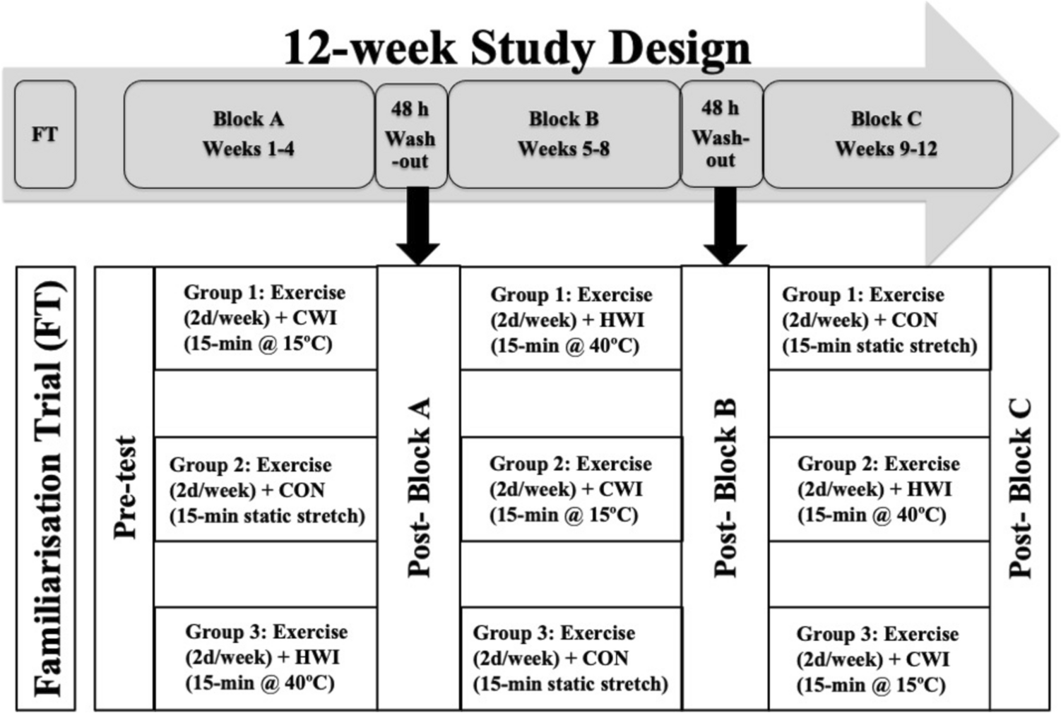
Fig. 1
Protocol schematic showing time course of sweat patch application and removal
Sweat sample handlingA total of 96 sweat samples (8 subjects × 4 sites × 3 time points) were collected. However, because of low sample volumes for some subjects (especially from the RT), fewer than 96 samples were analyzed for each analyte (IL-6, n = 92; EGF, n = 91; IL-1ra, n = 90; IL-1α, n = 90; IL-1ꞵ, n = 88; IL-8, n = 85; cortisol, n = 81; IL-10, n = 76, glucose, n = 56). The final sample sizes for each anatomical location and time point are shown in Figures S1-10. Once ready for analysis, sweat samples were removed from the freezer and allowed to thaw at room temperature. After thawing, sweat samples were vortexed and/or centrifuged depending on the requirements of the chosen analytical method.
ELISA assaysELISA was used for the detection of cortisol (Invitrogen, ThermoFisher Scientific) and glucose (Caymen Chemical Co.) concentrations. All ELISA assays were performed according to the manufacturer’s instructions. For cortisol, 12.5 μL of sweat sample (1:4 dilution), in duplicate, was assayed. Assay buffer (for detecting non-specific binding) cortisol conjugate, and cortisol antibody were added to the samples in each well and incubated for 1 h at room temperature. After manual washing of wells 4 times, tetramethylbenzidine (TMB) substrate was added and incubated for 30 min which produced a colorimetric signal. Stop solution was added for 10 min and the absorbance was read at 450 nm using a plate reader. Intra-assay coefficient of variation was 1.2%. For glucose, 15 μL of sweat sample, in duplicate, was added to assay buffer in each well. The reaction was initiated by adding enzyme mixture, and the plate was covered and incubated for 10 min at 37 °C. Absorbance was read at 520 nm using a plate reader. Intra-assay coefficient of variation was 2.0%. Both assays were read using Bioteck Cytation 3 Multi-Reader (Bioteck, Winooski, VT) with Gen5 software.
Multiplex assayEpidermal growth factor (EGF), interferon (IFN)-γ, interleukin (IL)-1β, IL-1α, IL-1 receptor agonist (ra), tumor necrosis factor (TNF)-α, IL-6, IL-8, and IL-10 were measured using magnetic bead-based immunoassay Multiplex. The Multiplex assay was performed according to the manufacturer’s instructions (Milli-Q, Millipore Sigma, Burlington, MA). Briefly, 25 μL of sweat sample, in duplicate, was assayed. The Multiplex assay involved beads of defined spectral properties conjugated to protein-specific capture antibodies and added with the samples (including standards of known protein concentration and control and test samples) into the wells of a microplate. The target proteins bound to the capture antibodies for an overnight incubation. After manual washing of the beads using a magnetic plate, protein-specific biotinylated detector antibodies were added and incubated with the beads for 1 h. Then, excess biotinylated detector antibodies were removed, and a streptavidin-conjugated fluorescent protein, R-phycoerythrin (SAV-RPE), was added and incubated for 30 min. SAV-RPE bound to the biotinylated detector antibodies, forming a four-member, solid-phase sandwich. After washing to remove unbound SAV-RPE, the beads were analyzed, and standard curves (EGF = 0.81% CV; IFN-γ = 0.83% CV; IL-1β = 0.41% CV; IL-1α = 0.23% CV; IL-1ra = 0.17% CV; TNF-α = 0.16% CV; IL-6 = 0.21% CV; IL-8 = 0.02% CV; IL-10 = 0.62% CV) along with individual well concentrations were determined using the Magpix multiplex system with xPonent software (Luminex, Austin, TX).
CalculationsLSR was calculated from sweat mass over patch surface area (11.9 cm2) and exercise duration. Excretion rates were calculated as the product of analyte concentration and LSR. Whole-body sweating rate was calculated from the change in pre- to post-exercise nude body mass, corrected for fluid intake (difference in drink bottle mass from pre- to post-exercise to the nearest 0.01 kg using a digital scale; model PG802, Mettler Toledo, Columbus OH) estimated respiratory water loss, and estimated metabolic mass loss (Cheuvront and Kenefick 2017).
Statistical analysisStatistical analyses were performed using Minitab 19 Statistical Software (Minitab, Inc., State College, PA). Shapiro–Wilk tests were used to assess the normality of the residuals. The Box–Cox transformation technique was used to identify the optimal data transformation where needed. Analysis of variance (ANOVA) was used to determine site, time, and site*time interaction effects on LSR, sweat analyte concentrations, and sweat analyte excretion rates. ANOVA was followed by Tukey’s HSD post hoc comparisons, where main effects were found. The significance level was set at P < 0.05. Data are presented as Least Squares (LS) mean ± SE. Due to the exploratory nature of this study, a post hoc analysis was performed for cortisol (primary analyte of interest), which determined an effect size of 0.29.
留言 (0)