General Reagents
Complete Freund’s adjuvant (CFA) is provided by Chondrex Company (Woodinville, USA). PE anti-mouse CD4, FITC anti-mouse CD8a, and PeCy7 anti-mouse OX40 antibody were supplied by BioLegend (San Diego, CA). InVivo MAb anti-mouse OX40 (clone: OX-86) and InVivoPlus rat IgG2a isotype control (clone: 2A3) were supplied by BioXCell (West Lebanon, USA). IRDye® 680RD NHS Ester Infrared Dye was supplied by Li-cor (Lincoln, USA). Fixable Viability Stain 780 was purchased from BD Pharmingen (San Diego, USA). Dimethyl sulfoxide (DMSO) Hybri-Max (TM) sterile-filtered, BioReagent was purchased from Sigma (St. Louis, MO, USA). AbC™ total antibody compensation bead kit was acquired from Thermo Fisher (Waltham, MA, USA). Paraformaldehyde, 4% and phosphate buffered solution (PBS) was supplied by Solarbio (Beijing, China). Fetal bovine serum (FBS) was purchased from Invitrogen (New York, America).
AIA Model
All animal studies were carried out in accordance with protocols approved by the Institutional Animal Care and Use Committee of Harbin Medical University’s Second Affiliated Hospital. For AIA model generation, 8–10 weeks male BALB/c mice (20–25 g) were given a single injection of 0.5 mL/kg CFA into the plantar subcutaneous tissue of RP, whereas equal amount of PBS was injected into RP of control mice [8]. As a control, equal amounts of PBS were injected subcutaneously into each animal’s left hind paw (LP). Animals were monitored every day after injection. Paw thickness was considered as the measurements of paw swelling related to arthritis. A caliper was used to gauge paw thickness.
Flow Cytometry
On day 11 after CFA injection, RP and spleen were collected. Spleens were cleaned and fat was removed before passing through a 40 µm filter with FACS buffer (PBS containing 2% FBS). The preceding approach was followed to create a single cell suspension [16]. The following antibodies were used in FACS staining: PE anti-mouse CD4, FITC anti-mouse CD8a, and PeCy7 anti-mouse OX40. Data was collected using a BD FACSCanto II Flow Cytometer, and version 10.7.1 of FlowJo was used for analysis.
Histological Analysis
For HE staining, RP was harvested on day 7, fixed with 4% paraformaldehyde for 24 h, and decalcified with 10% ETDA for 20 days, then embedded in paraffin and cut into sections of 5 μm thick. To determine infiltration of immune cells from both the AIA and control groups, every paw section was stained with hematoxylin and eosin (H&E; Solarbio, Beijing, China). Specimens were observed under a BX53 microscope (Olympus, Tokyo, Japan) and measured with Software (Image J).
Synthesis and Characterization of IRDye-680RD-OX40 mAb, T Cell Isolation, and Cell Uptake Study
The IRDye-680RD NHS ester was dissolved in DMSO to a final concentration of 5 mg/mL. OX40 mAb was diluted in sterile PBS to 1 mg/mL. IRDye-680RD NHS ester was mixed with OX40 mAb, at a molar ratio of OX40 mAb to IRDye-680RD of 1:10. The mixture was incubated at 4 °C overnight. For purification, a Vivaspin2 50 KDa cutoff MWCO spin filter (GE Healthcare, Piscataway, USA) was used. The NanoDrop 2000 UV–vis spectrophotometer (Thermo Fisher, Waltham, MA, USA) was used to determine the final concentration of IRDye-680RD-OX40 mAb.
BALB/c mice were anesthetized with 2% isoflurane. Spleens were collected immediately after euthanasia and smashed through a 40 μm cell strainer. T cells were isolated using the EasySep Mouse T cell Isolation Kit’s standard protocol (STEMCELL Technologies, Canada) and incubated in Iscove’s Modified Dulbecco’s Medium (IMDM, Thermo Fisher) with Cell Stimulation Cocktail (Thermo Fisher) at 37 °C. Both resting and activated T cells were collected, counted, and incubated with IRDye-680RD-OX40 mAb for 1 h at 37 °C 72 h later. All samples were analyzed using a BD flow cytometer after three washes with sterile PBS.
In Vivo and Ex Vivo NIRF Imaging
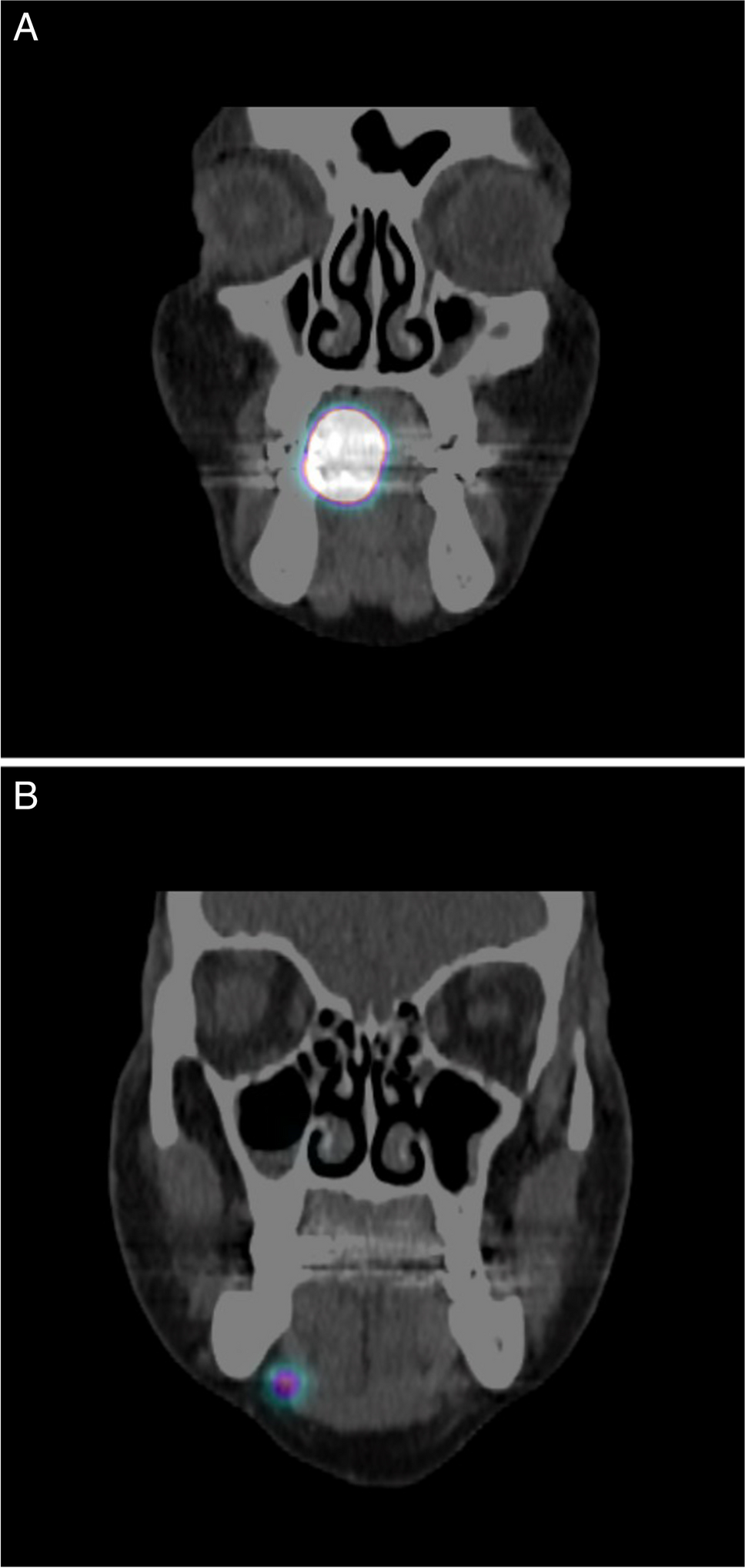
Bruker InVivo FX PRO in vivo fluorescence imaging was performed and analyzed using Bruker Molecular Imaging Software (IB5438150 Rev. B 12/12, Bruker, USA). On day 7, mice were anesthetized with 2% isoflurane and injected with 20 μg IRDye-680RD-OX40 mAb via tail vein. Images were taken on days 8, 9, 10, and 11 using the following settings: (f-stop 2.5, FOV 200 mm, 750 nm WA Emission Filter, 72 mm, 670 nm Excitation Filter, 25 mm). After the final scan, mice were euthanized, and organs (heart, liver, lung, spleen, kidney, right paw, left paw, femur, muscle, and intestine) were collected and imaged under the same conditions. Bruker Molecular Imaging Software was used to analyze all of the data. The quantification was normalized as p/sec/cm2/sr.
Statistical Analysis
The PRISM 9 platform was used for all data analysis (GraphPad). Data analysis methods included unpaired 2-tailed Student’s t-tests and one- or two-way analyses of variance (ANOVA). Statistics were considered significant for P values under 0.05.


留言 (0)