Data collection and analysis
Raw data from 70 OA samples were extracted from 7 GEO databases (https://www.ncbi.nlm.nih.gov/geo/) (Supplementary Table S1) and preprocessed with RMA algorithm normalization using the “affy” R package. Potential batch effects were removed using “removeBatchEffect” from the “limma” R package. A total differentially expressed genes (DEGs) were identified between N-CDH+ and N-CDH− OA synovial samples (with stricter screening criteria P < 0.05, |LogFC| > 0.5). Then the intersection of these DEGs and receptors involved in EMT signaling were analyzed with Venn diagrams and PPI network. The list of receptors involved in EMT was taken from a review of EMT molecular mechanisms (Lamouille et al. 2014) (Supplementary Table S2).
Tissue collection and cell isolation
Human synovium and cartilage samples were collected during total knee arthroplasty (advanced OA, n = 10), and amputation (non-OA, n = 10) (Supplementary Table S3). The cartilage was obtained from the tibial plateau and distal femur of the knee, and the harvested FLSs and chondrocytes were isolated and cultured by a previously described method (Cao et al. 2022).
Co-culture assay
A co-culture system was established using six-well Transwell plates (3428, Corning, NY, USA) in which 3 × 10⁵ chondrocytes in the same batch were cultured in the lower compartments and 3 × 10⁵ FLSs derived from N-CDH+ and N-CDH− OA groups (n = 3) were cultured in the upper compartments in DMEM with 10% FBS. Chondrocytes were cultured alone as a control, and all co-cultures were maintained for 7 days before evaluation. The co-cultures were conducted in biological triplicate for each assay.
Cell transfection and infection
The plasmids (pCDNA-GSK3β wild type and pCDNA-GSK3β S9A mutant) and lenti-virus (pCDNA-Ctrl, pCDNA-C-kit, plko-EGFP-shCtrl, and plko-EGFP-shC-kit) used in this experiment were purchased from Shanghai Tsingke Biotechnology Co., Ltd. All plasmids were constructed with a puromycin resistance. For cell transfection, FLSs from the same batch were transfected with various plasmids using Lipofectamine 3000 (L3000-015, Invitrogen, CA, USA) reagent according to the manufacturer’s protocol. For cell infection, FLSs from the same batch were infected with the lentivirus in the presence of polybrene (5 µg/ml) with centrifugation at 1,800 rpm for 40 min at 30 °C. Finally, the knockdown efficiency of C-kit was detected by immunoblotting with anti-C-kit antibody (18696-1-AP, Proteintech, Wuhan, China). The specific shRNA sequences are shown in Supplementary Table S4.
Animal experiments
All Sprague Dawley (SD) rats used in animal experiments were provided by the Department of Laboratory Animals of Central South University. The OA rat model was established by performing left knee joint surgery using a Hulth method (Ma et al. 2017). 12-week-old female rats were divided into operated sham group + sh Ctrl injection (Sham), Hulth’s model + sh Ctrl injection (Control), Hulth’s model + sh C-kit injection. Briefly, after administering anaesthesia, rat’s left knee was skin perpetrated and sterilized, and then an anteromedial incision was made to expose the articular cavity. After joint-space opening, anterior cruciate ligament transection (ACLT) was performed to cut off two-thirds of the medial meniscus. Sham surgery was performed by making a skin incision at the same location in the left knee. After this procedure, we injected 20 µl 1 × 10⁸ TU/ml lenti-virus packaged empty vector (Sham group and Control group, n = 6 for each group) or EGFP-shC-kit (Hulth + shC-kit group, n = 6) intra-articularly once a week until 14 days after surgery. After 3 and 6 weeks, the rats were randomly selected for sacrifice, and cartilage and synovial samples were collected from each group for cell culture, RT-qPCR, western blot, and histological evaluation (see the corresponding sections of Material and Methods below).
Histological evaluation
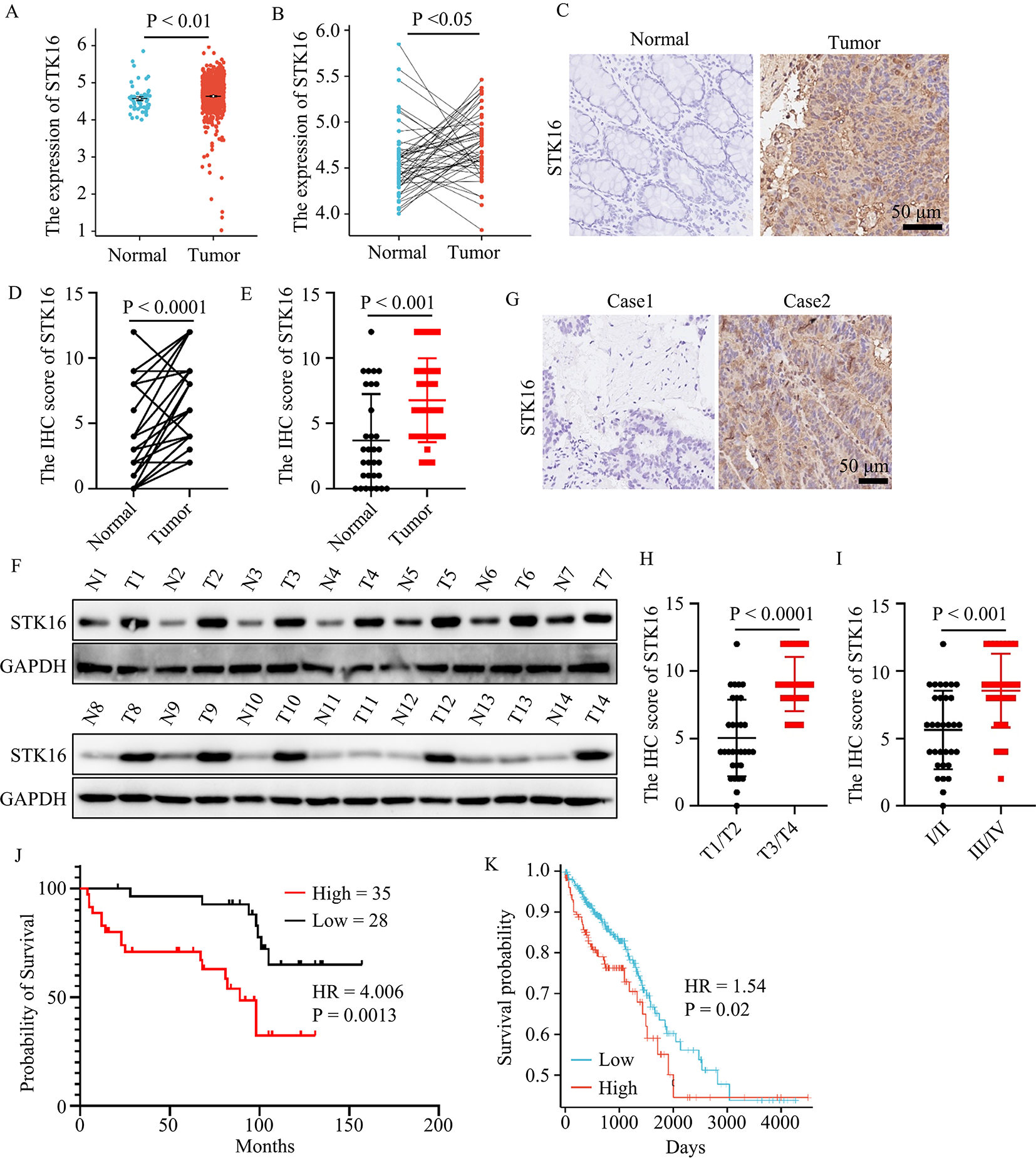
For the histological evaluation, the joints samples derived from rats were scanned by magnetic resonance imaging (7.0T MRI Biospoin GmbH, BRUKER, USA) and graded by their MOAKS score (Hunter et al. 2011). All the samples were then decalcified in 0.5 M EDTA ( G1105, Servicebio, Wuhan, China) for 4 weeks, and cut into sagittal Sect. (5 μm). Immunohistochemistry and immunofluorescence were performed using anti-N-cadherin (66219-1-lg, Proteintech, Wuhan, China), anti-MMP13 (#41,154, Signalway antibody, CA, USA), or anti-C-kit antibodies. Slices of rat knee joints were also stained with safranin O/fast green (G1371, Solarbio, Beijing, China). The Osteoarthritis Research Society International (OARSI) scoring system including cartilage, subchondral bone, osteophyte and synovitis was used to evaluate the OA cartilage pathology (Glasson et al. 2010).
Western blot
Total proteins obtained from cells and tissues were subjected to SDS-PAGE, then transferred and blocked in 5% skimmed milk for 30 min. The membranes were incubated overnight at 4 °C with primary antibodies against IL-6 (#53,904, Signalway antibody, CA, USA), IL-8 (27095-1-AP, Proteintech, Wuhan, China), MMP-13, N-cadherin, E-cadherin (20874-1-AP, Proteintech, Wuhan, China), Vimentin (10366-1-AP, Proteintech, Wuhan, China), Col I (bs-0578R, Bioss, Beijing, China), Col II (GB11021, Servicebio, Wuhan, China), Col X (DF13214, Affinity, NJ, USA), C-kit, p-AKT (66444-1-AP, Proteintech, Wuhan, China), AKT (51077-1-AP, Proteintech, Wuhan, China), p-STAT3 (#11,045, Signalway antibody, CA, USA), STAT3 (#41,464, Signalway antibody, CA, USA), p-Erk1/2 (#12,082, Signalway antibody, CA, USA), Erk1/2 (#29,162, Signalway antibody, CA, USA), p-GSK3β (67558-1-Ig, Proteintech, Wuhan, China), GSK3β (22104-1-AP, Proteintech, Wuhan, China), p-Snail (63,568, Abcam, Cambs, UK), Snail (13099-1-AP, Proteintech, Wuhan, China) and GAPDH (10494-1-AP, Proteintech, Wuhan, China). Afterwards, they were incubated with HRP-conjugated secondary antibody (SA00001-1 or SA00001-2, Proteintech, China) at room temperature for 1 h and developed in electrochemiluminescence (ECL) Western blot detection reagents (BL520A, Biosharp, Beijing, China). The band was analyzed by UVP Chem studio PLUS 815 (Analytik jena, Germany). There are three biological replicates for Western blot.
RT-qPCR
Total RNA was isolated from synovium or cells by TRIzol reagent (15,596,026, Thermofisher, MA, USA) and tested by NanoPhotometer® spectrophotometer (IMPLEN, CA, USA). Next, the RNA was converted to cDNA following the manufacturer’s instructions (R223-01, Vazyme, Nanjing, China). ChamQ Universal SYBR qPCR Master Mix (Q711-02, Vazyme, Nanjing, China) was used for qPCR testing according to the manufacturer’s protocol, and gene transcription levels (N-cadherin, E-cadherin, MMP1, MMP3, MMP13, IL-1β, IL-6, IL-8, IL-32, CDK1, TIMP1, TNFa, SAA1, S100A8, S100A9 and Vimentin) were normalized to those of GAPDH or β-actin. The primer design is shown in Supplementary Table S4.
ELISA
The collected culture medium of OA-FLSs was analyzed by using the Human IL-6/IL-8 ELISA Kit (JM-03204H2, Jingmei Biological Technology, Shenzhen, China), according to the instructions given in the manual.
Statistical analysis
All experiments were repeated at least three times and the data were presented mean with ± SD by individual dot plots unless otherwise noted. We performed all our statistical analysis with GraphPad Prism 8. Statistical significance was determined by t tests (two-tailed) for two groups. For the observational experiment in vivo, one-way ANOVA was used for comparisons across multiple groups, and Dunnett’s test was used for post-hoc multiple comparisons. The time dependent experiments were calculated with Two-Way ANOVA followed by Tukey’s multiple comparisons test. Bioinformatic analysis and visualization was carried out using R version 4.0.3 (https://www.r-project.org/). Specifically, DEGs between two subclusters were calculated with the R package limma”, and heatmaps were produced with the “pheatmap” R package. Finally, we analyzed the PPI network STRING version 11.5 (https://cn.string-db.org/) and visualized it with Cytoscape version 3.6.0.


留言 (0)