Preparation of AAV vectors
AAV vectors were prepared in a large scale and harvested from culture supernatant (conditioned media), as previously described [3]. In brief, a 293EB cell line expressing adenoviral E1a, adenoviral E1b, and Bcl-xL [15] was expanded in two 500 mL flasks (HYPERFlask, Corning, Corning, NY, USA) or a 1 L bioreactor (iCELLis Nano Bioreactor, Pall, Port Washington, NY, USA) for 5 days or 4 days, respectively, in Dulbecco’s Modified Eagle Medium (DMEM high glucose, FUJIFILM Wako, Chuo-ku, Osaka, Japan) with 10% fetal bovine serum (Thermo Fisher, Waltham, MA, USA). Transfection was then performed with polyethylenimine max, (Polysciences, Warrington, PA, USA) using pAAV-ZsGreen1 (TaKaRa Bio, Kusatsu, Shiga, Japan), pRC9 (serotype 9), and helper plasmids in DMEM including 2 mM L-Alanyl-L-glutamine Solution(100x) (Nacalai Tesque, Nakagyo-ku, Kyoto, Japan), 0.12% NaHCO3 (Nacalai Tesque), and 0.13% D-glucose (Nacalai Tesque) without serum. Five days post-transfection, culture supernatants were harvested and treated with 18.5 U/mL endonuclease (KANEKA CORPORATION, Minato-ku, Tokyo, Japan) with 5 mM MgCl2 (Nacalai Tesque) for 30 min at 37 °C. All cells were checked for mycoplasma contaminations resulting were reported negative.
Ultracentrifugation of AAV vectors with a zonal rotor
Five percent CsCl (FUJIFILM Wako) in HNE buffer (50 mM 4-(2-hydroxyethyl)-1-piperazineethanesulfonic acid (HEPES, FUJIFILM Wako), 0.15 M NaCl (Nacalai Tesque), and 25 mM ethylenediaminetetraacetic acid (EDTA, Nacalai Tesque), pH7.4) or HN buffer (50 mM HEPES and 0.15 M NaCl, pH7.4) were added to the culture supernatant, including AAV vectors (Table 1). A zonal rotor consists of a large cylindrical chamber subdivided into four sector-shaped compartments by vertical septa that radiate from the axial core to rotor wall. The entire chamber was used during centrifugation and loaded with a single density gradient, and each sector-shaped compartment served as a large centrifuge tube. The large chamber capacity of these rotors (1.7 L) eliminates the need for multiple runs and density gradients. A CsCl density gradient was generated in a zonal rotor (P32CT or P35ZT, Eppendorf Himac Technologies, Hitachinaka, Ibaraki, Japan) at 3000 rpm by loaded 200 mL HNE or HN buffer, AAV vector containing 5% CsCl, 300 mL of 25–27% CsCl in HNE or HN buffer, and 300 mL of 38–40% CsCl in HNE or HN buffer. AAV vectors were separated by ultracentrifugation (Himac CP 80NX, Eppendorf Himac Technologies) at 30,000–35,000 rpm for 4–10 h. After separation, 2 L of 42–45% CsCl buffer was slowly added to the inside of the zonal rotor at 3000 rpm, and each fraction within the zonal rotor was pushed out from the outside (Tables 2, 3). RI were measured in each fraction using an refractometer NAR-1T LIQUID or RX 5000i (Atago, Minato-ku, Tokyo, Japan). Each fraction sample was dialyzed with 20 kDa molecular weight cut-off dialysis cassettes (#66003 Thermo Fisher) in 0.5 mM MgCl2 (Nacalai Tesque) in water for ~2 h at 4 °C, and 0.5 mM MgCl2 in PBS (#27575–31, Nacalai Tesque) overnight at 4 °C.
Table 1 Conditions of zonal ultracentrifugation.Table 2 RI and volume of each fraction in experiment #Z1.Table 3 RI and volume of each fraction in experiments #Z3, #Z5, #Z7.Evaluation of genome copies, capsid proteins, and transduction efficiency of AAV vectors by quantitative polymerase chain reaction (qPCR), western blotting, and flow cytometry
After ultracentrifugation with a zonal rotor, AAV genome copies of each fraction were evaluated using the AAVpro Titration Kit (for Real Time PCR) Ver.2 (TaKaRa Bio, Kusatsu, Shiga, Japan) in a QuantStudio 3 Real-Time PCR System (Applied Biosystems, Waltham, MA, USA). The sample size was n = 3 minimally needed for statistically significance.
The AAV capsid proteins in each fraction were evaluated by western blot analysis. The samples were degraded with NuPAGE LDS sample buffer (Thermo Fisher) and NuPAGE Reducing Agent (Thermo Fisher), electrophoresed on a 4–15% (v/v) gradient polyacrylamide gel (Criterion TG Precast Gels, Bio-Rad, Hercules, CA, USA) with SDS running buffer (Nacalai Tesque), transferred to a PVDF membrane (Trans-Blot Turbo Midi PVDF Transfer Packs, Bio-Rad), and detected using anti-AAV VP1/VP2/VP3 mouse antibody (clone B1, Progen, Heidelberg, Germany) and Amersham ECL Mouse IgG, HRP-linked whole Ab (Cytiva, Marlborough, MA, USA)5.
Transduction efficiency was evaluated using ZsGreen1-positive percentages (%ZsGreen1) in the transduced 293EB cells. 293EB cells (1 × 10⁵ cells) were cultured in 24-well plates overnight and transduced with each sample fraction (300 μL per well) in serum-free DMEM containing 2 mM L-glutamine, 12.1% NaHCO3, and 12.9% D-glucose (300 μL per well). The next day, 600 μL of the same culture medium was added to each well, and %ZsGreen1 was evaluated by flow cytometry (FACSMelody, Becton Dickinson, Franklin Lakes, NJ, USA) at 3 days post-transduction and analyzed using FlowJo Version 7.1 (Becton Dickinson).
Evaluation of full-genome and empty AAV particles by AUC
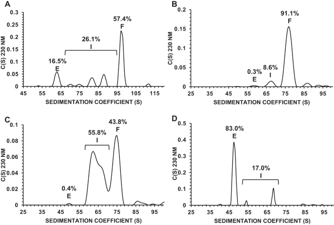
The purity of AAV vectors was analyzed using a Proteome Lab XL-I ultracentrifuge (Beckman Coulter, Indianapolis, IN, USA). Bulk AAV vector samples (400 μL) were applied to the Centerpiece on Cell Housing, and three cell houses with samples and one counterbalance were inserted into an AUC rotor. After equilibrating to 20 °C, samples were ultracentrifuged at 12,000 rpm at 20 °C, and the absorbance (260 nm) and interference were measured at 92 timepoints for 4–5 h. The percentages of full-genome, intermediate, and empty AAV particles were analyzed using SEDFIT (National Institutes of Health, Bethesda, MD, USA) [16] and visualized using GUSSI (UT Southwestern Medical Center, Dallas, TX, USA).
Evaluation of whole genome regions packaged in AAV vectors by droplet digital PCR (ddPCR)
Whole regions of the AAV vector genome were evaluated in each fraction of the samples from ultracentrifugation with a zonal rotor using ddPCR. 1.1 μL of sample less than 10,000 copies/µL (total 22 μL) was mixed with target primer/probe mixes (ddPCR Copy Number Assy, BioRad) (Table 4); 900 nM primers and 250 nM probe in droplets containing these materials were generated by an Automated Droplet Generator (BioRad) followed by PCR reactions in a C1000 Touch Thermal Cycler (BioRad). A QX200 droplet reader (Bio-Rad) using the QuantaSoft software package (Bio-Rad) was used to detect fluorescent signals in each droplet.
Table 4 A list of probe/primer mixture.Morphological analysis of AAV vectors by transmission electron microscopy (TEM)
Collodion membranes (Nissin EM, Shinjuku, Tokyo, Japan) were hydrophilized using an ion bombarder (Nisshin EM Co., type PIB-10), and 3 μL of AAV samples were placed to a hydrophilized grid for 1 min. After three times washing with 3 μL water, samples were stained with phosphotungstic acid (PTA) for 10 s. The samples loaded onto the membrane were analyzed using TEM (HT7800, Hitachi High-Tech, Minato-ku, Tokyo, Japan).
Polishing of AAV vectors by hydroxyapatite column
Chromatography was performed using an ÄKTA avant 25 system (Cytiva, Marlborough, MA, USA) with a SuperloopTM 150 mL at a flow rate of 1.0 mL/min. A column (4.6 × 35 mm, Sugiyama Shoji Co., Ltd. Kanagawa, Japan) packed with CHT Ceramic Hydroxyapatite Type I, 40 m (Bio-Rad Laboratories Inc., Hercules, CA, USA) was equilibrated with 10 mM HEPES and 150 mM sodium chloride, pH 7.2. The samples were loaded onto the column and eluted with 50 mM sodium phosphate buffer and 150 mM sodium chloride at pH 7.2. The resulting eluate was monitored for ultraviolet (UV) absorbance at 260 and 280 nm and conductivity. The collected fractions were evaluated by qPCR, using primers and probes targeting ZsGreen1.
Data analysis
All values are expressed as means ± SEM. Statistical analysis of the data was conducted using a one-way ANOVA. For all statistical analyses, significance was defined as P < 0.01.

留言 (0)