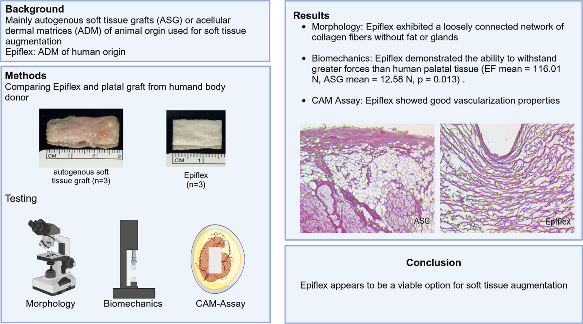
Posterior maxillary teeth loss due to periodontitis is a common clinical scenario. Compared to bone grafting, pterygoid maxillary implants provide an alternative solution in making the best use of residual bone of the maxillary-pterygoid complex. In contrast to the bone quality of the posterior maxilla, the implant engaged in the cortical layer of the pterygoid process makes it easy to obtain optimum primary stability [13,14,15]. Although this approach has been proposed for more than 30 years, its application is still limited. The anatomical complex of the maxillary-pterygoid complex constrains its clinical usage; moreover, clinicians are concerned about the entrance location of the implant for later maintenance.
To analyze the anatomical characteristics of this complex from a clinically feasible perspective, the present virtual study sought to provide information related to this complex from prosthetic-driven implant placement.
In previous anatomical measurements, the distance between the most concave point on the lateral surface of the pterygomaxillary junction and the greater palatine foramen was approximately 7 mm. With the horizontal and vertical absorption of alveolar bone, the distance between the aforementioned structures is reduced. The study set the entrance point of the virtual implant 10 mm away from the junction in the horizontal plane to mimic the entrance location of the second molar area and to proceed from the perspective of being more clinically feasible and maneuverable.
The inclination of pterygoid implants has been studied in previous radiographic and cadaver studies [16]. The range of 45–74° to the Frankfort plane in the anterior–posterior axis was proposed, and 45° was verified. Over a certain length with greater angles and more upright implants, the tip more easily reaches the pterygopalatine fossa, and the relationship with the maxillary artery will be closer. Undoubtedly, in clinical practice, implant placement is planned and driven by the individual anatomical character of each patient. However, compared to other angulations, tilted implants with 45° are easier to handle in clinical practice. Therefore, we set this criterion to virtually place the pterygoid maxillary implant.
In the assessment of buccal-palatal angulation, due to the fixed entrance and exit position of the virtually placed implant, it is easy to calculate the angulation in the software. A minor difference was detected between the present study and previous reports. In Rodriguez et al.’s study, an implant angulation of 81.09 ± 2.65° were found in the buccal-palatal axis [17]. To avoid the maxillary sinus cavity, every implant was placed according to individual anatomical structure in this study. Anatomically, the most concave cortical layer of the pterygoid process was in the palatal position of the alveolar ridge; when the implant entry moved more posteriorly, the angle of its buccal and palate reaching the most concave point became greater. In our study, the position of the implant entry point and the anterior–posterior inclination were determined, which explained the difference.
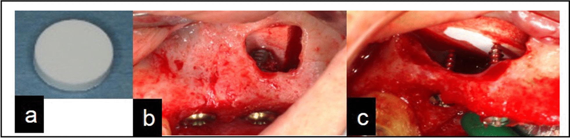
In the present study, the length of the implant beyond the pterygoid maxillary junction was the parameter of greatest concern. In some CBCT samples, the alveolar process of maxillary bone and pterygoid process of sphenoid bone are not completely fused; in this situation, the pterygoid maxillary junction is very easy to identify. Other samples showed a fused line of higher density compared to the surrounding structures. In contrast to large volume changes in the alveolar process and maxillary sinus cavity, the bone volume and density of the sphenoid pterygoid and palatine vertebrae are basically constant. These structures are precisely the most important structures that provide good stability of the implant during surgery.
Previous studies showed a mean bone column length (bone corridor) following the long axis of the implant of approximately 22 mm [1, 10, 18]. Some clinical studies established a minimal length of 13 mm for pterygoid implants. A deviation in implant length beyond the junction was detected in the study, and the range of 4–8 mm was the most common length.
We found that the limited anchorage of some implants in the pterygoid process is too short, usually when the sinus cavity is large. In addition, the maxillary tubercle is too small, and the posterior wall of the maxillary sinus is located behind the entrance of the pterygoid implant. When the implants had bone anchorage in the maxillary tuberosity, in category A, the mean virtually planned implant length was longer than the average length.
To establish a standardized entry point, the size of the maxillary tuberosity still affects the choice of implant length. Four patients could not complete the design because the vertical bone resorption of the alveolar ridge was too severe. After implantation at an oblique angle of 45°, the implant tip was at the root of the pterygoid process or close to the direction of the pterygopalatine fossa. This finding suggests that in clinical practice, when the alveolar bone is absorbed vertically instead of the lack of bone height caused by maxillary sinus pneumatization, oblique angle implantation is very risky. The slope should be increased. When the vertical defect of the alveolar ridge is not too serious, the inclination angle of the implant can be appropriately reduced, for example, between 45° and 70°.


留言 (0)