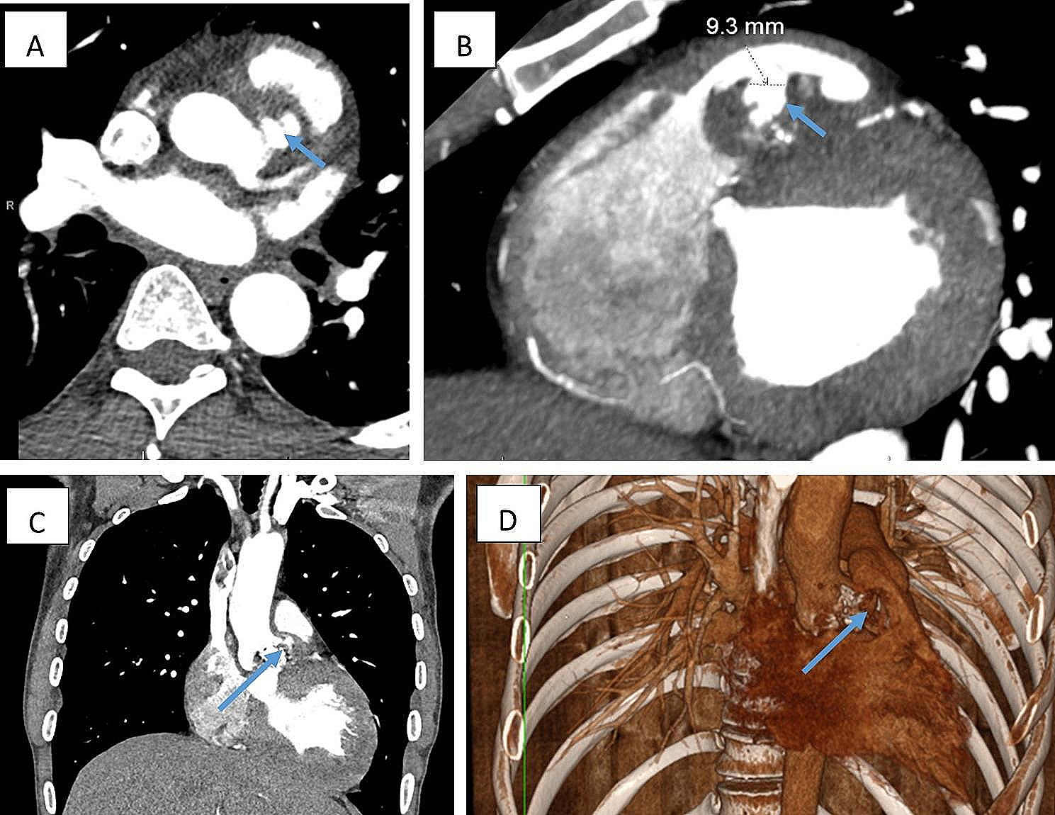
KD's management has different approaches depending on the patient's comorbidities, clinical presentation, anatomy, and the surgeon's expertise and personal preferences [1, 2, 23]. Some authors recommend surgical correction only in symptomatic patients since this population has been found to have an accelerated growth rate and a higher risk of rupture [1, 2]. It is also proposed to intervene in all patients with a KD diameter > 30 mm at the base of the KD or a distance from the apex of the KD to the opposite wall of the aorta of > 50 mm [1, 8].
After three years of follow-up, elective open and endovascular approaches demonstrated similar cumulative mortality; 16 and 18.2%, respectively. In this context, endovascular approaches may be an attractive alternative as they are associated with a lower incidence of postoperative pneumonia, shorter duration of invasive mechanical ventilation and shorter hospital-stay length. [2, 10, 24,25,26,27]. Evidence regarding hybrid approaches such as FET or thoracic endovascular aortic repair (TEVAR) plus supra-aortic debranching has been increasing recently, showing similar results in the short term compared to the endovascular management [18, 19, 23,24,25,26, 28, 29].
A systematic review of the literature compared the three approaches for KD. All are relatively safe and effective, showing no differences in outcomes, including 30-day mortality or stroke. The only significant difference was in the group of patients that underwent endovascular interventions, showing a higher number of re-interventions, mainly due to the appearance of endoleaks (11.6%) [30]. Each approach will be discussed in more detail in the following paragraphs, and their advantages and disadvantages are summarised in Table 3 [23, 27, 28, 30,31,32,33,34,35,36].
Table 3 Summary of the advantages and disadvantages of the different approaches and techniques for the correction of KD and KAGiven its low incidence, the evidence regarding the management of rKA needs more robust and extensive studies reporting long-term outcomes. There is even lower evidence about the most appropriate surgical approach for type-1 non-dissecting rKA. As a result, in the following section, we will describe the existing data about the different surgical approaches for all types of rKA.
Hybrid approach
Hybrid approaches are generally preferred as they are less morbid, avoid thoracotomy, and permit a posterior TEVAR if necessary. [28, 31]. Several techniques have been described. The most commonly performed are the FET and TEVAR + Supra-aortic debranching [30].
The TEVAR + supra-aortic debranching technique offers the possibility of performing a primary repair or alternative correction (embolization) of the AScA [31]. However, it does not solve the problem of the proximal landing zone and has been associated with complications such as branch occlusion, endoleaks, and stent-graft migration [31, 33]. One of its main advantages is that it can be performed in one or two stages, depending on the patient's surgical risk and general clinical condition [20].
The FET technique solves the issue of a limited proximal landing zone by performing a blood thigh proximal fixation and, therefore, decreases the risk of Type I and Type III endoleaks or stent-graft migration. This is achieved by approaching the arch through a sternotomy and suturing the stent-graft directly into the aortic arch. In addition, this approach allows direct access to the heart and the ligamentum arteriosum, which is highly useful when approaching patients with complex anatomies [28, 33]. It also decreases the potential risk of aortic rupture between procedures associated with the traditional elephant trunk, as it is a 1-staged procedure. [28, 32, 34]. The main disadvantage is that it requires longer cardiac arrest times, increasing the risk of cardiac, brain, and visceral ischemia [32, 33]. Likewise, it has been described that if the distal stent-graft is deployed below T7-T8 vertebral levels, the risk of spinal cord ischemia increases [32].
Ben Abdallah et al. [15] presented a 74-year-old male patient with a 81 mm contained rKA, who underwent a one-stage hybrid procedure. Initially, a TEVAR was performed associated with cervical debranching of the aLSA; then, a left carotid endarterectomy. This decision was related to the risk of haemorrhagic stroke due to cardiac arrest. Although the primary procedure was successful, the patient underwent two secondary procedures, one for vertebrobasilar insufficiency and the other for a right cervical lymphocele. After a four-month follow-up, the patient had fully recovered.
Another case report by Singh et al. [17] reported a 62‐year‐old African American male patient with acute onset tearing chest pain. An angio-CT identified a dissecting 40 mm Type I rKA with an intramural hematoma. The patient underwent a total arch replacement consisting of a right carotid-subclavian bypass, followed by open debranching of the bilateral carotid arteries and a zone II arch replacement with proximal intrathoracic ligation of aRSA and LSCA. Posteriorly, the patient underwent a FET. A retrograde TEVAR due to a type of IA endoleak was conducted in a second intervention. The patient presented a right lacunar infarct requiring a tracheostomy and percutaneous gastrostomy. After discharge, the patient persisted with mild residual left‐sided weakness.
Sica et al. [20] reported the case of a 74-year-old male patient with a saccular 70 mm Type II rKA associated with an anomalous course of the left brachiocephalic vein, which coursed posteriorly to the ascending aorta, and joined the right brachiocephalic vein to form the superior vena cava. Besides, a hematoma of the descending aorta was found, forming an extrapleural hematoma. The patient underwent a TEVAR, and a vascular plug was placed into the first segment of the aLSA. Twenty hours later, in a second surgical time, a left carotid-axillary bypass was performed. The length of hospital stay was 15 days. The patient presented transient tetraplegia. Nevertheless, at a 2-year follow-up, there were no residual or additional complications.
The evidence described above is related to our cases. We consider this approach promising for rKA as it allows an individualized approach for each patient. Furthermore, it is feasible in emergent situations as it is an effective, less invasive alternative compared to open approaches and does not require extensive planning or more specific resources like total endovascular approaches.
Open approaches
Historically, this has been the mainstay approach for rKA, as shown in a review by Cinà et al. [8]. Besides providing good access to the affected structures, it allows symptomatic relief and treatment of any related congenital or acquired cardiac abnormalities. It is particularly valuable for patients with complex anatomical variations as it allows complete visualization of the heart and great vessels, and in patients at high risk of stroke as it provides the possibility of securing brain perfusion. [23, 30, 36]. Nevertheless, it is highly invasive and morbid, requiring extensive surgical incision and multiple accesses strategies.
A study by Ikeno et al. [2] presented two patients with rKA who underwent emergent open surgery. The first patient included was a 77-year-old male with a dissecting rKA of 50.9 mm in diameter, who underwent an extensive aortic repair with selective anterograde cerebral perfusion. The authors reported no long-term postoperative complications in this patient. The second patient included was an 89-year-old male patient that presented a non-dissecting rKA with a diameter of 52 mm and underwent a total aortic arch replacement with selective antegrade cerebral perfusion. In-hospital mortality secondary to pneumonia was reported.
A case report by Kaki et al. [11] described a 73-year-old female patient with a 35 mm Type II rKA. An initial open proximal anastomosis was performed with a branched vascular graft. Then, the vascular graft was clamped, the aLSA was reconstructed, and reperfusion of the upper body was started through the rKA distal anastomosis. The patient presented pneumonia, which was treated adequately. The total hospital stay length was 70 days.
Endovascular approach
The evidence regarding the utility of this approach for rKA is null. This may be due to the requirement of extensive surgical planning since, for the complete treatment of the pathology, it is necessary to use chimney and periscope techniques that require surgeon-modified and custom-made thoracic endografts. Additionally, favourable anatomy with no evidence of anomalous supra-aortic branches or alterations of the descending aorta and a healthy landing zone with no calcifications, dilatations, or thrombi is required.
However, in centres with adequate experience and resources available, a total endovascular approach could be considered for select emergent cases, such as patients with contained rKA or those at high risk of surgical complications., However, its major role is in preventing rupture or dissection of KA. A proximal anterograde chimney technique with a retrograde approach to improving the proximal seal and a concomitant periscope technique should be performed to maintain the AScA patency [35, 36].
TEVAR with or without embolization of the AScA have also been described, but with complications such as arm claudication, predisposition to spinal ischemia, subclavian steal syndrome, and vertebrobasilar insufficiency [35]. Vascular plugs or coil embolization can prevent retrograde blood flow, and sometimes it has been related to decompression of the aneurysmal sac [31]. Additionally, given the sharply-curved distal arch (small radius curvature) when a KA is present, there is a predisposition to kinking, collapse of the thoracic endograft, or aortic wall injury by stent fractures [31, 35]; In this setting, some authors suggest the use of bare metal stents to decrease the risk of mechanical complications associated with stent fracture [36]. These techniques may be suitable in patients with a Type III KA.
It should be noted that endovascular techniques are contraindicated in the presence of compressive symptoms as the failure of clinical symptoms to improve has been described in the setting of complete vascular rings or if aneurysmal size reduction is not successful [23, 28, 30, 34].
In conclusion, technique selection must be individualised for each patient, depending on various factors such as the availability of resources, the patient's status (symptoms, hemodynamic condition, surgical risk, comorbidities), the different anatomical variations that may be present, and the surgeon's ability and preference. In our experience, FET can be a feasible procedure for patients with rKA requiring emergent surgical repair. However, other hybrid or open procedures could also be successfully performed. More robust studies are required to assess each approach's quality and long-term results to establish the most effective and safe for rKA.


留言 (0)