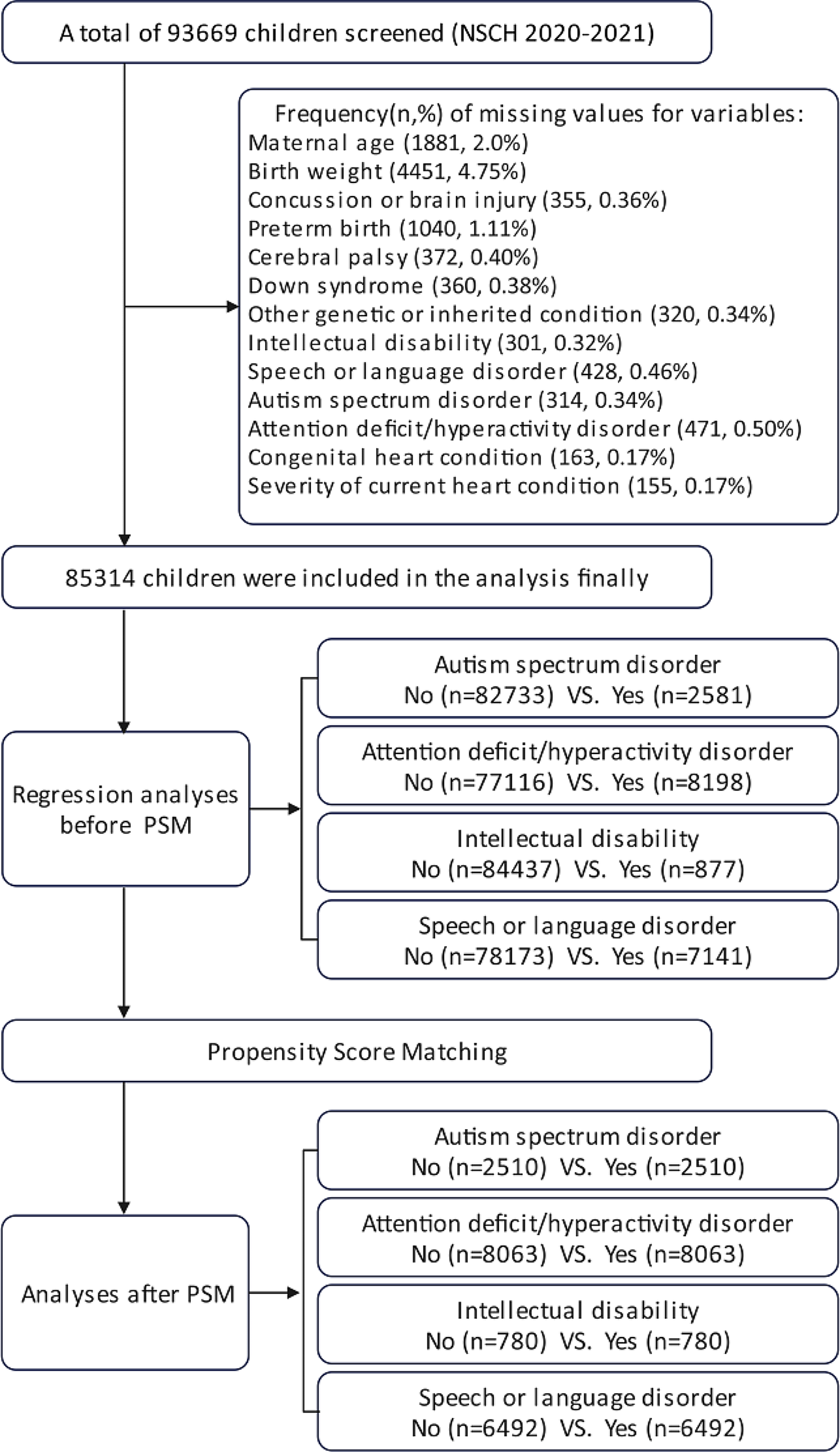
The present study, carried out on several groups of pediatric patients with CD, was aimed to analyze the age differences at diagnosis in relation to different specific autoantibody titers. We found that children diagnosed using the “biopsy-sparing protocol”, with the highest serum TGA-IgA titers (≥ 10xULN), were significantly younger than the other CD groups.
To date there is still little data focusing on the association between the age at presentation of children with CD and titers of serum specific autoantibodies. Our study confirms previous results showing that CD children tend to have high TGA-IgA titers, particularly at a younger age [11]. The dynamics of autoantibody development are still unclear, and it is unclear why the levels of autoantibody markers are so different in the pediatric CD population. In 2005, Salmi et al. [17] found lower titers of TGA-IgA autoantibodies in EMA-negative adult CD patients: the authors suggested a possible entrapment of autoantibodies in the intestinal mucosa which would prevent them from entering the blood, due to a higher tissue avidity of autoantibodies in a long-standing disease.
Interestingly, our data on higher titers of TGA-IgA autoantibodies in the youngest children appear to parallel the study by Marine et al. [3] showing for the first time that children, mainly the youngest, have a higher CD prevalence compared to adults. We are tempted to speculate that the difference in serum autoantibody titers might be due to a polarization of the immune system towards overproduction of TGA-IgA autoantibodies, in parallel with a concomitant reduced immunological tolerance. As is known, upon recognition of foreign antigenic peptides presented by MHC native T cells are activated and clonally expanded. Based on a recent study by Yao et al. [18], we believe that different types of lymphocytes could be activated by differential clonal expansion as well as autoantibody production. In the same way, cytokine expression could modulate the production of autoantibodies since they can selectively affect intraepithelial cytotoxic T cells [19].
However, this specific mechanism needs to be studied in detail before this hypothesis can be confirmed. The high prevalence of seronegative CD in adults [15, 20] supports our speculation.
The relationship between age and autoantibodies at onset in other autoimmune diseases has been studied. In fact, as reported in type 1 diabetes [21], children with an early age at onset of the disease have higher levels of autoantibodies and more autoimmune diseases. Interestingly, among children at high genetic risk for type 1 diabetes, those with late onset islet autoimmunity tend to develop diabetes in adolescence or early adulthood [21, 22]; in addition, there is widespread agreement that in various autoimmune diseases (e.g. systemic lupus erythematosus, type 1 diabetes mellitus) an early age at onset can act as a negative prognostic factor for the course of the disease [23]. Interestingly, data from studies of our group of CD patients suggest that age at diagnosis is a strong predictor for the occurrence of organ-specific autoantibodies and the development of additional autoimmune diseases [24]. We cannot determine whether the dynamics of other autoimmune diseases would differ between CD children who underwent EGD and those with the highest levels of serum autoantibodies diagnosed by a biopsy-sparing protocol. However, this could be an important research topic to fully understand autoantibody’ “autonomy” with respect to other autoimmune diseases.
Interestingly, we observed no statistical difference in patient age (p = 0.1665) between the low and the moderate titer groups: this may indicate that autoantibodies in a large proportion of subjects do not continuously increase over time. This could also bring further focus to clinical scenarios with positive low-to-moderate serum TGA-IgA levels, for which a clear and linear diagnostic work-up is not yet defined by guidelines [25].
Notably, potential CD children were significantly younger than those with biopsy-proven histological defects. This finding can be explained by the “progression of mucosal damage” [26]: indeed, due to the patchy damage to the small bowel mucosa in CD [27], it is conceivable that the damaged areas are not initially identified in some children. However, as the number of “damaged areas” increases over time, these patients can later be identified as “overt CD patients”, despite non-elevated autoantibody titers. Undoubtedly, age is one of the variables to consider when assessing the risk of progression from potential to overt CD [28]: since potential CD patients appear to be younger, a rigorous follow-up is remarkably important to intercept the potential transtition to overt CD [29].
It is worth noting that the median age was not statistically different between the biopsy-sparing and the potential CD groups, suggesting that, in autoimmunity, the peak of autoantibodies would resemble a sudden, uncontrolled storm manifesting unexpectedly and with an unpredictable autoantibody titer.


留言 (0)