
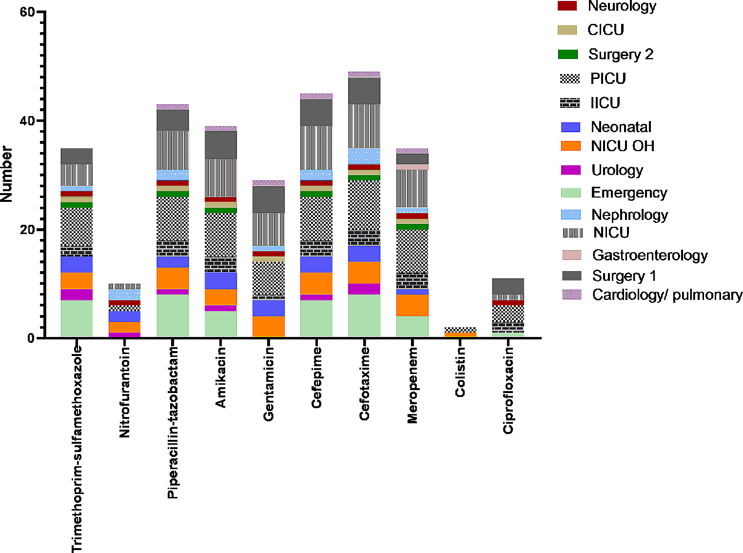
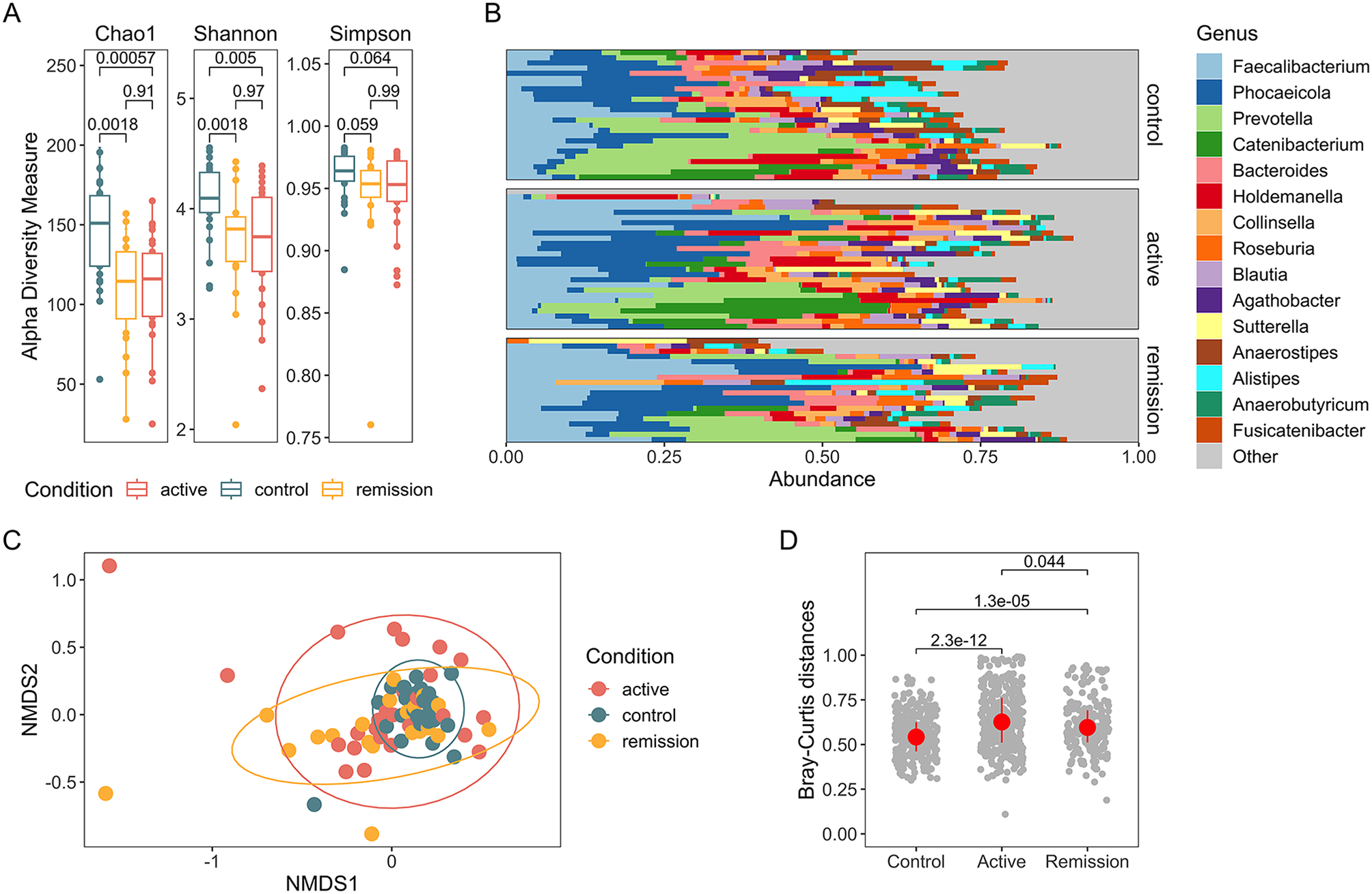
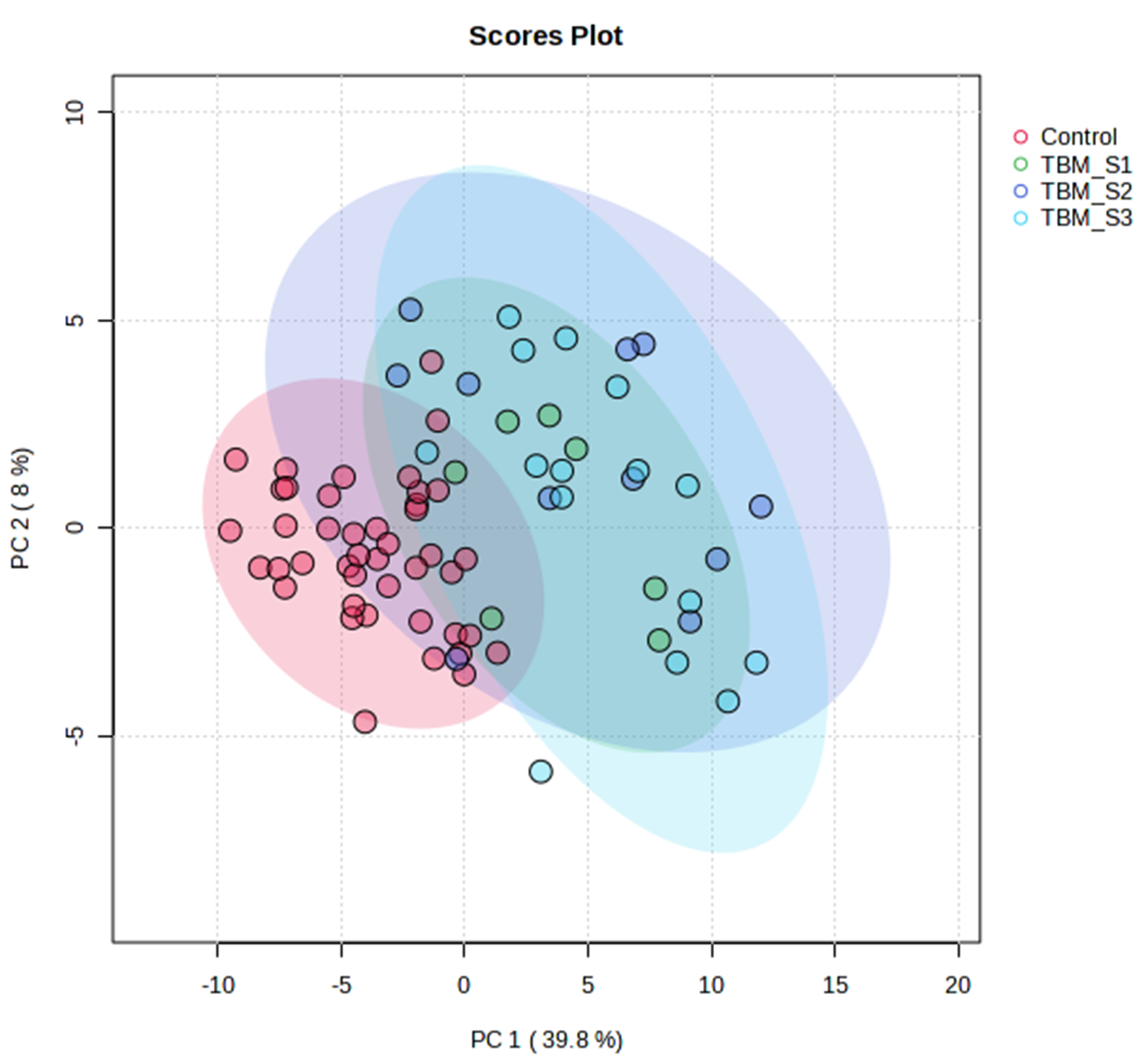
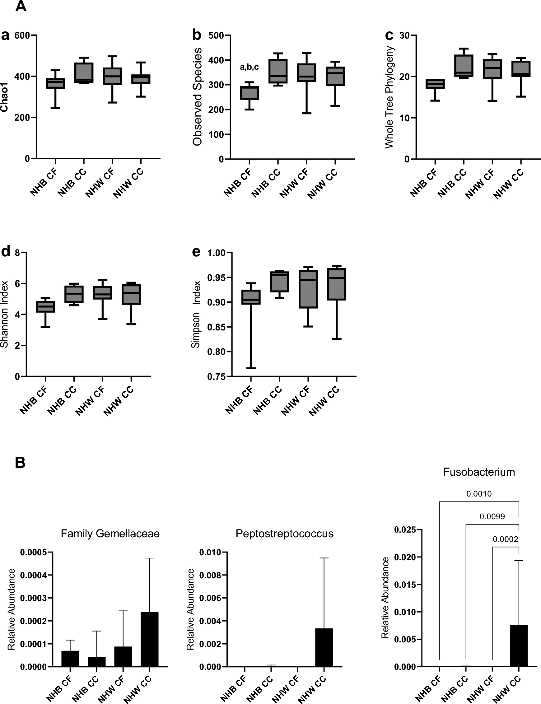
記住我

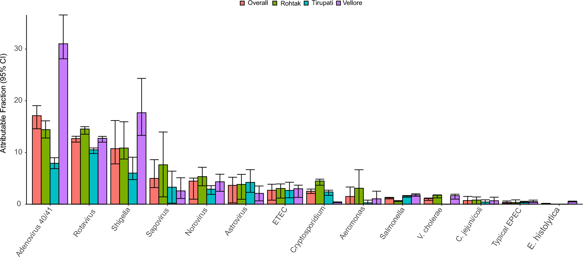

We administered CCl4 or corn oil to 8-week-old BALB/c mice. Figure 1A shows pathological images of the liver with Masson trichrome staining. Compared to the 8-week administration of CCl4 in the preliminary experiment, the 12-week administration showed more severe fibrosis in the Desmet-Scheuer scoring Stages 2–3 [27].
Fig. 1
Study design. A Pathological images of the liver with Masson trichrome staining. Control mice showed no fibrosis. After 8 weeks of CCl4 administration, slight bridging fibrosis was observed between the portal area (preliminary experiment). After 12 weeks of CCl4 administration, bridging fibrosis was more significant and showed the Desmet-Scheuer scoring Stages 2–3. Rifaximin treatment for 2 weeks did not improve the fibrosis. B Mice in the control group were treated with corn oil and Tween-80. Mice in the CCl4 group were treated with CCl4 and Tween-80. Mice in the rifaximin group were treated with CCl4 and rifaximin. C FITC-dextran gut permeability test. Average and 95% confidence interval are shown. CCl4, carbon tetrachloride. *p < 0.05, compared with the CCl4 group
Based on these results, we analyzed three groups (Fig. 1B): (1) a control group in which mice were treated with corn oil for 12 weeks followed by 2 weeks of Tween-80 (n = 9), (2) a CCl4 group in which mice were treated with CCl4 for 12 weeks followed by 2 weeks of Tween-80 (n = 13), and (3) a rifaximin group in which mice were treated with CCl4 for 12 weeks followed by 2 weeks of rifaximin (n = 13). Two weeks of rifaximin administration did not alter the pathological liver fibrosis (Fig. 1A).
To examine the effect of CCl4 and rifaximin on intestinal permeability, the FITC dextran intestinal permeability test was performed. The dextran concentration, estimated from the fluorescence of FITC in plasma, is shown in Fig. 1C. Plasma dextran concentrations were significantly higher in the CCl4 group compared to those in the control (control vs CCl4, average 0.11 vs 0.17 µg/mL, p = 0.03), while the rifaximin group did not show a significant decrease (average 0.15 µg/mL, p = 0.36).
Sufficient reads were derived even in the duodenum and small intestineWe then performed next-generation sequencing of the V3 and V4 regions of the 16S rRNA gene for the intestinal components and stool. To evaluate the number of reads, we checked the alpha rarefaction curve (Additional file 1: Figure S1). The median reads were 20,301 (interquartile rage [IQR], 14,849–26,607). The duodenum and small intestine had relatively low numbers of reads assigned to bacteria due to contamination with host DNA because of their low bacterial abundance, but the minimum number of reads was 1387, which was considered sufficient for the analysis of bacterial flora.
Rifaximin-induced changes in alpha and beta diversityFirst, we analyzed the alpha and beta diversity of samples with > 3000 reads where the operational taxonomic units (OTUs) had not reached a plateau from the diversity analyses (duodenum, n = 1; jejunum, n = 4; ileum, n = 1). This exclusion of the data did not significantly affect the below results.
The Shannon index is shown in Fig. 2A. In the jejunum, the Shannon index tended to decrease in the CCl4 group (control vs CCl4, median [IQR], 5.1 [4.6–5.6] vs 4.3 [3.9–5.1], p = 0.10), and tended to increase in the rifaximin group (4.5 [4.4–5.2], p = 0.16). In the ileum, the Shannon index was significantly higher in the control group than in the CCl4 group (control vs CCl4, 5.9 [5.5–6.7] vs 5.2 [4.4–5.6], p = 0.04). In the cecum and stool, the Shannon index was significantly decreased by rifaximin administration (CCl4 vs rifaximin, cecum, 7.6 [7.4–7.8] vs 7.2 [7.1–7.4], p = 0.003; stool, 7.1 [6.9–7.2] vs 6.6 [6.4–6.7], p < 0.001).
Fig. 2
Alpha and beta diversity of each intestinal site. A Shannon index of each intestinal site. B Principal coordinate analysis of each intestinal site. Green triangles are the control group. Red squares are the CCl4 group. Blue circles are the rifaximin group. CCl4, carbon tetrachloride; PC, Principal Coordinate. *p < 0.05, compared with the CCl4 group
A principal coordinate analysis plot is shown in Fig. 2B. Weighted Unifrac analysis showed that the rifaximin and CCl4 groups had different flora structures in the duodenum (p = 0.04), jejunum (p = 0.006), cecum (p = 0.02), and stool (p = 0.003), but not in the ileum (p = 0.78). Comparing the control and CCl4 groups, only the stool showed difference (p = 0.002).
The duodenum and small intestine had fewer bacterial families than the cecum and stoolThe relative abundance of bacterial families in each sample is shown in Fig. 3. Compared to the cecum and stool, the duodenum and small intestine contained fewer bacterial families and had much greater variation among samples even in the same group. Bacterial families with a median relative abundance greater than 1% in at least the duodenum, jejunum, or ileum were extracted; the results showed that only six bacterial families accounted for a median of 95.9% (IQR 88.1–98.5%) of the duodenal microbiota, 94.9% (IQR 89.3–97.6%) of the jejunal microbiota, and 86.9% (IQR 77.8–94.2%) of the ileal microbiota: S24_7, Lactobacillaceae, Lachnospiraceae, Streptococcaceae, Enterobacteriaceae, and Desulfovibrionaceae.
Fig. 3
Relative abundance of bacterial families in each sample. CCl4, carbon tetrachloride; c, class; f, family; o, order; p, phylum
On the contrary, more bacterial families were observed in the cecum and stool. S24_7, Bacteroidaceae, Rikenellaceae, Paraprevotellaceae, Deferribacteraceae, Lactobacillaceae, Lachnospiraceae, Ruminococcaceae, unassigned Clostridiales, Erysipelotrichaceae, Desulfovibrionaceae, Helicobacteraceae, and F16 had a relative median abundance greater than 1% in the cecum or stool. These bacterial families accounted for a median of 94.4% (IQR, 93.4–96.0%) of the cecal microbiota, and a median of 94.4% (IQR, 92.2–95.8%) of the stool microbiota.
Rifaximin significantly decreased Lactobacillaceae abundance in the duodenum and jejunumOwing to the large variation in relative abundance among samples in the duodenum and small intestine, we illustrated the box plot (median [IQR]) of bacterial relative abundance at the phylum and family levels, as shown in Fig. 4. All bacteria with statistical significance in the linear discriminant analysis effect size (LEfSe) analysis are shown in Additional file 2: Table S1–S10.
Fig. 4

The box plot of bacterial phylum and family with a high relative abundance. The bacterial phyla and families with a median relative abundance of > 1% in at least the duodenum, jejunum, or ileum are shown in A to F. The bacterial families with a median relative abundance of > 1% in at least the cecum or stool, are shown in G to J. A microbiota of the duodenum at the phylum level. B microbiota of the duodenum at the family level. C microbiota of the jejunum at the phylum level. D microbiota of the jejunum at the family level. E microbiota of the ileum at the phylum level. F microbiota of the ileum at the family level. G microbiota of the cecum at the phylum level. H microbiota of the cecum at the family level. I microbiota of the stool at the phylum level. J: microbiota of the stool at the family level. * p < 0.05, and LDA > 3.5, compared with the CCl4 group. CCl4, carbon tetrachloride; LDA, linear discriminant analysis
In the duodenum (Fig. 4A and B), although there was no statistically significant change after CCl4 administration, an abundance of Firmicutes, especially Lactobacillaceae, significantly decreased in the rifaximin group (CCl4 vs rifaximin, 79.4% [52.9–90.8%] vs 19.0% [15.4–75.4%], p = 0.006).
In the jejunum (Fig. 4C and D), the abundance of Lactobacillaceae significantly increased in the CCl4 group (control vs CCl4, 67.0% [32.2–78.6%] vs 87.8% [67.5–90.6%], p = 0.03) and significantly decreased in the rifaximin group (61.3% [33.7–77.4%], p = 0.03); similar results were observed in the duodenum. In contrast, S24_7 demonstrated a decreasing trend in the CCl4 group (control vs CCl4, 10.8% [0.2–64.2%] vs 1.7% [0.2–7.4%], p = 0.18) and increasing trend in the rifaximin group (3.4% [0.1–20.6%], p = 0.50).
In the ileum (Fig. 4E and F), Lactobacillaceae abundance tended to increase in the CCl4 group (control vs CCl4, 29.5% [24.5–71.0%] vs 52.0% [34.3–70.2%], p = 0.43) while the rifaximin group showed little change (50.6% [29.3–64.5%], p = 0.69). Although the relative abundance was low, Clostridiaceae (CCl4 vs rifaximin, 0.03% [0–3.5%] vs 0% [0–0%], p = 0.008) and F16 (CCl4 vs rifaximin, 1.4% [0.1–4.3%] vs 0% [0–0.8%], p = 0.05) were significantly less in the rifaximin group compared to the CCl4 group (Additional file 1: Table S6).
The effect of rifaximin on the cecal and stool microbiota was minimalIn the cecum (Fig. 4G and H), there were only changes within the same phylum (e.g., Bacteroidaceae and Rikenellaceae in the phylum Bacteroidetes) or in bacteria with low relative abundance. Ruminococcaceae abundance was significantly increased in the rifaximin group (CCl4 vs rifaximin, 9.4% [5.8–11.8%] vs 12.6% [11.0–14.5%], p = 0.03), and F16 was significantly decreased in the rifaximin group as in the ileum (CCl4 vs rifaximin, 1.3% [0.8–2.2%] vs 0.3% [0.02–0.9%], p = 0.009).
In the stool (Fig. 4I and J), the CCl4 group showed some changes in bacterial proportions; however, the changes were only within the same phylum (e.g., S24-7, Paraprevotellaceae, Rikenellaceae, and Bacteroidaceae) and bacteria with low relative abundance (Additional file 1: Table S9). Rifaximin did not alter the microbiota remarkably but an increased abundance of the genus Oscillospira in Ruminococcaceae (CCl4 vs rifaximin, 5.1% [3.1–7.4%] vs 10.6% [8.2–12.1%], p = 0.004) and decreased F16 (CCl4 vs rifaximin, 4.0% [2.5–6.4%] vs 0.7% [0.2–2.6%], p = 0.007) as in the cecum.
The relative abundance of bacterial families in the duodenum and small intestines poorly correlated with that in the stoolTo assess whether the microbiota of the duodenum and small intestine can be predicted from the microbiota of the stool, we correlated the proportion of bacteria between sites of the intestinal tract (Fig. 5A–F).
Fig. 5
Correlation between bacterial families. Pearson’s correlation coefficient between sites of the intestinal tract is shown in each bacterial family. A Lactobacillaceae, B S24_7, C Lachnospiraceae, D Streptococcaceae, E Enterobacteriaceae, F Desulfovibrionaceae. G The correlation between Lactobacillaceae and other bacterial families in the duodenum, jejunum, and ileum. *p < 0.05, **p < 0.001
The relative abundance of most bacterial families correlated between the duodenum and jejunum (Lactobacillaceae, r = 0.64, p < 0.001; S24_7, r = 0.65, p < 0.001; Lachnospiraceae, r = 0.68, p < 0.001; Streptococcaceae, r = 0.93, p < 0.001; Enterobacteriaceae, r = 0.45, p = 0.006), and between the jejunum and ileum (Lactobacillaceae, r = 0.59, p < 0.001; S24_7, r = 0.58, p < 0.001; Lachnospiraceae, r = 0.59, p < 0.001; Streptococcaceae, r = 0.86, p < 0.001; Desulfovibrionaceae, r = 0.78, p < 0.001). However, the relative abundance of duodenal and small intestinal bacterial families did not significantly correlate with that of the stool.
S24_7 was inversely correlated with Lactobacillaceae in the duodenum, jejunum and ileum,Correlation analysis was performed between Lactobacillaceae, the most significantly altered bacterial family, and other bacterial families (Fig. 5-G). S24_7 had the strongest inverse correlation with Lactobacillaceae in the duodenum (r = − 0.61, p < 0.001), jejunum (r = − 0.72, p < 0.001), and ileum (r = − 0.64, p < 0.001). Lachnospiraceae had a weak inverse correlation with Lactobacillaceae in the duodenum (r =− 0.49, p = 0.003) and jejnum (r = − 0.43, p = 0.01). Desulfovibrionaceae (r = − 0.44, p = 0.008), and Enterobacteriaceae (r = − 0.38, p = 0.03) had a weak inverse correlation with Lactobacillaceae only in the duodenum. Streptococcaceae did not have significant inverse correlation in any of the sites.
The amount of Lactobacillaceae increased in the CCl 4 group and decreased in the rifaximin groupTo evaluate the bacterial amount in the intestinal tract, quantitative polymerase chain reaction for the 16S rRNA gene was performed on 15 jejunal (control, n = 4; CCl4, n = 5; rifaximin, n = 6) and 20 cecal (control, n = 4; CCl4, n = 7; rifaximin, n = 9) samples.
Although the sample size was small, there was an overall increasing trend in the CCl4 group and a decreasing trend in the rifaximin group (Fig. 6). The most significant difference was in the amount of Lactobacillaceae in the jejunum (control vs CCl4, 7.0 [1.6–13.0] vs 16.9 [16.6–20.9] × 106/g, p = 0.06; CCl4 vs rifaximin, 16.9 [16.6–20.9] vs 3.2 [0.6–6.5] × 106/g, p = 0.13).
Fig. 6
Quantitative 16S rRNA PCR. The number of 16S rRNA gene copies per intestinal contents (g) is shown. Fifteen jejunal (control, n = 4; CCl4, n = 5; rifaximin, n = 6) and 20 cecal (control, n = 4; CCl4, n = 7; rifaximin, n = 9) samples were analyzed. A total bacteria in the jejunum. B Lactobacillaceae in the jejunum. C total bacteria in the cecum. D Lactobacillaceae in the cecum. CCl4, carbon tetrachloride
留言 (0)