
記住我

This observational study was conducted between April 2021 and January 2022. The investigation was performed in compliance with the Declaration of Helsinki and the Guidelines for Good Clinical Practice (Prot. n. 0,000,208, 07/02/2022). All participants provided written informed consent to undergo surgery and follow-up. The inclusion criteria were Caucasian patients of either sex, with early T1 stage of oral cancers, precancerous lesions, or revision of previous surgical cancer treatment, that were treated with the dermal regeneration template either during the primary treatment or on secondary revision surgery. None of the patients in this study underwent radiotherapy or chemotherapy after DS placed surgery. The only patient with a DS placed for neo-fornix creation had previously undergone RT. All patients were treated at the maxillofacial surgery department of the “Policlinico Umberto I” hospital in Rome for ten months. The following data were gathered for each patient: demographic data, lesion site (palate, tongue, alveolar crest, trigonous and cheek), histology, staging, surgery procedure, comorbidities, healing time and complications.
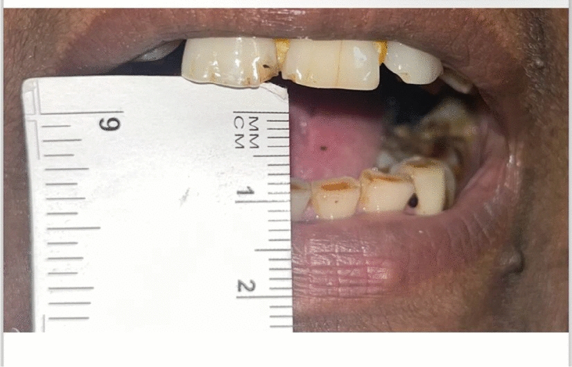
Surgical ProtocolAll patients with malignant lesions in need of primary surgery whose tumor did not exceed an early T1 cancerous stage (AJCC 8th Edition 2017) were previously discussed at the tumor board, where the indication for transoral resection was given; all patients underwent a transoral resection of the neoformation in wide free margins, performing intraoperative frozen sections of the margins. All tongue cancers underwent partial glossectomy in order to obtain a full-thickness excision of the lesion; cheek cancers underwent total thickness excision to the muscular plane, which was preserved with the intraoperative frozen section of the deep plane of resection resulting negative for neoplasm infiltration; retromolar trigon and alveolar crest lesions were treated via complete thickness excision including the periosteum. As for palatal cancers, one underwent the same treatment as the retromolar trigon lesions, while the other underwent a Brown I maxillectomy due to signs of bone infiltration; the same patient had previously undergone a Brown IIB right maxillectomy to treat a right superior alveolar crest G2 squamous cell carcinoma. Subsequent reconstruction of the gap using a 5 × 5 cm dermal substitute membrane from Integra® (Fig. 1) followed the demolitive surgery for all patients above. Patients who had previously undergone oral cancer treatment and came back to our department to solve the scar retraction from previous surgery were also included in this study; the neofornix was created using a 5 × 5 cm dermal substitute membrane from Integra® in order not to form new adherences and to make the fornix heal properly. The membrane shape and size were customized each time according to the gap using a template to adequately fit without excess or a tent-like effect. The layer was then accurately sutured in place using a Vicryl 3.0 suture. A compressive medication consisting of paraffin gauze anchored to the surrounding mucosa with a Silk 2.0 was used not to elevate the membrane from the gap. To ensure nutritional support, avoid contact between food and the membrane, and keep the site as clean as possible, enteral nutrition, managed by placing a nasogastric tube (NGT) at the end of the operative session, was set up in all but two cases, due to the impossibility for the patients to tolerate the NGT. In this case, a liquid diet was prescribed. Seriated medications were planned twice a week to check on the site and, when necessary, replace the gauze. The silicone layer of the membrane was removed between the 13th and 21st days post-surgery. In order to better evaluate the results, the patients were divided into groups based on the lesion location, and the outcomes were analyzed accordingly to highlight differences that might be related to the site, its mobility, and the ease of keeping the compressive medication in position.
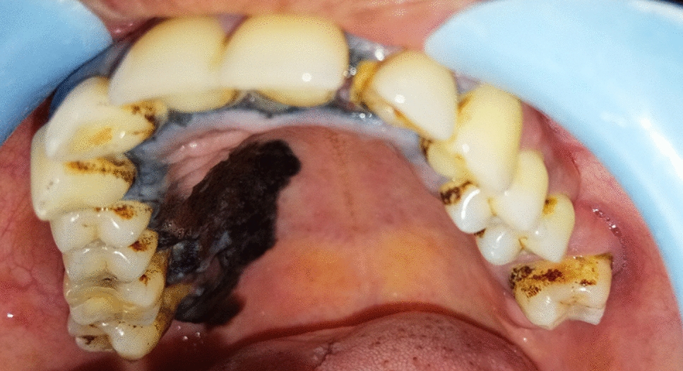
Fig. 1
Palatal lesion. A Preoperative image; B Intraoperative image; C Integra® placement; D 9 months follow-up image
Integra® Bilayer Wound MatrixIntegra® Bilayer Matrix Wound Dressing (Integra LifeSciences, Princeton, NJ, USA) is a manufactured acellular dermal regeneration template made of a bilaminate sheet of cross-linked bovine tendon collagen and shark glycosaminoglycans (chondroitin-6-sulfate) with a silicone sheet cover [7]. Integra® acts in wound healing by stimulating natural recovery processes promoting localized inflammation, infiltration of neutrophils, macrophages, fibroblasts, and keratinocytes and neovascularization of the scaffold [6]. Integra® was initially created out of necessity to provide temporary coverage for patients with extensive burns. Those who benefited most from this technology were patients with severe full-thickness burns in whom donor sites were severely limited or nonexistent. However, since its first applications, the use of this biosynthetic substitute has widely increased. In particular, Integra® bilayer wound matrix is very effective in intra-oral reconstructions [1]. Its inner porous layer works as a scaffold guiding cellular migration and capillary invasion; the migration of blood vessels and other cells into the matrix allows the formation of a new layer of the dermis, while the outer silicone layer has a role in covering the wound surface, controlling moisture loss, and increasing tear strength of the matrix. Studies showed that the first layer is usually replaced within 14 to 21 days while the second, non-absorbable, is removed to allow epithelial growth [7]. When removing the silicone layer, remove the sutures and staples holding it in place, then with forceps and a spatula or other blunt instrument, lift the layer starting from the edge and gently peel back. After the removal of the silicone layer, eventually, a graft can also be added to the neodermis.
留言 (0)