
記住我

In Indonesia, the first case of COVID-19 was diagnosed in March 2020; since then, more than 2 million people have contracted COVID-19, with more than 21,000 testing positive as of the end of June 2021.1 As the number of patients admitted to hospitals increases, particularly ICU admissions, a greater number of individuals are vulnerable to pressure injury (PI) as a result of inactivity, immobility, and the use of artificial airways.2–5
Pressure injury is a type of local trauma caused by constant pressure on the skin, most commonly over bony prominences. This pressure is high enough to interfere with blood flow to the capillaries, reducing oxygen supply to the tissues. This results in ischemia and necrosis of the afflicted tissue.6 The sacrum, heel, sciatic tuberosity, greater trochanter, and lateral malleolus are frequently impacted.7 Advanced age, immobility, poor nutrition, excessive moisture, incontinence, altered state of consciousness, poor perfusion, specific skin diseases, and concomitant disorders (eg, respiratory failure, anemia, diabetes, and septicemia) are all risk factors.8 Patients who develop PIs tend to be older and less mobile and have longer hospital stays than patients who do not.9 Gedamu et al10 reported that patients who were hospitalized for 7 to 20 days had a higher rate of PI than those who were hospitalized for fewer than 7 days. Slow-healing wounds may diminish patients’ quality of life.11
A “cytokine storm” may arise as COVID-19 infection develops. This unregulated immune response causes immune cells, lymphocytes, and macrophages to infiltrate and produce a substantial amount of proinflammatory cytokines.12 The cytokines interleukin 6 and tumor necrosis factor α13 are both involved in PI development14,15 and are essential components of the cytokine storm. The rise in d-dimer values in COVID-19 indicates that interleukin 6 and tumor necrosis factor α are related to a mix of systemic inflammatory processes and hypercoagulability situations.16
Because of the urgency of the issue and the increased risk of PI in patients with COVID-19, this study was conducted at an infectious disease hospital to describe the clinical characteristics of patients with COVID-19 and PI.
METHODS EthicsOn June 22, 2021, the Clinical Research Ethics Committee accepted this study with ethical approval number 157/KEP/2021. Because this was a retrospective research study based on anonymous and deidentified data, no consent was sought. The data were extracted from hospital medical records in August 2021; the raw data from hospital medical records were only accessible to the primary researcher and not shared with others.
Study Design and SettingThis was a descriptive and retrospective study undertaken at a single site. The study was conducted at Airlangga University Hospital, a referral hospital for COVID-19. It is located in Surabaya, Indonesia's second largest city. With 307 beds, Airlangga University Hospital is the largest university hospital in East Java. Between March 2020 and June 2021, samples were taken from each patient at the hospital who had been diagnosed with PI and COVID-19.
ParticipantsParticipants were chosen from medical records by their polymerase chain reaction-confirmed result for COVID-19 after being admitted to the COVID-19 referral hospital. The study included patients who were at least 18 years old and had a diagnosis of PI by the attending plastic surgeon in their medical records. Only PIs induced by supine position were considered, such as those on the sacrum, occipital, temporal, heels (calcaneus), gluteus, scapula, and trochanter according to the European Pressure Ulcer Advisory Panel, National Pressure Ulcer Advisory Panel, and Pan Pacific PI Alliance.17 Medical device-related PIs were excluded.
Variables and Data SourcesThe secondary data drawn from patient medical records included sex; age; body mass index (BMI), categorized as follows: (1) underweight (BMI <18.5 kg/m2), (2) healthy weight (BMI 18.5–22.9 kg/m2), (3) overweight (BMI 23–24.9 kg/m2), (4) obese I (BMI 25–29.9 kg/m2), and (5) obese II (BMI >30 kg/m2), according to the WHO recommendations for Asian populations;18 symptoms related to COVID-19 on admission; coexisting disorder (hypertension, diabetes mellitus, cerebrovascular disease, coronary artery disease); type of oxygen therapy used when the patient was consulted for PI (room air, nasal cannula, simple oxygen mask, mechanical ventilation); laboratory results of leukocyte, total neutrophil, total lymphocyte, neutrophil-to-lymphocyte ratio (NLR), platelets, albumin, and d-dimer values dated less than or equal to 3 days prior to the PI consultation; and PI location and stage. Pressure injury stages were classified in accordance with guidelines from the European Pressure Ulcer Advisory Panel, National Pressure Ulcer Advisory Panel, and Pan Pacific PI Alliance.17 Length of stay was the number of days a patient spent in the hospital before being discharged.
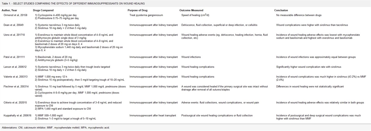
Data AnalysisInvestigators conducted a descriptive analysis of the data, reporting medians, percentages, and interquartile ranges (Table).
Table - CHARACTERISTICS OF PATIENTS WITH COVID–19 AND PRESSURE INJURY Characteristic Total (N = 12) Stage 2 (n = 5) Stage 3 (n = 7) Demographic characteristics Median age, y (interquartile range) 60 (51–71) 65 (57.5–66) 63,5 (52.5–68) Men, n (%) 8 (66.7) 4 (80.0) 4 (57.1) Women, n (%) 4 (33.3) 1 (20.0) 3 (42.9) Body mass index, n (%) Underweight (<18.5 kg/m2) 1 (8.3) 0 1 (14.3) Healthy (18.5–22.9 kg/m2) 1 (8.3) 0 1 (14.3) Overweight (23–24.9 kg/m2) 2 (16.7) 2 (40.0) 0 Obese I (25–29.9 kg/m2) 6 (50.0) 3 (60.0) 3 (42.9) Obese II (≥30 kg/m2) 2 (16.7) 0 2 (28.8) Symptoms, n (%) Cough 7 (58.3) 3 (60.0) 4 (57.1) Fever 6 (50.0) 4 (80.0) 2 (28.6) Shortness of breath 6 (50.0) 1 (20.0) 5 (71.4) Fatigue 5 (41.7) 3 (60.0) 2 (28.6) Nausea or vomiting 4 (33.3) 3 (60.0) 1 (14.3) Diarrhea 1 (8.3) 1 (20.0) 0 Loss of taste or smell 1 (8.3) 0 1 (14.3) Sore throat 1 (8.3) 1 (20.0) 0 Nasal congestion 1 (8.3) 1 (20.0) 0 Ulcer location, n (%) Sacrum 8 (66.7) 2 (40.0) 6 (85.7) Gluteus 3 (25.0) 3 (60.0) 0 Temporal 1 (8.3) 1 (20.0) 0 Calcaneus 1 (8.3) 0 1 (14.3) Scapula 1 (8.3) 0 1 (14.3) Hip 1 (8.3) 0 1 (14.3) Comorbid condition, n (%) Hypertension 6 (50.0) 4 (80.0) 2 (28.6) Diabetes 5 (41.7) 2 (40.0) 3 (42.9) Cerebrovascular disease 5 (41.7) 1 (20.0) 4 (57.1) Coronary artery disease 3 (25.0) 1 (20.0) 2 (28.6) Median laboratory values (interquartile range) Leukocytes, per μL 14,265 (12,547.5–22,992.5) 14,830 (12,480–24,020) 13,700 (12,830–19,885) Differential count, per μL Total neutrophils 12,288.7 (10,830–21,012.9) 11,967.8 (10,886.9–21,401.8) 12,356.9 (11,439.9–18,479.5) Total lymphocytes 1,023.9 (782.3–1,442.7) 1,764.8 (1,335.4–1,969.4) 838.1 (698.9–1,023.9) Neutrophil/lymphocyte ratio 20.4 (10.6–24) 10.9 (7.7–36.6) 21.1 (15.5- 22.6) Hemoglobin (g/dL) 10.7 (10–11.8) 11.3 (10.6–11.4) 10.2 (8.8–11.3) Platelet count, per μL 260,500 (187,000–443,250) 241,000 (190,000–399,000) 280,000 (203,500–447,500) Albumin, g/dL 3.08 (2.9–3.1) 3 (2.8–3.1) 3.1 (3–3.1) d-Dimer, ng/mL 3,700 (1,500–8,400) 1,100 (600–1,700) 7,900 (5,200–11,200) Oxygen therapy, n (%) Room air 1 (8.3) 0 1 (14.3) Nasal cannula 1 (8.3) 1 (20.0) 0 Simple oxygen mask 2 (16.7) 1 (20.0) 1 (14.3) Mechanical ventilation 8 (66.7) 3 (60.0) 5 (71.4) Vasopressor support, n (%) 7 (58.3) 2 (40.0) 5 (71.4) Length of stay, d (interquartile range) 22 (9.8–40.3) 29 (26–41) 13 (8–29) Died during hospital stay, n (%) 6 (50.0) 2 (40.0) 4 (57.1)The authors collected data from patients with confirmed COVID-19 who were treated at their institution during the start of the pandemic, from March 2020 to June 2021. During that period, 1,070 patients were hospitalized for COVID-19 with varying severity; of those, 12 patients were also diagnosed with a PI. Two of the 12 patients had already experienced a PI before being admitted to the hospital. Eight of these patients (66.7%) were men. Five of the 12 patients (41.7%) had a stage 2 PI, and 7 (58.3%) had a stage 3 PI; none of the patients in this study had stage 1, stage 4, or unstageable PI or suspected deep tissue injury. Overall, these patients had a median age of 60 (range, 51–71) years. When looking at median age by PI stage, the median age of patients with stage 2 PI was only slightly older than the median age of those with stage 3 PI (65.0 vs 63.5 years, respectively). Equal numbers of men had stage 2 (n = 4) or stage 3 PIs (n = 4) in this study. Among women, three (75%) had a stage 3 PI, and one (25%) had a stage 2 PI. Two-thirds of the patients had obesity (n = 8).
SymptomsCough (58.3%), fever (50%), shortness of breath (50%), fatigue (41.7%), and nausea or vomiting (33.3%) were the most prevalent symptoms among the patients with both COVID-19 and a PI.
LocationSome of the 12 patients had numerous PIs. The sacrum (66.7% [n = 8]) was the most frequent PI site, followed by the gluteus (25% [n = 3]), calcaneus, scapula, temporal, and hip. Sacral wounds were more prevalent in patients with stage 3 PI (n = 6) than in those with stage 2 PI (n = 2).
Comorbid ConditionsOn admission to the hospital, 11 (91.7%) of the 12 patients who experienced PIs during treatment had at least one comorbidity, including hypertension (50%), diabetes (41.7%), stroke (41.7%), and coronary artery disease (25%). Most patients with a stage 2 PI had hypertension (80%), whereas most patients with a stage 3 PI also had diabetes (42.9%) and cerebrovascular disease (57.1%).
Median Laboratory ValuesDuring treatment, patients were slightly anemic, with a median hemoglobin of 10.7 (reference range, 12–16 g/dL), a median hypoalbuminemia of 3.1 (reference range, 3.4–4.8 g/dL), and an elevated leukocyte count with a median of 14,265 (reference range, 4,000–11,000) per μL of blood. The NLR median values were much higher among patients with stage 3 PI compared with the stage 2 group (21.1 vs 10.9). Platelet values were relatively normal across all patients with PI. Patients with a stage 3 PI had lower hemoglobin levels than those with a stage 2 PI (10.2 vs 11.3 g/dL). In these patients, the median d-dimer value was 3,700 (range, 1,500–8,400) ng/mL. Those with a stage 3 PI had a substantially greater median d-dimer value (7,900 ng/mL) than patients with a stage 2 PI (1,100 ng/mL).
Oxygen TherapyEight patients (66.7%) required the use of a ventilator, five of whom had stage 3 PIs and three of whom had stage 2 PIs. One patient (8.3%) used nasal cannula oxygen therapy and acquired a stage 2 PI. Two patients (16.7%) used a basic oxygen mask; one developed a stage 2 PI, and one developed a stage 3 PI. One patient did not receive oxygen therapy.
Vasopressor SupportBecause of low BP, the use of vasopressors contributes to poor peripheral tissue perfusion. Overall, 7 of 12 patients required vasopressor support. Five patients (71.4%) on vasopressors had stage 3 PI, whereas only two patients (40%) had stage 2 PI.
Length of StayThe median length of stay for these patients was 22 (range, 9.8–40.3) days. Patients with stage 2 PI were treated for 29 (range, 26–41) days, and patients with stage 3 PI were treated for 13 (range, 8–29) days.
DISCUSSIONAlthough all of these individuals received appropriate care, PIs developed throughout their hospitalization. In this study, the median age of patients with PI was 60 years, which was similar to the findings of a recent Chinese study.19 The median age difference between individuals with stages 2 and 3 PI was nonsignificant. However, a study on PI in patients with COVID-19 in Spain included more patients (37.3%) between the ages of 80 and 89 years.20 Because age is a determinant in the development of PIs,6 older patients made up the majority of the age group in COVID-19 hospitalized cases.21,22
Two-thirds of the patients diagnosed with COVID-19 had obesity. The majority of patients with PI also had obesity. Research suggests that patients who have a low BMI or severe obesity are more likely to develop PI.23,24 The present study likely included a high proportion of patients who were obese because almost all of the patients with a BMI of greater than 25 kg/m2 (87.5%) were using ventilators, thus putting them at higher risk of PI development.3
The most common symptoms seen in this research were cough, fever, and shortness of breath, followed by fatigue and nausea or vomiting. According to the literature, cough, shortness of breath, and fever are frequent complaints from patients with COVID-19,19,25 whereas diarrhea, loss of sense of taste or smell, and sore throat may be less common.25,26
The majority of patients in this study (66.7%) had PIs on their sacrum, followed by the gluteus (25%). Other research also found the sacrum to be the most common site of PI for patients with COVID-19.19,27 According to a study in Germany, the strongest predictor for sacral PI development was mobility.28 Because most of these patients were eventually mechanically ventilated, immobility would be a factor in their PI development.29,30
In older adults, atherosclerosis reduces blood circulation to vital organs such as the heart, brain, legs, and skin, increasing the risk of PI development. Hypertension was the most common comorbid condition in this study. Cardiovascular disease is frequently associated with PI. Reduced left ventricle ejection fraction predicts PI in patients who have had a myocardial infarction.31 These patients are more likely to have hypertension, but evidence of its consequences on PI development is conflicting.32
The second most common comorbid conditions in this study were diabetes and cerebrovascular disease. Diabetes-related peripheral vascular disease and neuropathy appear to be the root causes of PI in patients with diabetes.33 In a Turkish study, diabetes was revealed to be a significant risk factor for PI development in ICU patients.27 Patients with cerebrovascular disease are more likely to become immobile and acquire PIs.32
The patients in the present study were all anemic. Anemia lowers blood oxygen levels, resulting in a lack of oxygen flow to body tissues.32 This may enhance the likelihood of tissue ischemia and PI development. Two other investigations also reported lower-than-normal hemoglobin levels in ICU patients with PI.19,27
The NLR is considered a marker of physiologic stress34 but may also be a predictor for sepsis.35 An NLR value greater than 10 could also be a potential parameter for assessing sepsis severity.36 In this research, the median NLR value was higher among patients with stage 3 PI compared with that of patients with stage 2 PI (21.1 vs 10.9). The patients in this study also showed elevated levels of leukocytes, although their platelets were relatively normal. Leukocytosis and thrombocytopenia are commonly present during sepsis.34,37 Because sepsis impairs wound healing,38,39 these findings may indicate adverse effects related to PI development. One study on 104 patients admitted to the ICU suggested that NLR could be a marker for patients at increased risk of PI development.40
In this study, patients with stage 3 PI had a larger increase in mean d-dimer readings than did patients with stage 2 PI. COVID-19 stimulates an immune response, causing proinflammatory cytokines to be released and damaging the vascular endothelium. Following platelet aggregation activation in response to vascular damage, thrombosis and microemboli cause plasmin to promote fibrinolysis, resulting in an increase in d-dimer level.4,41 Although the mechanism by which COVID-19 affects the development of PI remains unknown, it has been proposed that the myalgia generated by COVID-19 may disguise the discomfort of a developing PI. Simultaneously, a cytokine storm could exacerbate inflammatory and ischemic tissue damage, as well as create oxygen-induced metabolic acidosis and microemboli.12,41 Yu et al19 found that patients with COVID-19 in the ICU who developed stages 2 and 3 PI had a higher d-dimer value than those with stage 1 PI.
The majority of these patients (66.7%) were in the ICU with acute respiratory distress syndrome and had to be on a ventilator, making them immobile, which contributed to their PI development.29,30,42 This conclusion is consistent with other studies of ICU patients with COVID-19 who developed PI.19,27 COVID-19 predominantly infects lung tissue, resulting in hypoxia due to decreased oxygen exchange. Low blood oxygen levels contribute to the development of PI.32 As pressure builds up on the skin, the interruption of blood circulation combined with a lack of appropriate oxygen delivery worsens the severity of ischemia.
Characteristics of multiorgan dysfunction syndrome might be detected in critically ill patients with COVID-19, such as dysregulation of the body’s response to infection characterized by hyperinflammation, alterations in coagulation, and dysregulation of the immune response.43 A weakened immune response makes the body vulnerable to opportunistic bacterial infections, which can result in septic shock.44 Vasopressors constrict blood arteries to help keep BP stable. However, the perfusion of smaller blood arteries may be reduced, thus putting the skin at risk of PI.45
In this study, the shorter hospital stays of patients with stage 3 PI (median, 13 days) compared with patients with stage 2 PI (median, 29 days) may be attributed to the quick progression of COVID-19, which led the patient to die before further progression of their PI. Fifty percent of patients died during their hospital stay: four of seven patients with stage 3 PI (57.1%) and two of five patients with stage 2 PI (40%).
The authors recommend that healthcare workers pay additional attention to cases of PI in patients with COVID-19. During the COVID-19 pandemic, Tang et al2 suggested that improvements in mobility and skin perfusion (eg, by antishock therapy), careful positioning, use of pressure-relieving devices, minimization of excess moisture, correction of malnutrition, and close daily monitoring would be helpful in preventing and managing PIs. According to one study, having a nurse who is skilled in wound and skin care assigned to high-risk patients reduces the likelihood of PI development by 93%.46
LimitationsThis study only reports on a single-center experience with a small group of patients, so it has limited generalizability. More analytical observational studies with larger sample sizes could help identify the risk variables for PI in patients with COVID-19.
CONCLUSIONSHealthcare professionals should pay close attention to cases of PI in patients with COVID-19, particularly those in the ICU because these patients have increased PI risk from immobility due to ventilator use. In patients with COVID-19 who develop PI, a rise in d-dimer and NLR values may indicate the severity of PI. Although PIs in these patients may not result in immediate mortality, an increase in morbidity may be prevented with the right care.
REFERENCES 1. Worldometer. Coronavirus: Country-Indonesia 2020. www.worldometers.info/coronavirus/country/indonesia. Last Accessed June 30, 2021. 2. Tang J, Li B, Gong J, Li W, Yang J. Challenges in the management of critical ill COVID-19 patients with pressure ulcer. Int Wound J 2020;17(5):1523–4. 3. Wu C, Chen X, Cai Y, et al. Risk factors associated with acute respiratory distress syndrome and death in patients with coronavirus disease 2019 pneumonia in Wuhan, China. JAMA Intern Med 2020;180(7):934–43. 4. Gibson PG, Qin L, Puah SH. COVID-19 acute respiratory distress syndrome (ARDS): clinical features and differences from typical pre-COVID-19 ARDS. Med J Aust 2020;213(2):54–6. 5. Karayurt Ö, Akyol Ö, Kılıçaslan N, et al. The incidence of pressure ulcer in patients on mechanical ventilation and effects of selected risk factors on pressure ulcer development. Turk J Med Sci 2016;46(5):1314–22. 6. Lyder CH, Ayello EA. Pressure ulcers: a patient safety issue. In: Hughes RG, ed. Patient Safety and Quality: An Evidence-Based Handbook for Nurses. Rockville, MD: Agency for Healthcare Research and Quality; 2008. 7. Bluestein D, Javaheri A. Pressure ulcers: prevention, evaluation, and management. Am Fam Phys 2008;78(10):1186–94. 8. Ahn H, Cowan L, Garvan C, Lyon D, Stechmiller J. Risk factors for pressure ulcers including suspected deep tissue injury in nursing home facility residents. Adv Skin Wound Care 2016;29(4):178–90. 9. Lindgren M, Unosson M, Fredrikson M, Ek AC. Immobility—a major risk factor for development of pressure ulcers among adult hospitalized patients: a prospective study. Scand J Caring Sci 2004;18(1):57–64. 10. Gedamu H, Hailu M, Amano A. Prevalence and associated factors of pressure ulcer among hospitalized patients at Felegehiwot Referral Hospital, Bahir Dar, Ethiopia. Adv Nurs 2014;2014:767358. 11. Putri IL, Adzalika LB, Pramanasari R, Wungu CDK. Negative pressure wound therapy versus conventional wound care in cancer surgical wounds: a meta-analysis of observational studies and randomised controlled trials. Int Wound J 2022;19(6):1578–93. 12. Gefen A, Ousey K. COVID-19: pressure ulcers, pain and the cytokine storm. J Wound Care 2020;29(10):540. 13. Ragab D, Salah Eldin H, Taeimah M, Khattab R, Salem R. The COVID-19 cytokine storm; what we know so far. Front Immunol 2020;11:1446. 14. Kurose T, Hashimoto M, Ozawa J, Kawamata S. Analysis of gene expression in experimental pressure ulcers in the rat with special reference to inflammatory cytokines. PLoS One 2015;10(7):e0132622. 15. Jiang L, Dai Y, Cui F, et al. Expression of cytokines, growth factors and apoptosis- related signal molecules in chronic pressure ulcer wounds healing. Spinal Cord 2014;52(2):145–51. 16. Jose RJ, Manuel A. COVID-19 cytokine storm: the interplay between inflammation and coagulation. Lancet Respir Med 2020;8(6):e46–7. 17. European Pressure Ulcer Advisory Panel, National Pressure Ulcer Advisory Panel, Pan Pacific Pressure Injury Alliance. Prevention and Treatment of Pressure Ulcer/Injuries. Quick Reference Guide. Haesler E, ed. EPUAP/NPIAP/PPPIA; 2019. 18. World Health Organization. Regional Office for the Western Pacific. The Asia-Pacific perspective: redefining obesity and its treatment. 2000. https://iris.wpro.who.int/handle/10665.1/5379. Last accessed June 1, 2021. 19. Yu N, Li Z, Long X, et al. Pressure injury: a non-negligible comorbidity for critical COVID-19 patients. J Plast Reconstr Aesthet Surg 2021;74(3):644–710. 20. Nieto-García L, Carpio-Pérez A, Moreiro-Barroso MT, Ruiz-Antúnez E, Nieto-García A, Alonso-Sardón M. Are there differences between COVID-19 and non-COVID-19 inpatient PIs? Experiences in internal medicine units. PLoS One 2022;17(2):e0263900. 21. Pouw N, van de Maat J, Veerman K, et al. Clinical characteristics and outcomes of 952 hospitalized COVID-19 patients in the Netherlands: a retrospective cohort study. PLoS One 2021;16(3):e0248713. 22. Liu J, Zhang S, Wu Z, et al. Clinical outcomes of COVID-19 in Wuhan, China: a large cohort study. Ann Intensive Care 2020;10:99. 23. Hyun S, Li X, Vermillion B, et al. Body mass index and pressure ulcers: improved predictability of pressure ulcers in intensive care patients. Am J Crit Care 2014;23(6):494–501. 24. Ness SJ, Hickling DF, Bell JJ, Collins PF. The pressures of obesity: the relationship between obesity, malnutrition and PIs in hospital inpatients. Clin Nutr 2018;37(5):1569–74. 25. Stokes EK, Zambrano LD, Anderson KN, et al. Coronavirus disease 2019 case surveillance—United States, January 22-May 30, 2020. Morb Mortal Wkly Rep 2020;69(24):759–65. 26. da Rosa Mesquita R, Francelino Silva Junior LC, Santos Santana FM, et al. Clinical manifestations of COVID-19 in the general population: systematic review. Wien Klin Wochenschr 2021;133(7-8):377–82. 27. Baykan H. Evaluation of pressure ulcer development and follow-up in COVID-19 patients followed in pandemic intensive care units. Fam Pract Palliat Care 2021;6(2):86–91. 28. Lechner A, Lahmann N, Neumann K, Blume-Peytavi U, Kottner J. Dry skin and pressure ulcer risk: a multi-center cross-sectional prevalence study in German hospitals and nursing homes. Int J Nurs Stud 2017;73:63–9. 29. Alderden J, Rondinelli J, Pepper G, Cummins M, Whitney J. Risk factors for PIs among critical care patients: a systematic review. Int J Nurs Stud 2017;71:97–114. 30. Mordiffi SZ, Kent B, Phillips NM, Choon Huat GK. Assessing PI risk using a single mobility scale in hospitalised patients: a comparative study using case-control design. J Res Nurs 2018;23(5):387–403. 31. Komici K, Vitale DF, Leosco D, et al. PIs in elderly with acute myocardial infarction. Clin Interv Aging 2017;12:1495–501. 32. Jaul E, Barron J, Rosenzweig JP, et al. An overview of co-morbidities and the development of pressure ulcers among older adults. BMC Geriatr 2018;18(1):305. 33. Spittle M, Collins R, Conner H. The incidence of pressure sores following lower limb amputations. Pract Diab Int 2001;18:57–61. 34. Farkas JD. The complete blood count to diagnose septic shock. J Thorac Dis 2020;12(Suppl 1):S16–21. 35. Jang JY, Yoo G, Lee T, Uh Y, Kim J. Identification of the robust predictor for sepsis based on clustering analysis. Sci Rep 2022;12:2336. 36. Drăgoescu AN, Pădureanu V, Stănculescu AD, et al. Neutrophil to lymphocyte ratio (NLR)—a useful tool for the prognosis of sepsis in the ICU. Biomedicines 2021;10:75. 37. Agnello L, Giglio RV, Bivona G, et al. The value of a complete blood count (CBC) for sepsis diagnosis and prognosis. Diagnostics (Basel) 2021;11(10):1881. 38. Sommer K, Sander AL, Albig M, et al. Delayed wound repair in sepsis is associated with reduced local pro-inflammatory cytokine expression. PLoS One 2013;8(9):e73992. 39. Davis FM, Schaller MA, Dendekker A, et al. Sepsis induces prolonged epigenetic modifications in bone marrow and peripheral macrophages impairing inflammation and wound healing. Arterioscler Thromb Vasc Biol 2019;39(11):2353–66. 40. Adiyeke E, Adiyeke L. Neutrophil to lymphocyte ratio and mean platelet volume may predict the development of the pressure ulcers. J Surg Med 2020;4:578–81. 41. Gefen A, Ousey K. Update to device-related pressure ulcers: SECURE prevention. COVID-19, face masks and skin damage. J Wound Care 2020;29(5):245–59. 42. Team V, Team L, Jones A, Teede H, Weller CD. Pressure injury prevention in COVID-19 patients with acute respiratory distress syndrome. Front Med (Lausanne) 2021;7:558696. 43. Mehta P, McAuley DF, Brown M, et al; HLH Across Speciality Collaboration, UK. COVID-19: consider cytokine storm syndromes and immunosuppression. COVID-19: consider cytokine storm syndromes and immunosuppression. Lancet 2020;395:1033–4. 44. Delano MJ, Ward PA. The immune system's role in sepsis progression, resolution, and long-term outcome. Immunol Rev 2016;274(1):330–53. 45. Cox J, Roche S. Vasopressors and development of pressure ulcers in adult critical care patients. Am J Crit Care 2015;24(6):501–10. 46. Johnson C, Giordano NA, Patel L, et al. Pressure injury outcomes of a prone- positioning protocol in patients with COVID and ARDS. Am J Crit Care 2022;31(1):34–41.
留言 (0)