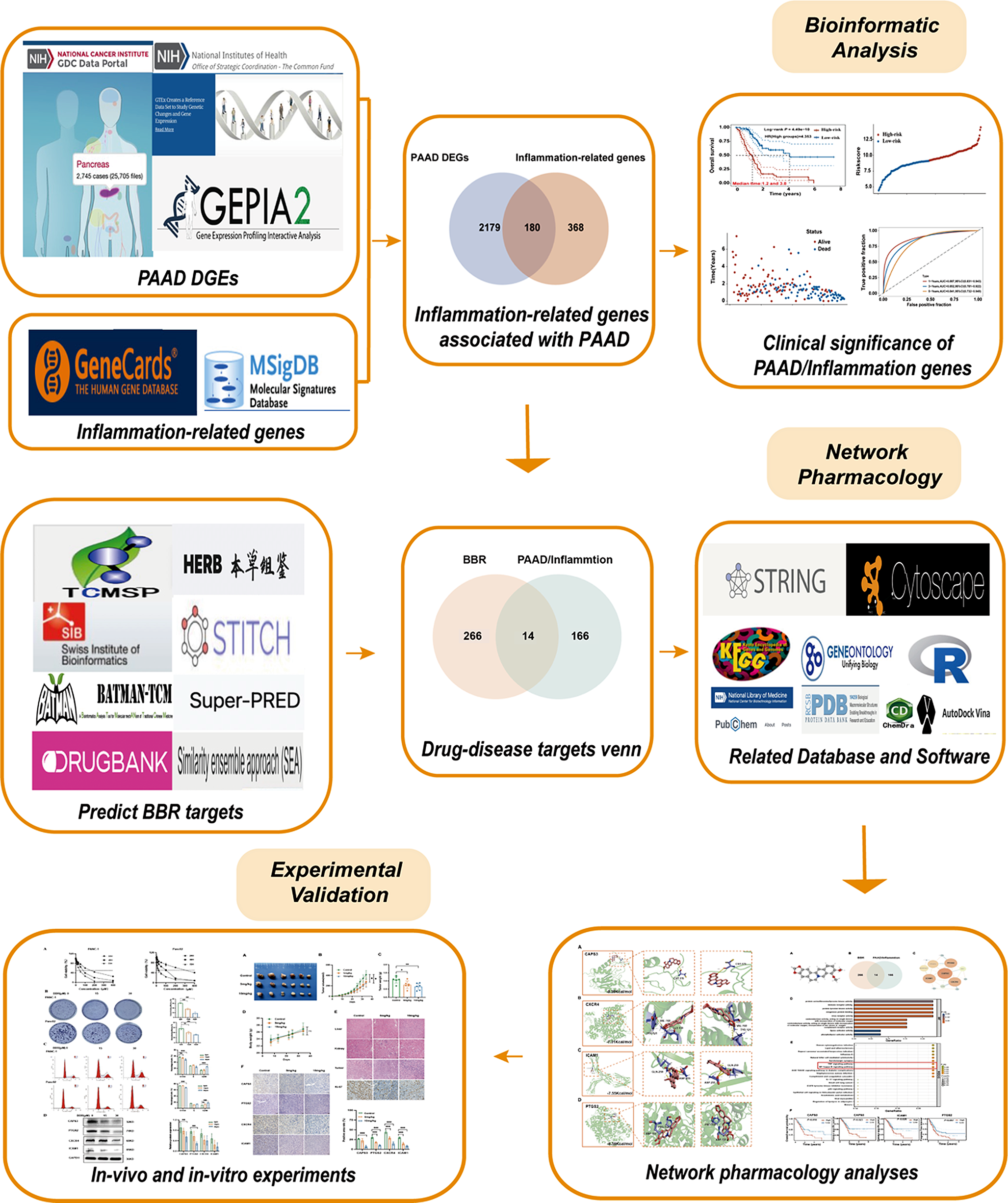
Chemicals and reagents
Emetine hydrochloride and positive agent 5-Fu were acquired from a commercial source. The antibodies against β‐catenin, LEF1, Cyclin D1, Axin 2, GSK3 beta, YAP1 and Survivin were obtained from Abcam. The antibodies against p-p38 MAPK (T180/Y182), p-p44/42 MAPK (ERK1/2 T202/T204), p‐JNK (T183/Y185), non-phospho (active) β‐catenin (S37/T41), p38 MAPK, ERK1/2, JNK, AKT were purchased from Cell Signaling Technology. Antibodies for p‐AKT (Ser473), CTGF and the secondary antibody were obtained from Proteintech.
Cell culture and reagents
The human MGC803 and HGC-27 cells were acquired from the National Platform of Experimental Cell Resources for Sci-Tech (China) and were grown in the recommended medium supplemented with 10% fetal bovine serum (FBS) and 1% penicillin/streptomycin. All cell lines were cultured at 37 ̊°C in an incubator supplied with 5% (v/v) CO2 and experimented within 3 months of receipt or resuscitation.
Cell viability assay
MGC803 and HGC-27 cells were seeded in 96-well plates at 2000–3000 cells/well and cultured overnight in the incubator. Then, the cells were treated with emetine or 5-Fu at different concentrations for 72 h. After treatment, MTT reagent (5 mg/mL, 20 μL/well) was added to the plates, which were then incubated at 37 °C for another 3 h. The formazan crystals were dissolved overnight with 50 μL of acidified SDS (20%, w/v). The absorbance was detected at 570 nm with SpectraMax iD5-multifunctional microplate reader, and IC50 values were calculated using GraphPad Prism v9.0 software.
Colony formation assay
MGC803 and HGC-27 cells were seeded in 12-well plates at a density of 5000 cells/well and cultured overnight. The next day, different concentrations of emetine were added to the plates. Cells were then incubated at 37 °C, and the medium containing emetine was replaced every 3 days. After treatment for 10 days, the cells were washed with PBS and then fixed with methanol for 20 min and stained with crystal violet solution (0.05%, w/v) for another 20 min. Pictures were taken with a camera after natural drying.
Edu incorporation assay
MGC803 and HGC-27 cells were cultured in 96-well plates. Serial dilutions of emetine were added to the plates when the cells reached about 80% confluence. After treatment for 18 h, cell proliferation detection was conducted according to the instruction of BeyoClick™ EdU Cell Proliferation Kit with DAB. Pictures were taken using an OLYMPUS light microscope.
Flow cytometry for apoptosis analysis
MGC803 and HGC-27 cells were seeded in 6-well plates and treated with indicated concentrations of emetine the next day for 24 h. The cells were then harvested and washed twice with precooled PBS, followed by staining with Annexin V-fluorescein isothiocyanate (FITC) and propidium iodide (PI) according to the manufacturer’s instructions. The cell suspension was incubated for 15 min in the dark at room temperature. Cellular apoptosis was analyzed by flow cytometry (NovoCyto 2070R) within 1 h.
Wound healing assay
MGC803 and HGC-27 cells were seeded in 12-well plates and cultured to a density of about 90%. Then a straight scratch was made on the monolayer cells using a sterilized pipette tip, and the floating cells were softly rinsed with PBS. Serial dilutions of emetine were added to the plate to treat GC cells for 12 h. The wound healing was observed and photographed with an OLYMPUS microscope.
Transwell invasion assay
The Corning transwell chambers precoated with diluted matrigel were inserted into a 24-well plate and placed at 37 °C to allow the matrigel to solidify. Then, GC cells resuspended with serum-free medium were seeded in the upper chamber and treated with emetine. The complete medium was added to the outer bottom of the chamber to induce cell invasion. After treatment for 12 h at 37 °C, the invaded cells were fixed with methanol for 20 min and stained with crystal violet (0.05%, w/v) for another 20 min. Pictures were taken under an OLYMPUS upright microscope.
RNA sequencing and differentially expressed genes analysis
MGC803 cells seeded in a 6-well plate were treated with 0.1 μM emetine for 18 h and then harvested. Total RNA was extracted according to the protocol of mirVana miRNA Isolation Kit (Ambion). The libraries were constructed with TruSeq Stranded mRNA LTSample Prep Kit (Illumina, USA) and sequenced on Illumina HiSeq X Ten sequencing platform. The DEGs between emetine-treated and control groups were analyzed using the DESeq R package. We set the adjusted p value < 0.05 and fold change > 2 or fold change < 0.5 as the threshold of significant difference in gene expression. Hierarchical cluster analysis of DEGs was performed to analyze the gene expression pattern in different groups. KEGG enrichment analysis of DEGs was carried out using R based on the hypergeometric distribution.
Western blotting
GC cells were treated with indicated concentrations of emetine for 18 h and then lysed with RIPA buffer (Solarbio, China) containing phosphatase and protease inhibitors. The proteins extracted from cells were separated using 10% SDS-PAGE, and then transferred to PVDF membranes (Millipore, USA). After blocking in 5% nonfat milk for 2 h, the PVDF membranes were incubated with the diluted specific primary antibodies overnight at 4 °C and then incubated with the secondary antibody for 1 h at 37 °C. Then the membranes were washed 3 times with TBST and developed using ECL Enhanced Kit (Biosharp, China). Specific proteins were detected by the ChemiScope 6000 Fluorescence Chemiluminescence Imaging System (CLiNX, China).
Xenograft studies
All animal studies were approved and guided by the Animal Care and Use Committee of Sichuan Academy of Medical Sciences and Sichuan Provincial People’s Hospital (Chengdu, Sichuan, China). Five-week-old BALB/c nude mice were adaptively fed for 1 week. MGC803 cells were collected and washed 3 times with serum-free medium, and then subcutaneously injected into the posterior axillary region of each mouse at a concentration of 5 × 10⁶ cells/100 μL. When the xenograft tumors grew to about 100 mm3, the mice were randomly divided into three groups (n = 5 each): vehicle treatment group, emetine 10 mg/kg treatment group, and 5-FU 30 mg/kg treatment group. All agents were administered by intraperitoneal injection every other day. The tumor volumes and body weights of the mice were also monitored every 2 days. After treatment for 3 weeks, mice were killed, and tumors as well as main organs were obtained for further analysis. Tumor volume was calculated as (a2 × b)/2 (a = width, b = length). The tumor growth inhibition rate was calculated using this formula: 100% × .
H&E staining and immunohistochemistry staining
The tumors and major organs obtained from the animal experiment were fixed in 4% paraformaldehyde and then embedded in Paraffin. Sections of the main organs were subjected to H&E staining according to the standard protocols. Immunohistochemical analysis was performed on tumor tissue sections to detect the expression of Ki67 and TUNEL. Representative images were captured under a Leica microscope.
Statistical analysis
GraphPad Prism v9.0 was used for statistical analysis. All experiments were repeated at least three times and data were shown as mean ± standard deviation (SD). For statistical analysis, Student’s t test was used to assess comparison between two groups, and one-way ANOVA was performed to assess comparison among multiple groups. Statistical significance was presented as *p < 0.05, **p < 0.01, ***p < 0.001.

留言 (0)