
記住我


The incidence of foot ulcerations in patients with diabetes has been increasing and is linked to high morbidity and mortality. Reportedly, diabetic foot ulcers (DFUs) are more prevalent in patients with type 2 diabetes than type 1.1 Generally, these patients experience recurrent foot infections and tissue damage along with lower limb peripheral neuropathy and/or peripheral artery disease, leading to amputation in most cases.2 The probability of reamputation is almost double in these patients because it is difficult to control infection once limb tendons and tissues are severely damaged. Notably, seropurulent discharge and heavy odor can accompany severe DFU, and these injuries require immediate medical attention.
Extensive debridement followed by advanced wound management remains the first line of treatment after the diagnosis of DFU; however, orthopedic surgery is considered if the wound becomes severely painful and is not controllable by dressings and medications alone.3 Often, individuals with DFU receive revascularization surgery to reconstruct the vascular network and promote blood circulation in the affected limb to minimize the risk of amputation. Once the wound is properly managed, DFU healing can be accelerated by split-thickness skin grafting, which has shown 78% success in closing 90% of wounds by 8 weeks during follow-up treatment.4
Given that strict monitoring and controlling blood glucose levels are the key to the success of these types of treatments, endocrinology providers closely monitored the blood glucose levels of these patients during their hospital stay and provide personalized therapy whenever needed. Recent studies have highlighted the significance of multidisciplinary team (MDT) approach in improving the clinical outcomes of DFU treatment.5–8 This MDT approach is considered the criterion standard to expedite diagnostic procedures and minimize unwanted diagnostic testing and medication, while increasing treatment accuracy. Here, the authors report a case study of a patient with DFU involving MDT-based treatment. The patient provided written, informed consent for this case report and permission for the images to be published.
Case ReportThe patient was a 65-year-old retired woman diagnosed with type 2 diabetes 16 years ago with fluctuating blood glucose levels above the threshold level due to blood glucose control treatment in another hospital. She had a history of repeated fungal infections of the foot longer than 10 years.
She developed second toe swelling starting in September 2020 that progressed to gangrene in January 2021. On February 25, she was admitted to the Department of Vascular Surgery. Her blood glucose level was greater than the reference range. A computed tomography angiography detected arteriosclerotic occlusion in the right lower extremity. She underwent balloon angioplasty of the femoral artery, popliteal artery, and anterior tibial artery, as well as an amputation of the second toe on her right foot on March 4, 2021, and was discharged 5 days later on March 9.
After the operation, the patient’s entire right foot became ulcerative, red, and swollen. Accordingly, the patient visited the authors’ wound care clinic on March 12, 2021. The patient scored 2 points on the Digital Pain Scale and 18 points on the General Anxiety Disorder-7 psychological evaluation, indicating moderate anxiety. She had good medical insurance and family support without any history of substance use disorder.
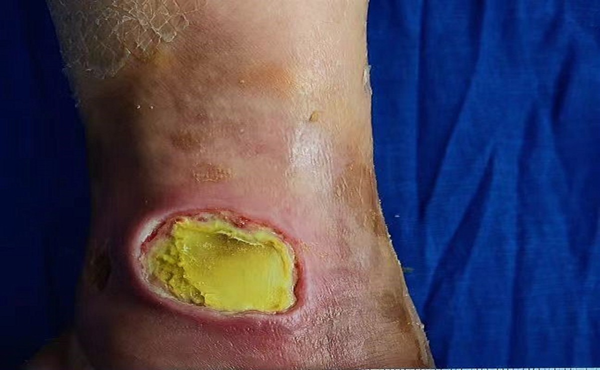
The right foot wound measurements were approximately 3.5 × 2 × 1.5 cm. In one wound, infected tissue was 100% yellow, whereas in the other wound, tissue was 100% black. Both wounds had foul-smelling exudate with brown viscous discharge. The wounds were deeply penetrating, and the skin around the wound area was inflamed and swollen with increased skin temperature. Some of the patient’s skin had begun to separate and fall off.
Providers began negative-pressure wound therapy (NPWT) to care for the wounds. Continuous suction mode was selected, with a negative pressure value of −120 mm Hg. Offloading was achieved by applying foam dressings extending a minimum of 1 inch beyond the edges of the wound. The process of wound care is presented in the Figure 1.
 Figure 1:
 THE PROCESS OF WOUND HEALINGA, March 12. B, March 15. C, March 18. D, March 24. E, April 15. F, April 19. G, April 26. H, May 4.
The skin of the right foot darkened progressively and her skin sensation subsided. On March 29, the patient was admitted to the Department of Trauma Orthopedics, where the patient received radical debridement to remove all devitalized, necrotic, and infected tissue plus tenolysis for the plantar flexors. Bone cement was applied to packing wounds.
Meanwhile, the patient continued to receive wound care in the clinic. The skin flap on the stump had been healing gradually. In this case, there was a significant amount of pus in the wounds. Because pus does not serve a wound-healing function and can increase the risk of local infection, the authors performed therapeutic wound cleansing and debridement, which aims to disrupt biofilm; prevent its reformation; and facilitate removal of necrotic, nonviable, or infected tissue.9 Pus was removed by irrigation or wound swabbing with normal, room-temperature saline at every dressing change. In May 2022, in the Department of Plastic Surgery and Burns, granulation tissue of 6 × 2 cm was seen. A G test and endotoxin test were negative. On May 12, the patient received an autologous skin graft. Then, she was followed up and given health education in the wound care clinic.
In this case, the MDT involved experts from vascular surgery, the wound care center, trauma orthopedics, endocrinology, plastic surgery, and pharmacology to treat the patient successfully. With nursing care, the wound condition progressively improved, accompanied by a reduction in redness, swelling, and odor and improved periwound skin condition. The patient and her family members were quite satisfied with the treatment effects and affirmed the treatment plan many times. The patient and her wound care nurse specialists said they gained knowledge about DFU onset, preventive measures, and efficient treatment options.
DiscussionThe mechanisms underlying DFUs are multifactorial, including neuropathy, infection, ischemia, and abnormal foot structure. Their management is a complex clinical problem requiring an MDT approach.10 Given that sometimes it is impractical to have several specialists from various departments available at the same time and under one roof caring for the needs of each individual patient, the authors’ facility established an MDT Joint Clinic for Diabetic Foot Disease. The Joint Clinic comprises specialists in endocrinology, orthopedics, wound care, vascular surgery, plastic surgery, nutrition, and infectious diseases, including senior physicians as well as nurse specialists.
Vascular surgeons support revascularization by ensuring adequate perfusion to the limb to avoid ischemia. Orthopedic surgeons can perform radical debridement and necessary amputation (which is done by vascular surgeons at the authors’ facility). Dietitians play a role in nutrition assessment and intervention to help patients maximize their nutrition status and promote wound healing. Wound care nurse specialists optimize the wound healing response via direct patient care, consultation, and patient and family education. Endocrinologists aim to control blood sugar, which is essential for a successful treatment outcome. Infectious disease specialists provide appropriate antimicrobial regimens to prevent the aggravation of the foot ulcers that may result in limb loss. Plastic surgeons can enhance healing via soft tissue manipulation, following a reconstruction algorithm to manage and salvage DFUs.
In the authors’ hospital, wound care nurse specialists also play the role of health educator. Health education can improve the relevant knowledge level, self-efficacy, and self-protection ability of patients with DFU, leading to a better quality of life. Health education can also enhance family members’ and carers’ ability to help the patient recover more efficiently without imposing stress and anxiety about the disease. Primarily, the wound care nurse specialists educate patients and families about basic wound care so they can perform the steps properly at home, without waiting for admission, and regularly update the physician about wound status.
Complete recovery from DFU can take a long time because of the condition’s complex and chronic nature; thus, family members and wound care nurse specialists should provide adequate mental support, company, and encouragement to the patient so that the patient can strictly adhere to the treatment guidelines. Wound care nurse specialists may also share success experiences of other patients with DFU to boost the patient’s willingness to recover. Wound care nurse specialists should also pay attention to patients’ complaints and coordinate with other providers if therapeutic evaluation is warranted. If needed, wound care nurse specialists should encourage the patient to pursue psychological counseling to prevent the negative effects of depression on healing. The patient’s family can also ask their care team about setting up an online chat group for rapid medical assistance from home through telemedicine.
Providers had many treatment considerations throughout the patient’s course. First, it was imperative to sterilize the wound area with an appropriate disinfecting solution and water prior to any procedure. The debridement methods were carefully and aseptically applied to completely remove the necrotic tissues and toxic exudate, allowing the normal growth of healthy tissue to fill the wound gap. Providers paid considerable attention to infection management following the debridement procedure. Standardized antibiotic therapy can be initiated under such conditions if the patient’s blood infection index improves.
Because the periwound skin condition plays a crucial role in the healing process, providers consistently assessed skin dryness and any sign of skin flaking around the wound. A painless protective film can be applied to prevent skin from flaking. Further, the patient’s family should be properly educated to apply mildly acidic lotion to the lower limbs to prevent skin loss. Using these techniques, providers managed to keep the patient’s skin around the wound intact. Dressing selection was also key. Providers selected a silver alginate dressing to achieve hemostasis, promote auto-debridement, inhibit bacterial growth, and promote exudate absorption. The secondary dressings were boundless external foam dressings, used to improve exudate absorption and achieve decompression. The elastic bandage was simply fixed and changed regularly.
The use of NPWT provides a moist environment for the wound, which can stimulate tissue growth and enhance collagen production and revascularization to heal the wound. In this case, providers removed excess pus to avoid local infection. Negative pressure helps remove pus and other toxic liquids, as well as necrotic tissue debris, thus preventing pathogen growth and edema formation in and around the wound. During NPWT use, when the transparent foot film stuck to the skin, the seal was considered complete. In practice, full-foot wrapping can ensure a complete seal of the wound. Foam dressings can also prevent stress damage between the toes.
ConclusionsRapidly changing lifestyle and eating habits have substantially influenced the increasing incidence of diabetes among the global population. Accordingly, the rate of DFU and secondary complications has also steadily increased, although treatment options have not improved to match.11 In cases of severe DFU, death may also occur secondary to systemic infection. Therefore, understanding the pathogenesis of diabetes and comorbid diseases such as hypertension and coronary heart disease is crucial for the prognostic assessment of the disease.
The general principle of DFU treatment is to clear debris from the wound area and transform the chronic wound into an acute wound to speed up the healing process. In this case, the MDT approach to treatment doubtless increased the clinical efficacy of the treatment course. Negative-pressure wound therapy is also routinely practiced for DFU treatment.12 Compared with traditional treatment procedures, NPWT facilitates wound healing, and some evidence suggests that NPWT reduces the need for surgery.13 In combination with MDTs, NPWT may significantly reduce rehospitalizations and lengths of hospital stay, thereby reducing the socioeconomic burden on the patient and family.14
Further, health education and facilitating patient-provider interaction are very important to improve patient care after discharge. Health education can be offered both face-to-face and online, and modalities such as online chat groups enable the MDT to closely monitor the implementation of the follow-up treatment strategy via telemedicine. This case shows that prompt interaction between the patient’s wound care nurse specialists and medical practitioners can dramatically improve treatment outcomes by routinely reassessing the treatment strategy as the wound evolves.
REFERENCES 1. Zhang P, Lu J, Jing Y, Tang S, Zhu D, Bi Y. Global epidemiology of diabetic foot ulceration: a systematic review and meta-analysis (dagger). Ann Med 2017;49(2):106–16. 2. Perez-Favila A, Martinez-Fierro ML, Rodriguez-Lazalde JG, et al. Current therapeutic strategies in diabetic foot ulcers. Medicina (Kaunas) 2019;55(11):714. 3. Wang A, Lv G, Cheng X, et al. Guidelines on multidisciplinary approaches for the prevention and management of diabetic foot disease (2020 edition). Burns Trauma 2020;8:tkaa017. 4. McCartan B, Dinh T. The use of split-thickness skin grafts on diabetic foot ulcerations: a literature review. Plast Surg Int 2012;2012:715273. 5. Lu Q, Wang J, Wei X, et al. Cost of diabetic foot ulcer management in China: a 7-year single-center retrospective review. Diabetes Metab 2020;13:4249–60. 6. Ezeani IU, Ugwu ET, Adeleye FO, Gezawa ID, Okpe IO, Enamino MI. Determinants of wound healing in patients hospitalized for diabetic foot ulcer: results from the MEDFUN study. Endocr Regul 2020;54(3):207–16. 7. Blanchette V, Brousseau-Foley M, Cloutier L. Effect of contact with podiatry in a team approach context on diabetic foot ulcer and lower extremity amputation: systematic review and meta-analysis. J Foot Ankle Res 2020;13(1):15. 8. Jeyaraman K, Berhane T, Hamilton M, Chandra AP, Falhammar H. Mortality in patients with diabetic foot ulcer: a retrospective study of 513 cases from a single centre in the Northern Territory of Australia. BMC Endocr Disord 2019;19(1):1. 9. International Wound Infection Institute (IWII). Wound bed preparation: cleansing and debridement. In: Wound Infection in Clinical Practice: Principles of Best Practice. Wounds International; 2022:26–30. 10. Sumpio BE, Armstrong DG, Lavery LA, Andros G. The role of interdisciplinary team approach in the management of the diabetic foot: a joint statement from the Society for Vascular Surgery and the American Podiatric Medical Association. J Vasc Surg 2010;51(6):1504–6. 11. Armstrong DG, Cohen K, Courric S, Bharara M, Marston W. Diabetic foot ulcers and vascular insufficiency: our population has changed, but our methods have not. J Diabetes Sci Technol 2011;5(6):1591–5. 12. Hohendorff J, Drozdz A, Borys S, et al. Effects of negative pressure wound therapy on levels of angiopoetin-2 and other selected circulating signaling molecules in patients with diabetic foot ulcer. J Diabetes Res 2019;2019:1756798. 13. Liu X, Zhang H, Cen S, et al. Negative pressure wound therapy versus conventional wound dressings in treatment of open fractures: a systematic review and meta-analysis. Int J Surg 2018;53:72–9. 14. Atkins BZ, Wooten MK, Kistler J, Hurley K, Hughes GC, Wolfe WG. Does negative pressure wound therapy have a role in preventing poststernotomy wound complications? Surg Innov 2009;16(2):140–6.
留言 (0)