Surgery has been considered to be the only curative option for symptomatic diverticula [1]. Traditionally, thoracotomy and laparotomy are standard procedures for diverticula. With the advancement of technology, laparoscopy and thoracoscopy are increasingly used for treatment [2]. The most popular procedure is diverticulectomy. There are controversies regarding the application of myotomy and fundoplication [4]. In short, the method of treatment selected needs to be determined according to the specific situation of the patient. In the present case, the patient had no signs of esophageal motility diseases, and myotomy and antireflux procedures were not performed.
Full exposure of the neck is the basis for satisfactory resection of the diverticulum [3]. Inaccurate identification of the diverticulum neck may lead to incomplete resection of the diverticulum, resulting in the transformation of a large diverticulum into a small diverticulum, and may also lead to intraoperative iatrogenic esophageal rupture, which may increase the possibility of postoperative esophageal stenosis or leakage. We usually identify the neck under endoscopic intraluminal vision and the endoscopic light shining through the lumen of the diverticulum. Nonetheless, accurate identification of the diverticulum is a difficult task and requires extensive experience.
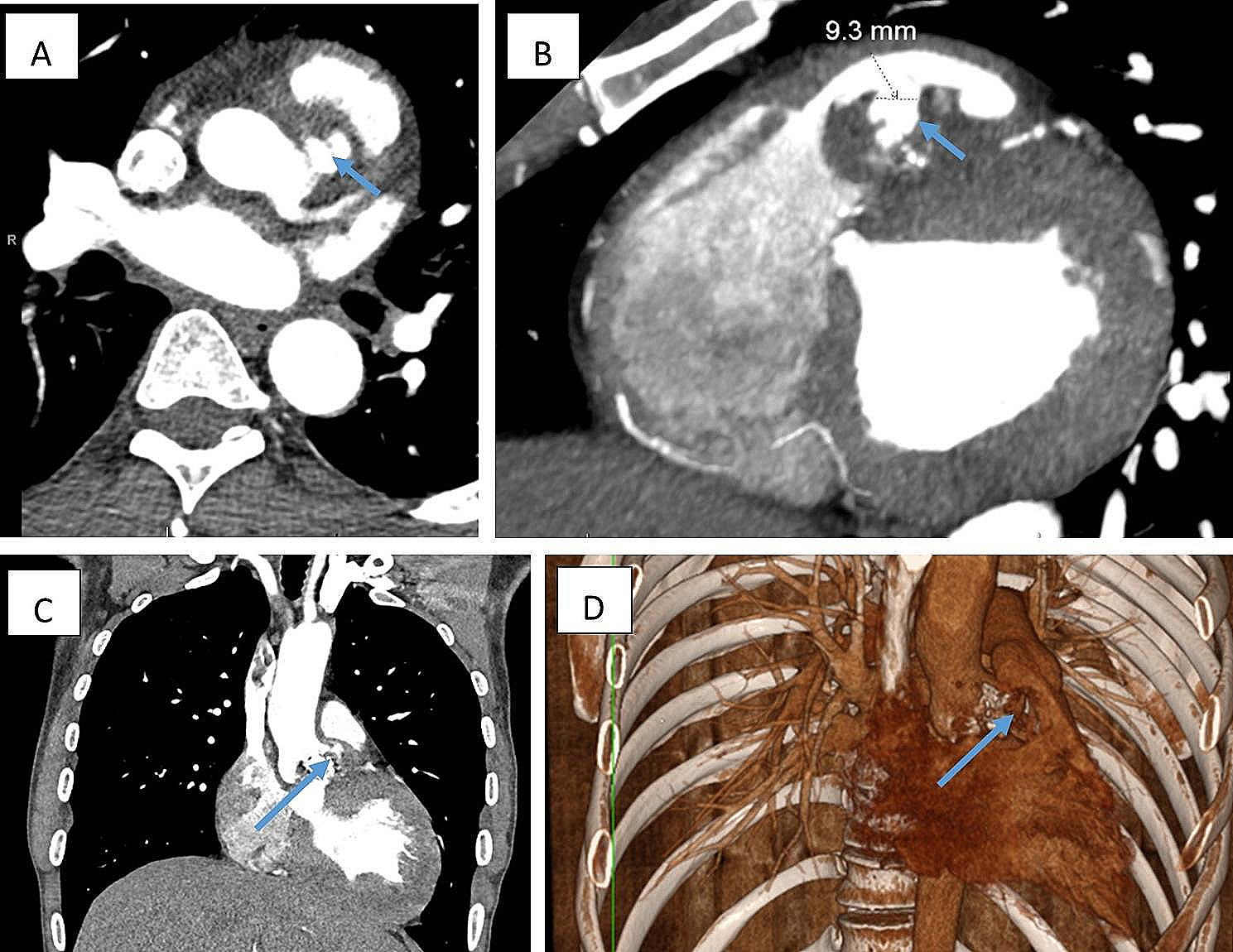
NIR fluorescence with ICG has been used clinically for decades. The application of this method includes the identification of thoracic ducts, segmental borders, pulmonary nodules and bullous lesions [5]. To the best of our knowledge, there are few reports of this technique being used for diverticulectomy [6]. To ensure that the diverticulum neck was sufficiently coated with ICG, we inserted the lens of the endoscope into the diverticulum before injecting the ICG. In this case, we injected approximately 30 ml ICG (2.5 mg/ml) into the diverticulum through the endoscopic channel. Since the fluid needs to fill the endoscopic channel before it can enter the diverticulum, the amount of ICG can be slightly higher. Immediately after injection of ICG into the diverticulum, the diverticulum wall and neck were clearly visible under NIR fluorescence. Since NIR fluorescence with ICG can only penetrate a few millimeters of tissue, even if ICG flows into the esophagus, the esophageal wall near the diverticulum cannot be seen due to the muscle covering (Fig. 2C), which means that this technique has high specificity in identifying the diverticulum neck. Different from the use of endoscopic light, the diverticulum neck stained with ICG was visualized as a whole and was very stable under NIR fluorescence, so the surgeon can perform the excision of the diverticulum neck calmly.
Since the NIR fluorescence of ICG can only penetrate a few millimeters of tissue, a limitation of this technique is that it may only be used for false diverticula or true diverticula with thin muscular layers. The pathology of this case suggests that the diverticulum is a true diverticulum with a thin muscular layer. When the diverticulum has adhesions to the surrounding tissue, especially when the epiphrenic diverticulum can sometimes be adherent, this method may help to accurately identify the diverticulum neck. The specific application scope and method of this technique need further research.
We applied ICG and NIR fluorescence during diverticulectomy to better identify the diverticulum neck. This method is safe, simple to perform, and effective and has the potential to increase the efficiency of diverticulectomy.


留言 (0)