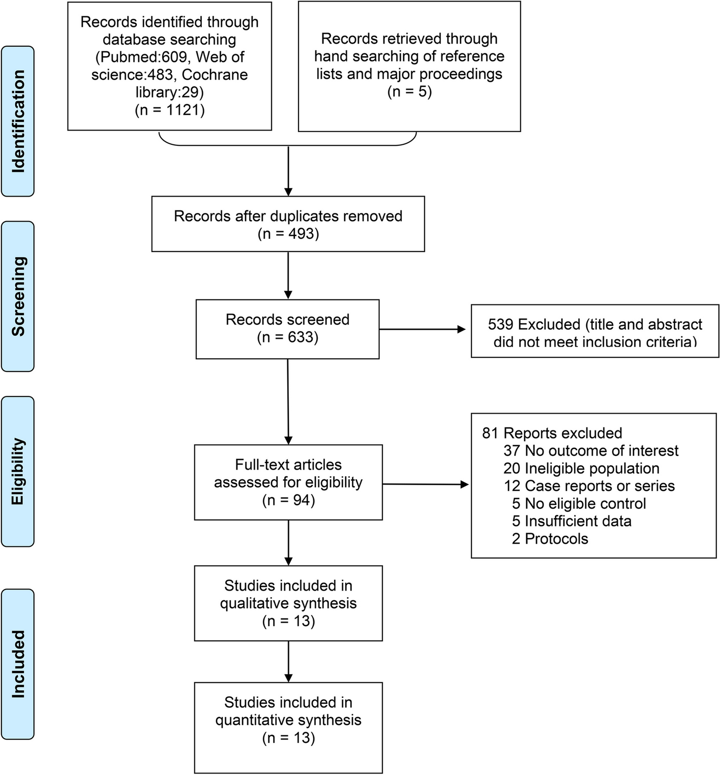
The pineal gland is a neuroendocrine organ that regulates daily body rhythm by the secretion of melatonin [1]. It is the principal seat of the soul and the place in which all thoughts are formed [2]. The pineal gland is the part of the epithalamus, located at the posterior wall of the third ventricle occupying the space between the corpus callosum and the superior colliculi [3,4,5,6]. Different studies report that the pineal gland grows in size from birth until 2 years of age and then remains constant between 2 and 20 years of age [5,6,7]. Unlike most parts of the brain, the pineal gland is located outside of the blood–brain barrier (BBB) [8, 9]. The pineal gland consists of different cell types in addition to pinealocytes, which include interstitial cells, perivascular phagocytes, pineal neurons, and peptidergic neuron-like cells [10, 11]. It plays an important role in regulating the light/dark circadian changes to synchronize the physiological activities [2, 4, 5, 8], cancer inhibition [4, 5], neuroprotector antioxidant [4, 8], and induction of the endocrine activity of the hypothalamus, pituitary, ovaries, and testis [3]. Melatonin act as negative feedback to the biological clock, the suprachiasmatic nucleus (SCN), that regulates the circadian rhythm of the body. The SCN sends signals to all the organs synchronizing the day-night cycle like sleep, lowering of body temperature, and blood pressure at night time, which leads them to function at the proper time [9, 12]. Pineal gland calcification (PGC) is the formation of corpora arenacea, composed of mainly calcium and phosphorus with a small contribution of magnesium and strontium [3, 5]. Pineal gland calcification presents histologically from fetal life to adulthood, which increases in number and size with aging [1, 13, 14]. There are two types of pineal gland calcification based on the affected area of the gland, intra-pineal calcification present within the parenchyma of the gland and extra-pineal calcification within the capsule of the gland common among old age groups [3, 15, 16]. Pineal gland calcification can also be pathological or physiological depending on the causes of calcification [16]. Physiological calcifications are unaccompanied by any evidence of disease, asymptomatic, and detected incidentally by neuroimaging [17, 18], whereas pathological calcifications are extremely rare and occurred due to any diseases or abnormality in the form of developmental, reactive, neoplastic, and vascular abnormalities [19]. Other pathological conditions like neurodegenerative diseases, including Alzheimer’s disease, autism, migraine, chronic primary insomnia, and stroke, can also be associated with pineal gland calcification [2, 9]. Genetic and environmental factors, which increases pineal gland calcification, are male gender, low altitude, and low intensity of sunlight exposure [5, 9, 14]. The prevalence of pineal glad calcification ranges from 58.8 to 76% across different countries [14, 16, 20, 21]. Currently, PGC is an important marker for neurologists and neuroradiologists and is an important radiological shadow suggesting the presence of a space-occupying lesion in an intracranial cavity [3]. Since pineal gland calcification had a strong association with different pathological conditions, genetic and environmental problems, it is important to have an appropriate information on its magnitude and geographic distribution. So, this study aims to assess the pooled prevalence of pineal gland calcification.


留言 (0)