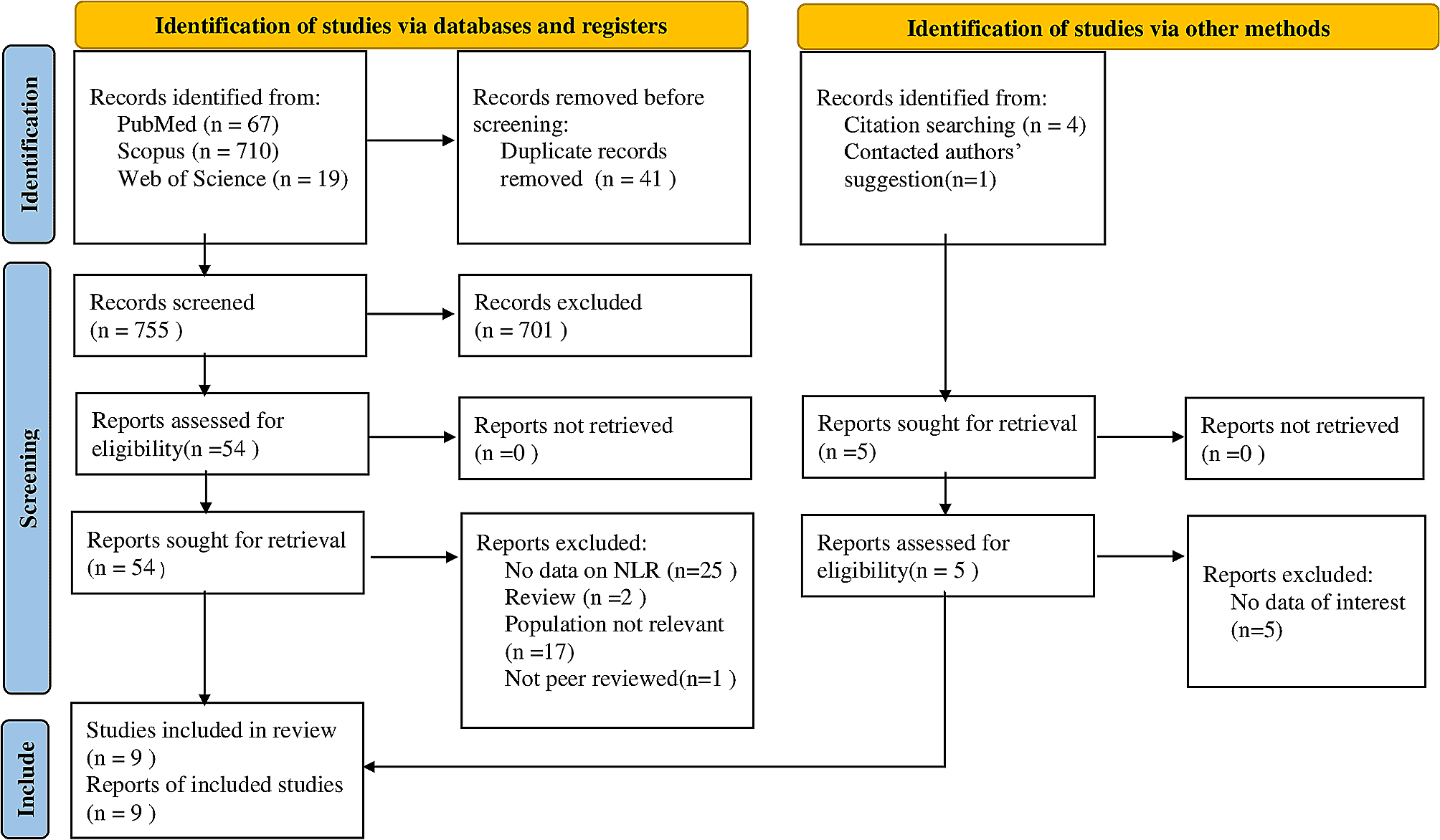
This systematic review aimed to determine whether there is sexual dimorphism in the odontometric parameters when assessed using CBCT. Of these published 23 studies, eighteen studies found a positive response that odontometric measurements could aid in sex estimation. The majority found sexual dimorphism in various odontometrics of permanent canines, especially with mandibular canines.
A recent systematic review on tooth crown mesiodistal measurements for estimating sexual dimorphism across a span of different people confirmed that canines reflect the greatest sexual dimorphism [10]. Several investigations found canines and first molars to be the most common teeth with a lot of morphological diversity between sexes [10, 34]. In the present analysis, sexual dimorphism was observed in all teeth across various populations, mostly in canines, followed by incisors, premolars, and molars. Tooth formation and development are controlled by sex-related genes, and structures of human permanent dentition exhibit sex differences. Accordingly, forensic researchers have evolved several methods to distinguish males from females. But still, experts choose canines for sex estimation [27].
Of the teeth in the permanent dentition using CBCT images, canines were analyzed in fourteen studies out of twenty-three studies. Tooth size or dimension is the most frequently assessed odontometric variable for sex estimation. The tooth size of the maxillary and mandibular canine displays the largest variation of sexual dimorphism [6]. Prolonged amelogenesis in males results in differences in enamel thickness between the sexes and, consequently, greater dimensions of canines in males than in females [35]. The ‘Y’ chromosome promotes mitotic activity in tooth germs and controls growth by enhancing amelogenesis and dentinogenesis, consequently greater dentin thickness in males [36]. In contrast, the ‘X’ chromosome controls only the enamel growth. This could explain the variation in size [36].
Few investigators consider that the sexual dimorphism of mesiodistal width is because of dentin deposition, which is in excess in men than in women. On the contrary, there is no difference in enamel thickness [32]. Few researchers contemplate the variation in the level of sex hormones in the course of tooth formation could influence tooth dimensions [9]. Parameters like crown width and length of mandibular canine and inter-canine width show highly significant sex differences [9]. The mesiodistal width of mandibular canines revealed statistically significant sexual dimorphism [5]. In permanent dentition, mandibular canines are known to show the greatest sex dimorphism; hence, it has become the tooth of choice for sex estimation studies. It has been considered that the mesiodistal width of mandibular canines is the simpler method for sex prediction with a better rate of accuracy [37]. A significant difference in the length of canines in both sexes and both jaws also have been reported [25].
Average tooth length is greater in men than in women [29]. In both women and men, the longest teeth turned out to be canines. The buccolingual root size on the mandibular canine also revealed significant differences between the sexes [30]. The average values of the canine pulp chamber were larger for males compared to females [3]. Dental volume showed a significant difference between sexes [32]. The dental volume observed a significant difference between the sexes, while different finding was noted for the volume of the pulp chamber and the ratio between the pulp chamber and dental volume. The lack of sexual dimorphism in the pulp chamber quantification is probably due to the effect of age on the pulp dimension [32]. Volumetric accuracy of the maxillary canine and mandibular canine for sex estimation were 74.4 and 79.5%, and the combined analysis of the maxillary and mandibular canines allowed an average accuracy of 83.7% [19].
While reviewing the literature on odontometric assessment of permanent incisors using CBCT for estimation, it was observed that the largest variation in the tooth dimension was found in the maxillary lateral, second premolars, and mandibular lateral incisors in men, whereas the maxillary canine and mandibular incisors in the women [6]. Males show a greater mean mesiodistal dimension of central incisors than females [22]. The mesiodistal dimension of both maxillary central incisors is significantly different in males compared to those in females. Though the form and shape of tooth structure are similar in both sexes, the size might differ, as tooth dimension is influenced by genetic, cultural, racial, and environmental factors [38].
Warnecki et al., 2021 found that mean tooth length is greater in men than in women [29]. In both men and women, the smallest difference in tooth length between women and men was found for the central incisors. In the same study, significant differences were found in tooth length between the sexes when evaluating the literature on odontometry of premolars using CBCT [29]. Males had significantly lengthy mandibular premolars than females. Llena et al., 2014 noted that the mean length of teeth and roots was significantly longer in males than in females [33]. Analysis of extracted premolars in the Jordanian population showed similar findings [39].
In the review of molar morphometry using CBCT in sex estimation, Paknahad et al., 2022 found that accuracy of sex estimation of the mandibular and maxillary first molar tooth was 84 and 77%, respectively [24]. The mesiodistal variables were more accurate in sexual dimorphism than the buccolingual ones. For sexual dimorphism, the most dominant variables for maxillary and mandibular first molar teeth were crown height and dentin thickness. Tobpas et al., 2021 found that sex was predicted by maxillary first molar volume ratio in 76.6% of females and 56.3% of males; it was observed that maxillary first molar tooth volume ratio provided more precise results in females’ sex estimation [27]. Salam et al., 2021 observed significant sex differences in mandibular first molar crown width and length and inter-molar width [9]. Chandler et al., 2003 found a significant difference between sexes and found that permanent first molar teeth pulps exhibit sexual dimorphism [40]. Pulp dimensions of the permanent first molar tooth in men are larger than that in women. Molars are the first permanent teeth to erupt in the mouth; hence, they are easily accessible for sex estimation at an early age when compared with other permanent teeth. It has the edge over canines, which have a greater propensity of being impacted and thus are not accessible for odontometric analysis [9].
The accuracy of sex estimation was assessed in eight investigations, which ranged from 47.8–92.3%. An accuracy of 100% for canine dimensions was reported and accepted that a small sample was responsible for the inflation of accuracy [41]. Considering the diversity in methodology, ethnicity of population and sample size, and age range, comparison of accuracies was not easy. Biochemical methods, DNA-PCR, fluorescent microscopy of the freshly extracted tooth, and analysis of Barr body provided 100% accuracy. Odontometrics on casts, skeletal remains, and pulp/tooth volume ratios on CBCT reported isolated accuracies of a cent percent [1]. Even the cascade of techniques reported a range of accuracy similar to individual methods.
Jaysinghe et al. 2022 found significant difference in all maxillary arch parameters using CBCT such as width of the alveolar ridge at the canine, first molar and second molar and the distance of the arch at the inter canine distance and junction between the hard and soft palate when assessed between the different genders [42].
There is some evidence of reverse dimorphism too in the literature related to dental structures. For example- Fernee et al. 2021 found larger surface areas and volumes of enamel and crown volumes in females unlike in the case of dentine and root, which were larger in males [43]. This was particularly seen in the case of upper canine. But none of these studies used CBCT for estimation. Further research is needed to establish the potential use of oral tissues for sex estimation in humans.
Strengths and limitations
Most of the studies included had a nearly equal distribution of both sexes. The majority of the participants were young adults between the second the fifth decade. This ensured that the dentition is less worn off in the case of odontometrics, morphologically unchanged, and shows adequate skeletal growth and development. The added benefits of CBCT are that it enables the assessment of a subject’s tooth dimensions and arch size. The analysis can be done directly using digitized images, and record maintenance is not an issue. CBCT imaging provides a platform to make linear and/or volumetric measurements of dentition. It is a better system to archive patient details and easy access to the records that will help in analyzing tooth dimensions at convenience directly from images using various software.
The present review did not separate studies based on the software used for taking measurements directly from CBCT images or software used to reconstruct the models to make odontometric analyses. As the difference between the two modes is unknown/ unclear, this could contribute to a risk of bias. Potential problems regarding sex estimation based on odontometric analysis using CBCT are diversity with respect to sample size, class of teeth assessed, odontometric parameters analyzed, and software used to analyze.
The reasons for not subjecting the cumulative data to meta-analysis are lack of uniformity in the data and specific protocol followed in these studies. The present analysis excluded studies related to anthropological skeletal remnants because the dimensions of the dentition might be withered; similarly, intrinsic and extrinsic variables are particular to specific populations. Hence, the results focused on adolescents and adults with permanent dentition.
The reasons for not subjecting the cumulative data to meta-analysis are lack of uniformity in the data and specific protocol followed in these studies. The present review did not separate studies based on the software used for taking measurements directly from CBCT images or software used to reconstruct the models to make odontometric analyses. As the difference between the two modes is unknown/ unclear, this could contribute to a risk of bias. Potential problems regarding sex estimation based on odontometric analysis using CBCT are diversity with respect to sample size, class of teeth assessed, odontometric parameters analyzed, and software used to analyze. Although the risk of bias assessment using The QUADAS 2 chart showed an overall low risk, few studies showed some unclear parameters and unclear risks. Small sample sizes, lack of power analysis in many studies, and insufficient explanation for calibrations are some of the major reasons. Grading of recommendations assessment, development and evaluation (GRADE) assessment was not performed in this systematic review to rate the certainty of evidences which is a limitation of the review.
Most recent reports about dental anatomy are with three-dimensional technologies, with exceedingly accurate results compared to previous studies, which focused on other methods of sex estimation using casts, extracted teeth, non-human teeth, skeletal remains, direct intraoral assessment, intraoral photography, and x-ray. Even with non-metric methods of sex estimation, some important, relevant information would have been lost. Therefore, a comparison of results becomes difficult to interpret. Included studies in this review exhibit the differences in the data sets, methodology, statistics applied, and results obtained. The consensus was challenging to achieve due to the diversity in linear and volumetric measurements applied in each class of teeth. The review included investigations conducted in different populations that utilized different data collection techniques but yielded similar results across studies.


留言 (0)