The anatomy of the sacrospinous ligament: how to avoid complications related to the sacrospinous fixation procedure for treatment of pelvic organ prolapse
Introduction and hypothesis
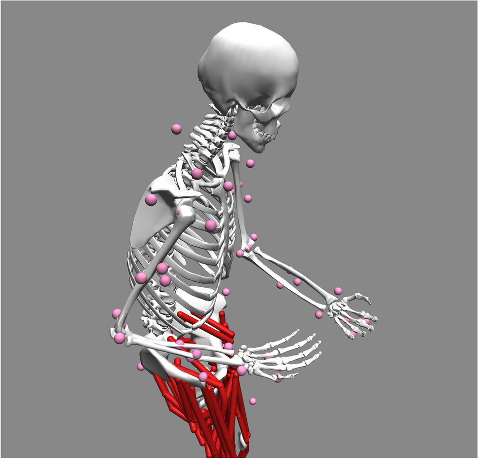
Historically, the sacrospinous ligament (SSL) has been used to treat POP in order to restore the apical compartment through a posterior or an anterior vaginal approach. The SSL is located in a complex anatomical region, rich in neurovascular structures that must be avoided to reduce complications such as acute hemorrhage or chronic pelvic pain. The aim of this three-dimensional (3D) video describing the SSL anatomy is to show the anatomical concerns related to the dissection and the suture of this ligament.
Methods
We conducted a research of anatomical articles about vascular and nerve structures located in the SSL region, in order to increase the anatomical knowledge and show the best placement of sutures to reduce complications related to SSL suspension procedures.
Results
We showed the medial part of the SSL to be most suitable for the placement of the suture during SSL fixation procedures, in order to avoid nerve and vessel injuries. However, nerves to the coccygeus and levator ani muscle can course on the medial part of the SSL, the portion of the SSL where we recommended to pass the suture.
Conclusions
Knowledge of the SSL anatomy is crucial and during surgical training it is clearly indicated to stay far away (almost 2 cm) from the ischial spine to avoid nerve and vascular injuries.


留言 (0)