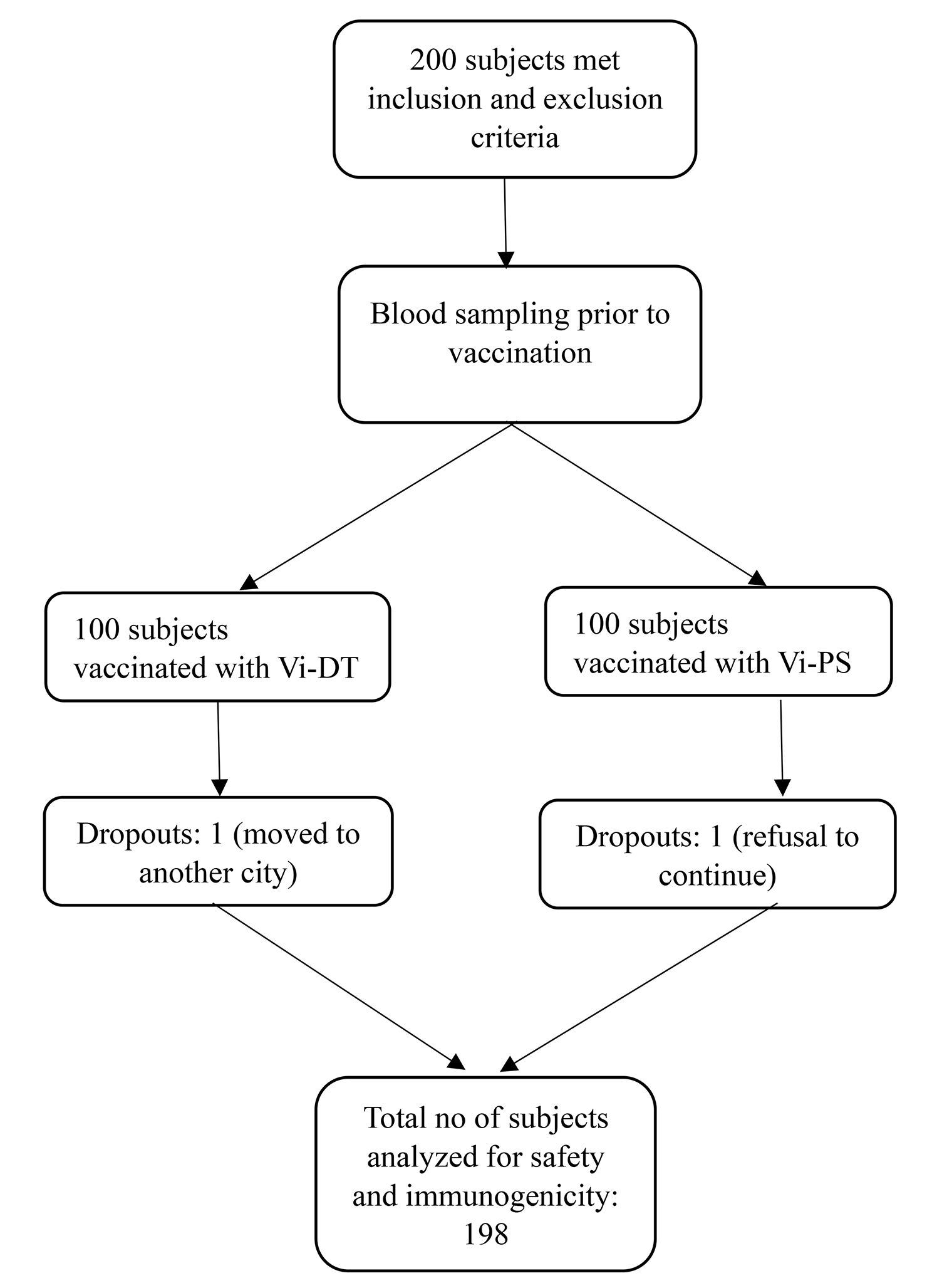
Placental and serum samples
Placental and serum samples were collected at the Tu Du Hospital, Ho Chi Minh City (Vietnam) and stored at -80 °C upon reception [23]. Only samples for which the placental tissue was available were included in the present study, for a total of 176 samples. As for the original study, samples analysed here were collected from January to December 2008. All patients included in the study gave their written consent and utilization of the samples was approved by the local ethical committees of the Tu Du Hospital, Vietnam (clinical part) and the Lausanne University Hospital, Switzerland (experimental part).
Flavivirus propagation and titration
Japanese encephalitis virus (JEV, CNS769_Laos_2009; GenBank accession number KC196115.1) was kindly provided by Remi Charrel, Aix-Marseille Université, France. West Nile virus (WNV, NY99–35, GenBank accession number DQ211652.1) was kindly provided by Martin Groschup, Friedrich-Loeffler-Institute, Germany. DENV-2 was kindly provided by Dr. Katja Fink, Singapore Immunology Network, Singapore). The low passage clinical isolate of Asian lineage ZIKV (PRVABC59, GenBank accession number KX377337) was obtained from Public Health England (PHE). Yellow fever virus (YFV, UVE/YFV/UNK/XX/Vaccinal strain 17D; GenBank accession number EU074025.1) was obtained at the European Virus Archive Global (EVAg). All flaviviruses were propagated in Vero cells (CCL-81, ATCC) cultured in DMEM (Gibco—Thermo Fisher Scientific, Reinach, Switzerland) supplemented with 10% FBS (Gibco) at 37 °C, 5% CO2. Flavivirus titers were determined in Vero cells using an immunoperoxidase assay using the anti-flavivirus group antigen antibody 4G2 (clone D1-4G2-4–15, ATCC, HB-112). Virus titers were calculated and expressed as 50% tissue culture infective dose per ml (TCID50/ml) using the Reed and Muench method [24].
Serum neutralization assay
Serial twofold dilutions of heat-inactivated sera (30 min, 56 °C) were prepared in serum-free DMEM medium, with a starting dilution of 1:4 for ZIKV and 1:8 for JEV, WNV and YFV. Serial serum dilutions were incubated at 37 °C for 60 min with an equal volume of the corresponding virus to provide 100 PFU per 100 μl (1000 PFU per 100 μl in the case of DENV2). Serum-virus mixture was added to monolayers of Vero cells in 96-well flat-bottom tissue culture-treated microtiter plates (TPP) and incubated at 37 °C with 5% CO2 for two hours, after which DMEM containing 2% FBS 1% PenStrep (Gibco) was added to the wells. After 72 h, the culture plates were washed with 300 μl/well of cold PBS (Gibco) and fixed for 20 min at RT with 4% PFA. Then, the plates were washed once with 300 μl/well of 0.1% saponin (AppliChem GmbH, Darmstadt, Germany). A 100 μl/well of primary antibody mix was added and incubated for 30 min at 37 °C. The plates were washed twice with 200 μl/well of 0.1% saponin. Secondary antibody mix (rabbit anti-mouse HRP, Dako) was prepared in 0.3% saponin with a dilution factor of 1:250. A 100 μl/well of secondary antibody was added for 30 min at 37 °C in the dark. The plates were washed twice with 200 μl/well of 0.1% saponin and 80 μl/well of AEC substrate solution was added and incubated for 15 min at RT in the dark. When the plaques appeared with the desired coloration, the reaction was stopped with 100 μl/well of tap water. The microtiter plates were scanned with an ImmunoSpot analyzer (Cellular Technology Limited).
Serological analysis
Serology for ZIKV was performed with the “Anti-Zika Virus ELISA (IgG)” kit (EI 2668–9601 G, EUROIMMUN Schweiz AG, Luzern, Switzerland), according to the manufacturer’s specifications. Confirmation of positive samples was performed using a custom designed flavivirus mosaic Indirect Immunofluorescence Test (IIFT) (EUROIMMUN Schweiz AG, Luzern, Switzerland), in which cells infected with Zika, Dengue (I-IV), West Nile, Yellow fever and Japanese encephalitis viruses were used to detect their antigens.
Detection of ZIKV in placental samples
For the analysis of the presence of ZIKV in placental samples, approximately 25 mgs of tissue were homogenized using the Molecular Grinding Resin (G-Biosciences, St. Louis, MO, USA) and subsequently processed with NuceloSpin RNA II extraction kit for total RNA extraction according to the manufacturer’s protocol. Complementary DNA (cDNA) was obtained with the SuperScript II Reverse Transcriptase kit (Invitrogen Life Technologies, Carlsbad, CA, USA) by using Random Primer Hexamers (Invitrogen Life Technologies) and following the manufacturer’s instructions. ZIKV RNA levels were assessed by quantitative RT-PCR using the iTaq Universal Probes Supermix (Bio-Rad, Reinach, Switzerland) and the Rotor Gene 6000 thermocycler (Corbett Research, Sydney, Australia) as previously described [25]. As positive controls, placental homogenates were spiked with different inocula of ZIKV strain PRVABC59.


留言 (0)