
記住我


From January to July 2022, >1000 cases of pediatric hepatitis were reported across 35 countries, almost half occurring in Europe.1,2 These cases, described as “acute hepatitis of unknown origin,” were characteristically severe, requiring hospitalization, and at least one child has required liver transplantation.1 The World Health Organization case definition comprises acute non-A-E hepatitis with serum alanine transaminase (ALT) or aspartate transaminase (AST) >500 IU/L in an individual ≤16 years of age. Cases have not been reported in Australia to date.3 A potential relationship with adenovirus infections has been proposed and 60%–70% of tested cases have identified the virus on PCR testing.3,4 Adenovirus-positive cases have had a greater risk of intensive care unit admission and liver transplantation.4 Adenovirus subtype 41 has attracted special attention as a potential cause.5 Other viruses were detected in some cases, including human coronavirus, rhinovirus, parainfluenza, sapovirus, norovirus and picornaviruses.1,2
To determine whether similar clusters of pediatric hepatitis have previously occurred in Victoria, Australia, we applied a unique spatiotemporal methodology previously validated in assessing viral association with febrile seizures.6 By applying this methodology to population-level data, we aimed to add insight regarding possible causes of severe acute hepatitis.
METHODS Study Design and CohortA retrospective ecologic cohort analysis drawing on data from July 1, 2011, to June 30, 2022, in independent, unlinked datasets to visualize and define associations between hepatitis diagnoses in children and virus activity.
The Victorian Agency for Health Information collates, analyses and shares health data from public hospitals across the Australian state of Victoria (population 6.5 million).7 Data are collated under one of 2 main datasets: the Victorian Emergency Minimum Dataset captures emergency department presentations in Victorian public hospitals, and the Victorian Admitted Episodes Dataset (VAED) contains data from Victorian public hospital admissions. Data are stored by ICD-10-AM codes and hepatitis cases were identified using a broad set of relevant codes. We extracted fields related to date of presentation; age (5-year bands); patient area of residence, aggregated to statistical area level 3 (SA3). (“SA3s are designed to provide a regional breakdown of Australia. They generally have a population of between 30,000 and 130,000 people. In regional areas, SA3s represent the area serviced by regional cities that have a population >20,000 people. In the major cities, SA3s represent the area serviced by a major transport and commercial hub. They often closely align to large urban local government areas (eg, Gladstone, Geelong). In outer regional and remote areas, SA3s represent areas that are widely recognized as having a distinct identity and similar social and economic characteristics.”8)
Monash Health (MH), based in southeast Melbourne, is the largest health network in Victoria, with >210,000 annual pediatric and adult presentations across 3 emergency departments.9 The Royal Children’s Hospital Melbourne (RCH) is the largest children’s hospital in Victoria, with care extending to children from Tasmania, New South Wales and other Australian states. RCH has almost 55,000 admissions annually, with various other non-admission services.1 We obtained respiratory multiplex polymerase chain reaction (PCR) test results during our study period from the 2 pediatric tertiary care hospitals in Victoria. PCR assays reported results for the following viruses: adenovirus, influenza A and B, parainfluenza 1, 2, 3 and 4, SARS-CoV-2, human metapneumovirus (hMPV), respiratory syncytial virus (RSV), parechovirus and picornavirus using commercial multiplex respiratory PCR panels (AusDiagnostics, Syndey, Australia). Data fields included date of test, patient area of residence aggregated to SA3 and PCR test result.
Our inclusion criteria were all respiratory multiplex PCR assays performed at MH and the Royal Children’s Hospital for patients of all ages with a Victorian residential SA3. All hepatitis presentations were included from the VAED, and Victorian Emergency Minimum Dataset between July 1, 2011, and June 30, 2022, if the patient was <19 years old on the day of presentation. Hepatitis A-E cases were excluded, as these cases had an identified viral etiology. Patient records were not available for analysis to ensure patient de-identification.
Statistical AnalysisTo account for change in testing behavior, with increased testing over the study period, all virus data were weighted by the number of monthly PCR tests.
We used SaTScan (Boston, MA, USA)10 to determine spatiotemporal clustering of hepatitis and adenovirus independently, and qualitatively assessed for similarities or overlaps. We used the program’s space-time scan statistic with a Poisson model to identify clusters, as described in the SaTScan user guide.11,12 The Australian Urban Research Infrastructure Network e-Infrastructure was used to access the “ABS—Regional Population—Population Estimates by Age and Sex (SA3) 2017” dataset on July 21, 2022.13 Cluster limitations were set at a 10-km radius and a maximum 3-month duration. We also limited the minimum adenovirus cluster as 1000/100,000 adenovirus cases and the minimum hepatitis cluster as 10 cases.
We then conducted negative binomial regression analysis to determine if any viruses were associated with increased hepatitis incidence, as in our temporal study of febrile seizures and respiratory viruses.6 Statistical analysis of temporal data was done with R (version 4.0.2),14 using the RStudio interface (version 1.2.5).15 Geographical areas and monthly virus rates were included as covariates to account for the spatial component. The first regression analysis assessed the relationships between respiratory viruses, except SARS-CoV-2, and pediatric hepatitis between 2011 and 2019. The second analysis assessed data between 2020-2022 and included SARS-CoV-2 as an independent variable. Significance was set at P <0.01.
Ethics ApprovalPrimary Human Research Ethics Committee approval was through MH, from July 24, 2019 (NMA/ERM Reference Number: RES-19-0000333L-53611).
RESULTSWe analyzed 5518 hepatitis cases reported in Victoria, Australia, from July 1, 2011, to June 30, 2022. Our space-time cluster analysis showed 16 statistically significant hepatitis clusters and we found 16 significant space-time adenovirus clusters (Tables 1 and 2).
TABLE 1. - Hepatitis Significant Cluster Locations, Time Period and Number of Cases Location Time No. Cases 1 Casey North, Dandenong February 1, 2021–April 4, 2021 280 2 Upper Goulburn Valley October 1, 2021–October 31, 2021 57 3 Cardinia January 1, 2021–February 28, 2021 65 4 Glen Eira February 1, /2021–March 31, 2021 57 5 Casey South October 1, 2021–October 31, 2021 48 6 Gippsland-South West November 1, 2021–November 30, 2021 26 7 Knox January 1, 2022–March 31, 2022 36 8 Maroondah December 1, 2021–January 31, 2022 19 9 Manningham East September 1, 2014–November 30, 2014 12 10 Tullamarine-Broadmeadows June 1, 2019–June 30, 2019 15 11 Wyndham June 1, 2016–June 30, 2016 16 12 Ballarat October 1, 2015–October 31, 2015 12 13 Frankston March 1, 2021–April 30, 2021 16 14 Yarra Ranges November 1, 2020–November 30, 2020 12 15 Brimbank August 1, 2018–August 31, 2018 12 16 Whittlesea-Wallan January 1, 2015–January 31, 2015 12The green border represents clusters occurring in October 2021.
Colored borders represent clusters occurring in the same time period. The green border represents clusters occurring between December 2015 and February 2016. The orange border represents clusters occurring between January and February 2013. The purple border represents clusters occurring between November 2019 and January 2020.
 FIGURE 1.:
 Hepatitis and Adenovirus clusters identified in Victoria, Australia throughout the study period using SaTScan's space-time scan statistic. A. Hepatitis clusters. B. Adenovirus clusters.
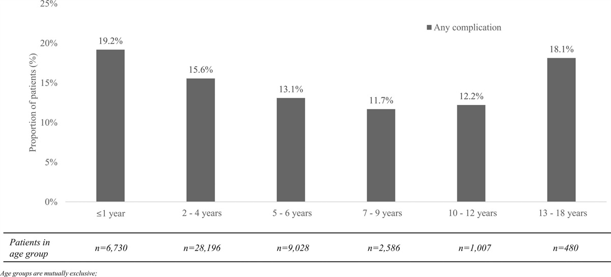
Nine of the 16 hepatitis clusters occurred in the most recent 2 years of the study from late 2020 to early 2022. Of these, 5 occurred from late 2021 to early 2022, coinciding with the global hepatitis outbreak. Most clusters (88%, 14/16) were in metropolitan Melbourne (clusters 1, 3–11, 13–16). No clusters were in the same location throughout the study period. There were 2 hepatitis clusters in different areas in the same time period (clusters 2 and 5); however, these clusters occurred in statistical areas approximately 120 km apart and are thus unlikely to be related. The largest cluster was in early 2021 and crossed two statistical areas in Melbourne’s Southeast, with 280 cases.
Respiratory adenovirus clusters spanned the entire study period, with the earliest cluster occurring between February and April 2012 and the latest in April and May 2021. Only one cluster occurred outside of metropolitan Melbourne (6). There was a narrower distribution of clusters of adenovirus rates of incidence, with the majority occurring in south-eastern Melbourne. Three clusters occurred in different locations during the same period (Table 2).
Visual comparison of the 2 cluster analyses showed no overlap of hepatitis and respiratory adenoviral clusters.
Negative binomial regression analysis showed significant associations between the included respiratory viruses and hepatitis (Table 3). This included a 0.77 risk ratio (RR) between adenovirus and hepatitis.
TABLE 3. - Risk of Pediatric Hepatitis Cluster Based on Virus Circulation Virus 2011–2019 RR (99% CI) P 2020–2022 RR (99% CI) P Adenovirus 0.77 (0.66–0.90)* <0.0001 1.04 (0.54–2.02) 0.88 SARS-CoV-2 - - 1.01 (0.99–1.03) 0.17 HMPV 1.44 (1.25–1.67)* <0.0001 0.98 (0.83–1.16) 0.79 Influenza A 1.42 (1.28–1.57)* <0.0001 1.00 (1.00–1.00) 0.58 Influenza B 1.23 (1.13–1.34)* <0.0001 1.00 (1.00–1.00) 0.35 Parechovirus 1.00 (0.00–1.00) 0.99 1.00 (1.00–1.00) 0.55 Picornavirus 1.90 (1.54–2.35)* <0.0001 6.70 (3.67–12.22)* <0.0001 Parainfluenza 1.48 (1.32–1.68)* <0.0001 1.12 (0.86–1.44) 0.27 RSV 1.90 (1.63–2.22)* <0.0001 1.44 (1.23–1.69)* <0.0001*Statistically significant RR.
This novel spatiotemporal analysis explored relationships between common respiratory viral pathogens and pediatric hepatitis, adding to the literature concerning a possible association between human adenoviruses and hepatitis. While we detected a rise in pediatric hepatitis presentations in late 2021 and early 2022, which is consistent with the reported outbreak, the negative association with respiratory adenoviruses stands in contrast to the infective agents identified in a number of cases.2 This is an important and relevant negative finding, suggesting that respiratory adenoviruses are an unlikely cause of pediatric hepatitis, noting that the adenovirus 41 identified in some clusters internationally is not a respiratory adenovirus. The PCR panels used in this study target respiratory viruses. Consequently, an association between all adenoviruses and hepatitis cannot be ruled out.
We explored patterns and potential associations between hepatitis and respiratory adenoviruses using 2 differing spatiotemporal analyses. The space-time analysis in SaTScan was primarily used to determine if peaks in hepatitis presentations had occurred throughout the study period but had passed undetected. Such findings could be relevant to understanding the recent clusters of acute hepatitis of unknown origin. This was seen with a third of all hepatitis clusters observed occurring in late 2021 to early, coinciding temporally with the global outbreak. These clusters ranged in size, consisting of between 19 and 57 cases. Further individual case details regarding the reason for presentation, severity and investigation results would be required to determine whether these cases fit the definition of severe pediatric hepatitis. In October 2021, there were 2 clusters found in geographically unrelated areas, supporting the possibility of a local Victorian hepatitis outbreak associated with the global outbreak. We found no overlap between adenovirus and hepatitis clusters detected on SaTScan. This suggests no association between respiratory adenoviruses and pediatric hepatitis incidence throughout the study period.
It is important to note that 3 sets of synchronous adenovirus clusters occurred in different locations. Given the proximity of these sets of clusters, it is likely that a relationship exists within each set. It is also important to note that all these clusters occurred within the key catchment areas for MH and RCH—in Melbourne’s south-eastern, and western and inner suburbs respectively. While these 2 hospital networks may capture data from patients who reside outside the key catchment areas, it is less likely to occur. As such, the implications of these clusters are limited by the skewed data sources.
Using a negative binomial regression analysis, we found numerous significant associations between hepatitis and respiratory viruses included in the multiplex PCR testing panels throughout our study period, including a negative association with adenovirus from 2011 to 2019. Before the COVID-19 pandemic, the strongest association was found between hepatitis and picornaviruses and RSV. From 2020 onward, the association between hepatitis and picornavirus was markedly greater with a 6.70 RR [99% confidence interval (CI): 3.67–12.22]. There was also a significant association between hepatitis and RSV in the second part of our analysis. Notably, there was no association between SARS-CoV-2 and pediatric hepatitis, another suggested viral etiology of the hepatitis outbreak.
The seasonal patterns of viruses may explain the associations found in our study. Enteroviruses and rhinoviruses are both picornaviruses, with enteroviruses predominant in summer months and rhinoviruses during winter, but also appearing throughout autumn and spring.16,17 This may explain the strength of association between picornaviruses and hepatitis. Similarly, this could explain the associations our analysis found with parainfluenza, whose subtypes have varying seasonal peaks.18 Associations were also found between hepatitis and viruses which have a winter seasonal peak, including HMPV, influenza and RSV. These associations were relatively small but may suggest a peak in hepatitis presentations in winter months before 2020. Throughout the study period, respiratory adenovirus rates appeared to peak annually in summer months, and this is supported by a study conducted in Japan.19 Ultimately, these findings suggest that the peak in hepatitis that has been observed in Europe, Asia and North America may have any number of infective triggers, but it is likely that another genetic or environmental factor is contributing to the recent spike.
A key limitation of our study is that the respiratory PCR panels used in our study do not include adenovirus type F, including subtype 41. As of May 2022, adenovirus has been detected in approximately 60%–70% of the global outbreak of pediatric cases of hepatitis of unknown origin, and various studies show a high proportion of adenovirus 41.3,20–22 Furthermore, gastrointestinal adenovirus may be more relevant to the current outbreak given the pattern of gastrointestinal presentations in affected children.3 It is likely that if the hitherto unrecognized local hepatitis clusters had been flagged by our Snotwatch spatiotemporal analysis platform operating near-to-real-time, as is intended in the future, that a more intensive diagnostic approach including F-type adenovirus would have been deployed.
Bias may have been introduced by hospital coding practices. Data collected from the VAED obtains all patient admissions with hepatitis-related codes, regardless of primary diagnosis. A secondary or tertiary hepatitis code may indicate a clinically relevant contribution for hepatitis to the primary reason for admission, or else may represent an administrative intervention for billing purposes reflecting a biochemical result of uncertain clinical relevance. In this way, the hepatitis coding data in this study may not accurately represent the true number of primary pediatric hepatitis presentations in Victoria.
Our approach in this study offers the unique and powerful capacity to rapidly assess a large number of hepatitis presentations in relation to detection of respiratory viruses and their spatial distribution. Significant associations were found in the present study, adding to the proof of concept for the rigorous spatiotemporal approach, and suggesting it may play an important role in public health investigations once it is deployed as a near real-time platform. Future directions could include application of this methodology to gastrointestinal PCR assays.
CONCLUSIONThis study has described pediatric hepatitis and adenovirus clusters in Australia from July 2011 to June 2022 and is the first evidence that the “global” pediatric hepatitis outbreak affected Australia. The spatiotemporal statistical analysis identified a negative association between pediatric hepatitis incidence and respiratory adenovirus circulation, and no association was found by qualitative analysis either. These findings suggest the current severe hepatitis outbreak is a novel phenomenon that requires further etiological investigation. Common circulating respiratory viral pathogens appear to be associated with pediatric hepatitis, and the particularly strong association with picornaviruses warrants further investigation, including by subtype—rhinoviruses and enteroviruses.
REFERENCES 1. Marsh K, Tayler R, Pollock L, et al. Investigation into cases of hepatitis of unknown aetiology among young children, Scotland, 1 January 2022 to 12 April 2022. Euro Surveill. 2022;27:3. 2. World Health Organization. Multi-Country—Acute, severe hepatitis of unknown origin in children. 2022. Available at: https://www.who.int/emergencies/disease-outbreak-news/item/2022-DON376. Accessed: September 7, 2022. 3. World Health Organization. Acute hepatitis of unknown aetiology in children—Multi-country: World Health Organization. 2022. Available at: https://www.who.int/emergencies/disease-outbreak-news/item/2022-DON400. Accessed September 7, 2022. 4. Romani Vidal A, Vaughan A, Innocenti F, et al. Hepatitis of unknown aetiology in children—epidemiological overview of cases reported in Europe, 1 January to 16 June 2022. Euro Surveill. 2022;27:10. 5. Baker JM, Buchfellner M, Britt W, et al. Acute hepatitis and adenovirus infection among children—Alabama, October 2021-February 2022. MMWR Morb Mortal Wkly Rep. 2022;71:638–640. 6. Sawires R, Kuldorff M, Fahey M, et al. An ecological analysis of the relationship between febrile seizures and respiratory virus activity. BMC Pediatr. 2022;22:359. 7. The Victorian Agency for Health Information. The Victorian Agency for Health Information State of Victoria, Australia: Department of Health. 2022. Available at: https://vahi.vic.gov.au. Accessed July 10, 2022. 9. Monash Health. Monash Health Annual Report 2018-2019. Victoria, Australia; 2018–2019. Available at: https://monashhealth.org/wp-content/uploads/2019/10/MH_AnnualReport2019_digital.pdf. Accessed August 31, 2022. 8. Australian Bureau of Statistics. 1270.0.55.001 - Australian Statistical Geography Standard (ASGS): Volume 1 - Main Structure and Greater Capital City Statistical Areas, July 2016 2021 [updated 20/07/2021]. Available at: https://www.abs.gov.au/ausstats/[email protected]/Lookup/by%20Subject/1270.0.55.001~July%202016~Main%20Features~Statistical%20Area%20Level%203%20(SA3)~10015. Accessed September 10, 2022. 10. Kulldorf. M, and Information Management Services. Inc. SaTScanTM v8.0: Software for the spatial and space-time scan statistics. 2009. 11. Kulldorff M, Heffernan R, Hartman J, et al. A space–time permutation scan statistic for disease outbreak detection. PLoS Med. 2005;2:e59. 12. Kulldorf M. SaTScanTM User Guide for version 10.1; 2022. 13. Government of the Commonwealth of Australia—Australian Bureau of Statistics. ABS—Regional Population—Population Estimates by Age and Sex (SA3) 2017. AURIN; 2018. 14. R Development Core Team. R: A Language and Environment for Statistical Computing. R Foundation for Statistical Computing; 2010. 15. Dempsy R, Teal T, Bowser P, et al. RStudio: integrated development environment for R Boston, MA. 2015. Available at: https://posit.co. Accessed May 2, 2022. 16. Morikawa S, Kohdera U, Hosaka T, et al. Seasonal variations of respiratory viruses and etiology of human rhinovirus infection in children. J Clin Virol. 2015;73:14–19. 17. Monto AS. The seasonality of rhinovirus infections and its implications for clinical recognition. Clin Ther. 2002;24:1987–1997. 18. Fry AM, Curns AT, Harbour K, et al. Seasonal trends of human parainfluenza viral infections: United States, 1990-2004. Clin Infect Dis. 2006;43:1016–1022. 19. Shike H, Shimizu C, Kanegaye JT, et al. Adenovirus, adeno-associated virus and Kawasaki disease. Pediatr Infect Dis J. 2005;24:1011–1014. 20. Wang H, Yang S, Liu J, et al. Human adenoviruses: a suspect behind the outbreak of acute hepatitis in children amid the COVID-19 pandemic. Cell Insight. 2022;1:100043. 21. The Lancet Infectious D. Explaining the unexplained hepatitis in children. Lancet Infect Dis. 2022;22:743. 22. Gutierrez Sanchez LH, Shiau H, Baker JM, et al. A case series of children with acute hepatitis and human adenovirus infection. N Engl J Med. 2022;387:620–630.
留言 (0)