
記住我


An 8-year-old boy was admitted with a fever of 4 weeks duration. Twelve days after fever onset, a blood test revealed elevated C-reactive protein (CRP) of 68.6 mg/L and procalcitonin (PCT) of 1.38 (ng/mL), with unremarkable white blood count and normal renal and liver function. He was treated with amoxicillin-clavulanate for 10 days, for presumed sinusitis, with no improvement. He then developed scarce petechiae on his legs and was admitted for further testing.
The boy was born to Bulgarian parents and lived in rural Spain. His grandfather was a farmer and owned lambs, turkeys, dogs, and cats, but the family denied consumption of unpasteurized milk. He had a history of congenital heart disease consisting of double outlet right ventricle (DORV) with ventricular septal defect (VSD) and pulmonary artery stenosis and underwent a Rastelli procedure when he was 2 years old. His latest surgery, 20 months prior to admission, consisted of closing the VSD with a bovine pericardial patch and placement of a prosthetic pulmonary valved conduit (Contegra®).
On admission, his weight was 18.8 kg, temperature was 38.7°C, heart rate was 102 beats per minute, respiratory rate was 25 breaths per minute, blood pressure was 92/57 mm Hg and oxygen saturation was 98%. On physical examination cardiac auscultation showed grade II-III/VI systolic murmur in pulmonary artery focus, and normal lungs auscultation. He had an enlarged liver and spleen (4 and 5 cm below costal margin respectively) and scarce petechiae on his legs.
His CBC, including blood smear, and renal and liver function tests were normal. His erythrocyte sedimentation rate was 48 mm/h, CRP 26.8 mg/L and PCT 0.6 ng/mL. Urine culture and throat swab culture as well as tuberculin skin test were negative. His chest radiograph was unremarkable aside from postsurgical changes. An abdominal ultrasound revealed homogeneous hepatosplenomegaly. A transthoracic echocardiography (TTE) did not show any evidence of endocarditis.
Four blood culture sets taken separately in two consecutive days were negative. He was empirically started on cefotaxime and vancomycin but continued to spike fever 3–4 times daily. Serologic tests for Brucella, Coxiella, Leishmania, Rickettsia, Cytomegalovirus and Epstein Barr virus were sent, and one of these results revealed the diagnosis.
DENOUEMENTFive days after admission, a serum sample was positive for IgG antibodies against Coxiella burnetii phase II antigen, by indirect chemiluminescent immunoassay (CLIA, Vircell, Spain), and this positive result, was confirmed and quantified by a commercial indirect immunofluorescence assay (IFA, Vircell), showing phase I IgG of ≥1:16384 and phase II IgG of 1:8192. Q-fever (QF) blood polymerase chain reaction (PCR) assay that amplifies the multicopy IS1111 insertion sequence was also positive. Considering these results, a repeat TTE revealed a 7x10 mm vegetation at the prosthetic valve without significant valvular dysfunction, mild pulmonary regurgitation, and mild pulmonary stenosis (Fig. 1). He was diagnosed with Q Fever (QF) endocarditis and was started on treatment with doxycycline at 4.4 mg/kg/d twice daily, becoming afebrile within the next 72 h. After a literature review and normal ophthalmological examination, hydroxychloroquine was added 4 days later at 200 mg/d but was stopped after 14 days due to nausea and vomiting and was replaced with cotrimoxazole at 8 mg/kg/d twice daily.
 FIGURE 1.:
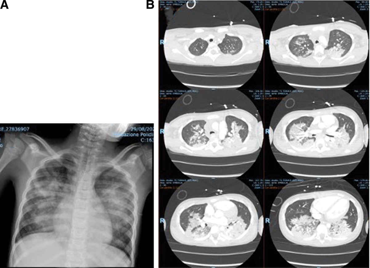
 Maximal intensity projection FDG-PET (A) and transaxial PET/CT (B and D), before treatment, show increased 18F-FDG uptake in the proximal valved conduit anastomosis in right ventricle (red arrows), as well as pulmonary infiltrate (D, yellow arrow), diffuse increased activity in the spleen (blue arrow) and reactive bone marrow (green arrow). Transaxial PET/CT (C) show normalization of metabolic activity after homograft replacement. 18F-FDGPET/CT = 18F-fluorodeoxyglucose positron emission tomography/computerized tomography. Transthoracic echocardiography (TTE): (E) Parasternal short axis (pulmonary valve at systolic phase) showing a 10 mm length vegetation attached to pulmonary valve; (F) Suprasternal view showing an axial view of pulmonary valve with the vegetation; (G) Parasternal short axis (pulmonary valve at diastolic phase) the arrow denotes the vegetation.
Within a week of the diagnosis a full body positron emission tomography/computed tomography (PET/CT) scan, performed to search for other infectious foci and establish baseline metabolic activity, revealed enhancement at prosthetic valve and spleen with lower lobe infiltrates in the left lung with no bone involvement (Fig 1). Two weeks into treatment, blood PCR for QF persisted positive and intermittent fevers continued. The case was presented in a multidisciplinary meeting, and the decision was to replace the pulmonary-valve conduit after 3 weeks of treatment. During the surgery, multiple vegetations were observed on the conduit mainly adhering to the leaflets, and an aortic homograft was placed. PCR for QF of the lesions on the removed conduit was also positive.
During follow-up, blood QF PCRs, tested on months 1, 4, 8, 20, 24, 28, and 36 after surgery were negative in all the visits and serology titers decreased and increased in subsequent follow-ups (phase I IgG of 1/16,384, 1/8192, 1/2048 and 1/8192 respectively on months 8, 20, 28, and 36 after surgery). The increase in phase I titers, tested routinely to monitor for relapse, happened while the child was asymptomatic with normal inflammatory markers, echocardiograms without evidence of vegetation, and negative blood QF PCRs and therefore serology titers were not helpful in making clinical decisions. Eighteen months into treatment, a repeat PET/CT showed clear improvement with no significant uptake around the valvular prosthesis, so it was decided to stop treatment after 19 months.
At the latest follow-up, 23 months from treatment discontinuation and 3 years from the diagnosis, he is asymptomatic and growing well.
Q fever (QF) is a worldwide zoonotic disease caused by Coxiella burnetii. According to the 2019 report from the European Center for Disease Prevention and Control (ECDC), the highest QF notification rate in Europe was observed in Spain (0.7 cases per 100,000 population).1 QF is usually transmitted from farm animals, mainly cattle, sheep, and goats, via inhalation of contaminated aerosols. Acute QF is usually asymptomatic in children. Endocarditis is the predominant form of chronic infection and has been observed in 1-16% of reported cases of QF, but few pediatric cases have been reported and even fewer have been associated with prosthetic material.2,3
The diagnosis of QF normally relies on serological methods. In acute infection, the immune response induces the production of anti-phase II and anti-phase I antibodies. Anti-phase II antibodies are predominant during primary infection and are detectable 7 to 15 days after clinical onset. The diagnosis of QF can be confirmed by a 4-fold increase in phase II IgG or IgM antibodies between two serum samples taken 3 to 6 weeks apart. Generally, titers of phase II IgG of ≥200 and/or IgM of ≥50 are considered significant for the diagnosis of primary QF. Anti-phase I IgG titers are associated with persistent infection and higher titers correlate with a higher positive predictive value for the diagnosis of Q fever endocarditis (QFE). It is recommended to investigate further when anti-phase I IgG titers are above 1:800 6 months after completion of treatment for acute QF. Residual IgG antibody titers may be detectable for years and even for life.4
Diagnosis of QFE typically requires demonstration of abnormalities on an echocardiogram; however, emerging data suggest that PET/CT could be a useful diagnostic tool. Unlike an echocardiogram, the diagnosis of endocarditis is essentially ruled out when PET/CT is negative. Conversely, an abnormal PET/CT can point to endocarditis in settings when there is a high index of suspicion of QFE and echocardiograms are negative for vegetations. PET/CT may also be also useful for clinical monitoring and guiding antibiotic therapy, especially when serology is misleading.5 However, there are concerns that FDG uptake around the prosthetic/periprosthetic area after heart surgery is common of PET/CT, and the uptake intensity and distribution pattern may remain stable during the first year after surgery.
The recommended treatment regimen for QFE is doxycycline and hydroxychloroquine or doxycycline with a quinolone. The combination of doxycycline and hydroxychloroquine requires a shorter duration of treatment and has demonstrated a lower risk of relapse, without evidence of lower mortality or the need for surgery.6 Limited data are available on treatment of chronic QF in children, and the safety of long-term hydroxychloroquine in children has not been determined. Alternative long-term treatment options in children include the use of a fluoroquinolone with rifampin or trimethoprim/sulfamethoxazole with doxycycline.
According to treatment recommendations, chronic QFE should be continued for at least 18–24 months and should be further prolonged until phase I IgG decrease by a fourfold. However, given that most patients treated appropriately for 18 months will clinically recover, they might not benefit from continued treatment despite high phase I IgG, as there is data suggesting a poor correlation between clinical and serological response.7 In our experience serologic monitoring created confusion as titers were erratic and increased and decreased without correlation to clinical events or evidence of relapse.
In conclusion, the diagnosis of QFE should be considered in any patient with blood culture-negative endocarditis and compatible exposure history. The patients should be closely monitored, basing medical decisions on clinical and inflammatory marker responses rather than on serological titers. Although promising, the role of PET-CT for decision making in follow-up needs to be further evaluated.
ACKNOWLEDGMENTSThank you to Dr. Arístides for clinical advice in this case.
REFERENCES 1. Jaltotage B, Ali U, Dorai-Raj A, et al. Q fever endocarditis: a review of local and all reported cases in the literature. Hear Lung Circ. 2021;10:1509–1515. 2. Raoult D, Tissot-Dupont H, Foucault C, et al. Q fever 1985-1998. Clinical and epidemiologic features of 1,383 infections. Medicine (Baltim). 2000;79:109–123. 3. Alhadhoud S, Vel M, Al Qbandi M. Q fever endocarditis after right ventricle to pulmonary artery conduit insertion: case series and review of the literature. Ann Pediatr Cardiol. 2018;11:60–63. 4. Fournier PE, Marrie TJ, Raoult D. Diagnosis of Q fever. J Clin Microbiol. 1998;36:1823–1834. doi:10.1128/JCM.36.7.1823-1834.1998. 5. Markowich A, Romani L, Leccisotti L, et al. Challenging case of chronic Q fever endocarditis: usefulness of 18F-FDG PET/CT in the diagnosis and follow-up. BMJ Case Rep. 2021;14:e243290. doi: 10.1136/bcr-2021-243290. 6. Raoult D, Houpikian P, Tissot Dupont H, et al. Treatment of Q fever endocarditis: comparison of 2 regimens containing doxycycline and ofloxacin or hydroxychloroquine. Arch Intern Med. 1999;159:167–173. 7. Anderson A, Bijlmer H, Fournier P, et al. Diagnosis and management of Q fever--United States, 2013: recommendations from CDC and the Q Fever Working Group. MMWR Recomm Rep. 2013;62:1–30.
留言 (0)