The use of near-infrared spectrometry (NIRS) in the management of critically ill patients with congenital heart disease has become almost ubiquitous for patients either in pediatric or pediatric cardiac ICUs, with about 90% of programs in the United States reporting using this device a decade ago and most recently, about 90% of surveyed programs in Europe reporting the same (1,2). How it is best used (all patients, neonates only, only babies with hypoplastic left heart syndrome [HLHS], single site vs multisite) and how it impacts outcomes remains unresolved after almost 2 decades of use.
In critical illness, occult regional hypoxia may occur much earlier than global tissue hypoxia. Global tissue hypoxia can be measured either by biomarkers such as whole blood lactate or by measuring mixed venous or central venous oxygen saturation directly. Theoretically, identifying regional hypoxia early and correcting it, might mitigate the development of global tissue hypoxia and therefore reduce morbidity and mortality in the critically ill. Doing it noninvasively and continuously, with a small, commercially available probe might be considered the Holy Grail of critical care medicine, the so-called canary in a coal mine. The hope was the NIRS monitor would offer just that.
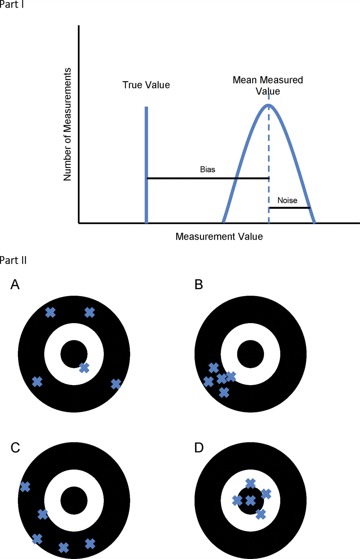
Jöbsis (3) described the potential for use of this novel technology in humans for both clinical and research purposes almost a half a century ago. NIRS monitoring was noninvasive, simple to use, and provided data under a myriad of abnormal physiologic conditions including pulseless physiology. On the other hand, regional tissue oxygenation is measuring physiologic derangements of a very small anatomic area. Redistribution or maldistribution of blood flow during low cardiac output states or shock would suggest that measuring such a small area could either underestimate or overestimate the global impact of these abnormal physiologic states. Correlation of NIRS data with more global indices of cardiovascular well-being such as systemic venous or jugular venous oxygen saturation as measured by co-oximetry has repeatedly shown that at times these measurements often lack correlation and may not be interchangeable (4). Such inaccuracy could lead to erroneous decision-making at the bedside. Because there is no gold standard technique to clinically measure regional oxygen saturation, at this time the data provided by NIRS becomes almost impossible to validate.
Ultimately, what NIRS monitoring promises is a noninvasive ability to measure the relationship between oxygen delivery (Do2) and oxygen consumption (Vo2). This relationship is arguably the single most important physiologic relationship in critically ill individuals and aberrations of this relationship result in cellular, tissue and ultimately organ system dysfunction and at the extreme end, death (5). In the ICU, accurate data reflecting this relationship therefore has a profound influence on our ability to prognosticate, and significant evidence exists suggesting optimizing this relationship leads to improved outcomes. During periods of low cardiac output or shock states, oxygenated blood is directed toward organ systems at need and away from those with low oxygen demands. NIRS can measure tissue oxygenation for multiple vascular beds with differing physiologic states at baseline and under duress. This would seem to be a significant advantage to the clinician.
While NIRS monitoring does offer insights to regional physiologic changes, problems associated with the inaccuracy of NIRS monitoring have been well described from its onset (6). Most recently, investigators from Boston compared a cerebral oxygen saturation index (as measured by NIRS) to co-oximetry values measured in the internal jugular vein in neonates recovering from stage 1 palliation. They found the sensitivity of NIRS cerebral values to be low, but most importantly, values below 30 were specific for low cerebral venous oxygen saturation (7).
Cerebral NIRS monitoring of neonates gained popularity in the late 1980s and early 1990s. Clinicians were looking for easy and accurate ways to measure cerebral blood flow and the relationship between Do2 and consumption in patients who might be at risk for brain injury (8). For patients with congenital heart disease, NIRS technology was initially employed in the cardiac operating room as a technique that allowed clinicians to monitor cerebral blood flow during periods of cardiopulmonary bypass (9).
In 2002, Tweddell et al (10) reported a cohort of patients with HLHS who underwent stage 1 palliation with outstanding results. Paramount to their postoperative management strategy was the use of an oximetric catheter placed in the superior vena cava at the time of the operation that allowed continuous measurement of systemic venous oxygen saturation, allowing clinicians to direct therapy at improving the relationship between Do2 and Vo2. Prior to this, estimates of Do2 in babies with HLHS were made by indirect measurements of overall well-being, such as heart rate, blood pressure, and acid-base status. Pulse oximetry, while universally used, could remain in the optimum range or even become elevated as derangements in the ratio of pulmonary to systemic blood flow worsened, rendering a very insensitive and often misleading indicator of tissue oxygenation (11).
The group in Wisconsin, understanding the value of being able to measure the relationship of Do2 to Vo2 globally, began to investigate how measuring regional tissue oxygenation might help gain insight into how to best protect the brain from derangements in brain oxygen transport (12). Their published work has led to the almost uniform use of NIRS monitoring for patients after stage 1 palliation of HLHS with hope that restoring a healthy relationship of tissue oxygenation would prevent end organ damage at both the regional and global levels.
In this issue of Pediatric Critical Care Medicine, Iliopoulos et al (13) present a well-designed study looking at NIRS monitoring data as it related to systemic venous oxygen saturation data and compared the performance of a new generation of NIRS monitors (FORESIGHT ELITE) to that of a widely used NIRS trend monitor (INVOS 5100C). The study by Iliopoulos et al (13) was performed on 36 infants and children undergoing planned cardiac catheterization for a variety of indications. Some of the patients were cyanotic and a secondary objective was to evaluate the performance of these probes in cyanotic patients. Each patient had one of the probes (FORESIGHT ELITE or INVOS) placed on either the right or left forehead and another on the ipsilateral flank. On the contralateral forehead a flank, a second set of probes, for the other manufacturer, was placed. Order of placement of each probe to right or left side appears to be appropriately randomized. The NIRS data were compared with venous oximetry data from the internal jugular vein (comparing sample to forehead probe) and the renal vein (comparing sample to flank probe).
Unfortunately, both monitors had what was considered poor to moderate concordance and poor to moderate correlation with the invasive venous oximetry. In fact, the authors report that interventions triggered by NIRS monitoring will either be delayed or triggered unnecessarily for almost 2/3 of the patients who are fully saturated. The authors conclude that the data are even worse for patients who are cyanotic. Their data suggest that the correlation of NIRS with jugular venous oximetry was very poor in this population of patients, patients who are at greatest risk for morbidity and mortality after cardiac surgery.
It is unclear just how to best use NIRS data when managing critically ill patients with complex congenital heart disease and physiology. Single site or multiple, single value or trend, absolute value or difference in cerebral and splanchnic NIRS values? There are many questions that remain to be answered. While adequate or acceptable NIRS values can be comforting, it can only be so if they correlate with other measures of cardiovascular well-being, we traditionally measure such as blood lactate values, acid-base status, heart rate, blood pressure, and the rest. At the very least, the article by Iliopoulos et al (13) should cause clinicians to give pause when making clinical decisions based on the NIRS-derived data alone. What is clear is that low values suggest your patient is at risk, and careful evaluation is warranted as to how best to correct this aberration in the critically important question regarding the relationship of Do2 to Vo2. It would be inappropriate to conclude that NIRS monitoring should be abandoned because of these deficiencies. Clinicians in ICU settings cognizant of alarm fatigue from noninvasive monitoring devices such as pulse oximetry giving spurious readings. But since there are decades of experience using pulse oximetry, clinicians are adept at assessing the data and making accurate decisions regarding its validity. The same will be true of NIRS monitoring. The clinician needs to use all available data to make the best clinical decision regarding the status of their patient. Only then will their ability to prognosticate improve, and use of the data to direct goal-oriented therapy optimized.
1. Rossi AF, Checchia PA, Lopez L, et al.: Web-based survey of current trends in hemodynamic monitoring after congenital heart surgery. World J Pediatr Congenital Heart Surg. 2012; 3:301–309
2. Hoskote AU, Tume LN, Trieschmann U, et al.: A cross-sectional survey of near-infrared spectroscopy use in pediatric cardiac ICUs in the United Kingdom, Ireland, Italy, and Germany. Pediatr Crit Care Med. 2016; 17:36–44
3. Jöbsis FF: Noninvasive, infrared monitoring of cerebral and myocardial oxygen sufficiency and circulatory parameters. Science. 1977; 198:1264–1267
4. McQuillen PS, Nishimoto MS, Bottrell CL, et al.: Regional and central venous oxygen saturation monitoring following pediatric cardiac surgery: Concordance and association with clinical variables. Pediatr Crit Care Med. 2007; 8:154–160
5. Pinsky MR: Oxygen consumption-delivery relationships: Physiology and pathophysiology. Semin Respir Crit Care Med. 1995; 16:372–381
6. Li J, Van Arsdell GS, Zhang G, et al.: Assessment of the relationship between cerebral and splanchnic oxygen saturations measured by near-infrared spectroscopy and direct measurements of systemic haemodynamic variables and oxygen transport after the Norwood procedure. Heart. 2006; 92:1678–1685
7. Rescoe E, Tang X, Perry DA, et al.: Cerebral near-infrared spectroscopy insensitively detects low cerebral venous oxygen saturations after stage 1 palliation. J Thorac Cardiovasc Surg. 2017; 154:1056–1062
8. Wyatt JS, Edwards AD, Azzopardi D, et al.: Magnetic resonance and near infrared spectroscopy for investigation of perinatal hypoxic-ischaemic brain injury. Arch Dis Child. 1989; 64:953–963
9. Fallon P, Roberts I, Kirkham FJ, et al.: Cerebral hemodynamics during cardiopulmonary bypass in children using near-infrared spectroscopy. Ann Thorac Surg. 1993; 56:1473–1477
10. Tweddell JS, Hoffman GM, Mussatto KA, et al.: Improved survival of patients undergoing palliation of hypoplastic left heart syndrome: Lessons learned from 115 consecutive patients. Circulation. 2002; 106(12 Suppl 1):I82–I89
11. Rossi AF, Sommer RJ, Lotvin A, et al.: Usefulness of intermittent monitoring of mixed venous oxygen saturation after stage I palliation for hypoplastic left heart syndrome. Am J Cardiol. 1994; 73:1118–1123
12. Hoffman GM, Stuth EA, Jaquiss RD, et al.: Changes in cerebral and somatic oxygenation during stage 1 palliation of hypoplastic left heart syndrome using continuous regional cerebral perfusion. J Thorac Cardiovasc Surg. 2004; 127:223–233
13. Iliopoulos I, Cooper DS, Reagor JA, et al.: Absolute Versus Relative Near-Infrared Spectroscopy in Pediatric Cardiac Patients. Pediatr Crit Care Med. 2023; 24:204–212


留言 (0)