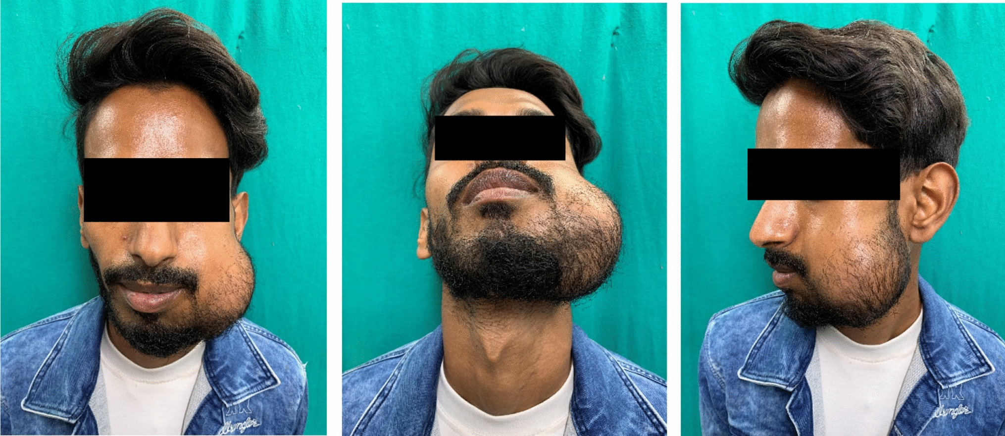
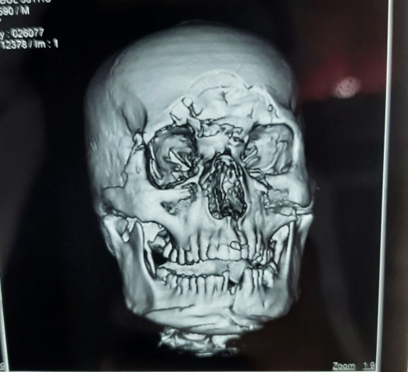
Diabetes-associated ROCM is the most prevalent manifestation of post-COVID-19 [3]. COVID-19 was declared a global pandemic by WHO. Because of poor understanding of the disease, its pathogenesis, and effective treatments, hypoxemic patients were treated with a variety of drugs (steroids, antibiotics, ivermectin, remdesivir, and tocilizumab), plasma therapy, oxygen therapy, and ventilation. Patients with DM are predisposed to COVID-19. A large meta-analysis of 851 ROCM cases found that DM was a risk factor [9]. The clinical signs of ROCM include central retinal artery occlusion and panophthalmitis. Other symptoms include proptosis and ptosis, facial swelling/discoloration, restricted ocular motility, visual loss, fixed pupils, and nasal or palatal eschar. Table 1 illustrates clinical signs of ROCM in our patients. Liposomal Amphotericin-B (5–10 mg/kg/day) or intravenous posaconazole or isavuconazole are used as medical line of treatment. [10] As a matter of course, a fungal disease such as this is staged as follows: stage 1 involves the nasal mucosa; stage 2 involves the paranasal sinuses; stage 3 involves the orbits (3a: nasolacrimal duct and medial orbit; 3b: diffuse orbit) stage 4 involves the central nervous system. The removal of the paranasal sinuses by turbinectomy, palatal resection, or medial orbital wall resection is required in stages 1, 2, and 3 of the disease. Stage 3c disease, with thrombosis of the central retinal artery or superior ophthalmic vein, and Stage 4 disease, with central nervous system involvement, will necessitate orbital exenteration/neurosurgical therapy. [11] After orbital exenteration, prosthetic rehabilitation was started in cases mentioned in Figs. 1 and 2. The definitive prosthesis in all cases was made from room temperature vulcanizing silicone. The cases as depicted in Fig. 1 were processed and completed by Analog method. Gunjan Pruthi et al. published a case series on prosthetic rehabilitation after orbital exenteration. In this case series the authors used different types of retentive aids for ocular prostheses like pin and socket, magnetic buttons, spectacles frame but in our case series, we have utilized anatomical undercuts and silicone based adhesives (Pro Bond) for retention [5]. In this case series the definitive prosthesis made by analog method had large defect sizes and all cases done by analog protocol had neurological involvement resulting in compromised manual dexterity so we could not use magnetic and pin socket attachment in these cases. 2 Cases as illustrated in Fig. 2 were processed by the digital protocol and completed by using analog method. In the digital protocol, we used 3D model of the patient’s face which was printed digitally using dental cad software (Exocad, Germany). The 3D printed model was used for fabrication of an acrylic conformer. Our digital protocol was different from an earlier published case report by Radhika A et al. in which they have described fabrication of an orbital prosthesis composed of a digitally produced acrylic resin conformer and traditionally produced silicon prosthesis [7]. Another case report published by Yunprn bi et al. describes digitally produced negative mold and packing of silicone done conventionally [8].
In this clinical case series Case number 5 had a large defect size and compromised bone support whereas case number 6 had a shallow defect, therefore to provide retention a spectacle-retained prosthesis was fabricated.
Usage of dental implants over adhesives and other mechanical retentive aids have been described in literature with advantages like better retention in large defects and convenient positioning of definitive prosthesis. [11,12,13,14] The advantages mentioned offer a greater acceptance by patient especially in younger individuals. In our case series, all the patients had a history of ROCM and Diabetes Mellitus II, in which the bone quality in the orbital region was compromised, and the overall general health of the patients also restricted the usage of implant retained ocular prosthesis in such patients. Shade matching of silicone with the patient’s skin, using intrinsic stains was challenging in all cases. Shade of the prosthesis turned out to be slightly darker in case 2 than the surrounding skin, whereas in rest of the cases shade matching was found to be satisfactory
.


留言 (0)