Insulin resistance (IR) refers to the reduced biological efficacy of insulin and the consequent decrease in glucose uptake in surrounding tissues, including the liver, skeletal muscle, and adipose tissues [1]. Mountains of evidence demonstrate that IR is an essential characteristic of chronic metabolic disorders (e.g., obesity, hyperlipidemia, and diabetes) [2]. Obesity is currently considered a global epidemic both in developed and developing countries. Excessive accumulation of body fat promotes obesity-associated metabolic dysfunctions, such as type 2 diabetes mellitus (T2DM), cardiovascular diseases, and neurodegenerative diseases [3]. A vast majority of obese individuals have increased plasma levels of free fatty acids (FFAs), which can result in a hepatic IR [4]. Excess accumulation of FFAs and their metabolites can interfere with insulin signaling and inhibit insulin-stimulated glucose uptake, which is closely linked to the pathogenesis of T2DM [5]. The underlying signaling pathways that FFA modulates hepatic IR have not been clearly elucidated.
Autophagy refers to the biological process in which damaged organelles, misfolded proteins, and other contents in cells are wrapped in double-membrane vesicles to form autophagosomes and transported to lysosomes for degradation [6]. As such, autophagy is dependent on both the rate of autophagic flux flow as well as the rate of substrate clearance by the lysosome [7]. Autophagy is the primary protein degradation system responsible for the turnover of bulky cellular constituents [8]. A variety of proteins are identified to be involved in the processes of autophagosome formation and autophagic flux flow. During autophagy, a cytosolic form of protein LC3 (microtubule-associated protein 1 light chain 3) is conjugated to phosphatidylethanolamine [9] to form LC3-II, which is recruited to autophagosomal membranes to elongate the phagophore membrane for autophagosome formation [10]. In addition, the receptor protein p62, also called sequestosome 1 (SQSTM1), directly conjugates with ubiquitinated protein aggregates for degradation via autophagy, which is featured to monitor autophagic degradation/flux [11]. Accumulation of p62 indicates impaired autophagic flux, whereas low p62 protein levels may represent efficient degradation. Hence, the combination of LC3-II and p62 thus is currently the most widely used autophagy monitor maker.
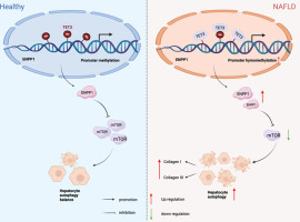
Ethanolamine phosphate phosphatase (ETNPPL, previously known as AGXT2L1) is a gene that encodes for ETNPPL protein and is abundantly expressed in the brain and liver [12]. ETNPPL is a metabolic enzyme that irreversibly degrades phosphoethanolamine (PETN) to acetaldehyde, phosphate, and ammonia. Currently, the physiological and pathological function of ETNPPL in mammals is still mysterious. A recent study in the brain shows that ETNPPL, as a phospholipid-precursor catabolizing gene, potentially mediates the brain's lipid metabolism [13]. ETNPPL has been reported to be upregulated in schizophrenia and bipolar disorder, a compensatory response to neurochemical imbalance [14]. Intriguingly, individuals who have schizophrenia are more susceptible to developing obesity, T2DM, and metabolic syndrome [15].
As a pivotal cellular housekeeping system, autophagy contributes to the maintenance of intercellular homeostasis [16]. Dysfunctional autophagy might contribute to the development of metabolic disorders, including IR, diabetes, and obesity [17]. The liver, a crucial issue in maintaining glucose homeostasis, acts as a target organ for insulin action [18]. Palmitic acid (PA), the most prevalent circulating saturated fatty acid, has been reported to induce hepatic IR by impairing cellular signaling pathways [19]. Little is known about ETNPPL, and a potential link with IR has yet to be established. Based on these, here we are prompted to investigate the role of ETNPPL and the signaling mechanisms in the regulation of autophagy and its contribution to the pathogenesis of PA-related IR in hepatocytes.

留言 (0)