Recent progress and challenges in single-cell imaging of enhancer–promoter interaction
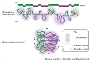
The question of how enhancers control the activity of promoters, across the thousands to millions of basepairs separating the two, has long fascinated biologists. In the last decade, chromatin conformation capture approaches, especially Hi-C, enabled mapping such 3D interactions across the genome. In particular, the discovery of topologically associating domains (TADs), and the correlation of these structural domains with several genetically defined enhancer–promoter (E–P) interaction domains, substantially advanced our understanding of E–P specificity [1]. Here, we discuss some innovative microscopy-enabled investigations from the last few years, which have illuminated TAD organization at the single-cell level, and begun to probe its dynamic nature. As the underlying technologies have been reviewed recently 1, 2, 3, 4, 5, 6, we focus on describing some implications of these emerging data for current models and explaining how some apparently contradictory key observations from recent works may be reconciled.


留言 (0)