
記住我

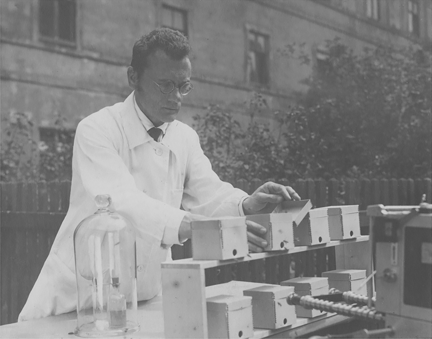


We collected Ecdyonurus sp. larvae end of April in Schlingenbach stream (Overrath, Germany). Some of the larvae were fixed in 70% ethanol (EtOH), while others were transported alive to a flow tank at the Kiel University. The larvae used in our study were middle to later instars. For identification, the key from Eiseler (2005) was used. The larvae in our study belonged to the Ecdyonurus venosus group (Ecdonurus cf. torrentis), but reliable identification on species level seems only possible in nymphs, so we refer to them in this study as Ecdyonurus sp.
Cross-sectional femur profileAlthough all legs will have an impact on the flow forces, we focused on the first femora, as they are typically most directly exposed to the current (Fig. 1a). Length and chord length of each foreleg femur from 10 different Ecdyonurus sp. specimens were measured under a dissecting microscope. For the preparation of cross sections, the right and left first femora of 10 Ecdyonurus sp. specimens were embedded in the Epon epoxy resin. For this purpose, we dehydrated the femora of fixed specimens in a series of ethanol (70%, 95%, 100%) and embedded them in Epon following the protocol after Luft (1961). To get semi-hard Epon resin, 15 ml Epon I (124 ml Epon 812, 200 ml Dodecenyl Succinic Anhydride) and 35 ml Epon II (300 ml Epon 812, 267 ml N-methyl Nadicanhyrdid) were mixed for 30 min, 0.75 ml Tris-2,4,5-dimethylaminomethylphenol added and mixed for another 30 min. 20 molds were filled with Epon half full and left to polymerise at 60 °C overnight. After infiltration (one part 100% Ethanol dehydrated by molecular sieve: one part Epon for 30 min, one part 100% Ethanol dehydrated by molecular sieve: two parts Epon for 90 min, pure Epon overnight, pure Epon 6–7 h), one leg was put into each mold. Then, the molds were filled up to the top with the pure Epon and left to polymerize at 60 °C for 48 h. The cross sections were prepared from the middle section of the first femur. A typical cross section (Fig. 1b) was chosen for the construction of the femur model.
Determination of the angle of attack (AOA)We took video recordings and photos of living Ecdyonurus sp. larvae in a flow tank (Fig. 1a). By analyzing video footage and photos, we estimated the AOA of the femora of the living larvae using the program ImageJ (NIH, https://imagej.nih.gov/ij/). As the perspective interferes with the determination of the AOA from 2D-images, we additionally determined the morphologically possible range of the femur’s AOA. Therefore, we placed rehydrated larvae on a polyvinylsiloxane layer (President Light Body, Coltene Whaledent, Hamburg, Germany) at the bottom of a Petri dish and pinned them with insect pins to the rubber-like polymer. To determine the AOA, we measured the possible tilt of the femur in relation to the body axes and the substrate. Three coordinates surrounding the larvae in a triangle were measured to determine the substrate surface plane. Along the longitudinal body axis, we measured three points and one more lateral point on each side of the first body segment to determine the mediolateral axis. The femur position was determined by three points, with point 1 at the femur–tibia joint, and points 2 and 3 outside the femur edge. Points were marked with a drop of red varnish and measured using Mitutoyo Measuring Microscope (MF-A Series, Series 176). 3D coordinates of all points were determined at the minimum and maximum AOAs of the femur while manually changing the position of the femur and securing the position with additional insect pins. The location of the measurement points is illustrated in Supplement 1.
Femur modelsAs the larvae often remain motionless in the same position, steady-state fluid dynamics and 2D-modelling are applicable. 2D-wing models were built from the foreleg femur pair of Ecdyonorus sp. The contour line of the cross section of the femur’s middle region was digitized using software Rhino 4.0 (McNeel, Seattle, USA), and the cross section was stretched to 250 mm in length. Different sizes of femur models were chosen to investigate not only at a range of the Re number comparable to the natural situation of Ecdyonurs sp. (M1), but also to investigate flow forces at higher Re numbers. The latter allowed the comparison with measurements described in the literature, which often have been performed at higher Re, and also can be important for potential technical applications. For all femur models, the cross section was enlarged (Table 1) to obtain Re from 1700 to 24,000 at wind speeds from 2.5 to 6.7 m/s. The femur models were printed using a Contex MX powder-based 3D printer (Contex A/S, Alleroed, Denmark). To smoothen the surface of the raw 3D prints, the surface was filled with body filler, polished with sandpaper and finished with an acrylic spray paint. We also included a flat plate as a reference.
Table 1 Geometrical variables and Reynolds numbers (Re) of the first femur of Ecdyonurus sp. and the profiles used in this study Wind tunnelDrag and lift forces perform comparably in various fluids, if determined for the same Re (Vogel 1994). For our experiments, we used a custom-made wind tunnel of Eiffel-type (open circuit) with an open test section, nozzle diameter 0.46 m, turbulence 0.3–0.6% (Dickinson and Götz 1993), and adjustable wind speed of 0.5–17.0 m/s (measured by a Pitot tube connected with a digital manometer EMA 200 with a range of ± 200 Pa (Halstrup-Walcher GmbH, Kirchzarten, Germany). The femur models were connected vertically to the force balance (Fig. 2). To limit spanwise flow and induced drag, an endplate on each side of the profile (distance < 0.5 mm) was placed.
Fig. 2
Schematic top view of the experimental setup in the wind tunnel. The artificial femur profile was mounted between two endplates on the two component force balance. The distance between the leading edge of the ground board and the leading edge of the femur profile was 100 mm. The distance to the ground (h) varied from 4–60 mm by moving the motorized translation stage mounted to a ground board
The Reynolds number is important to compare the flow conditions. It can be calculated from the equation:
where l is a characteristic length (here chord length), U the fluid velocity and ν the kinematic viscosity (water at 10 °C: 1.3063 × 10⁻⁶ m2 s−1, dry air at 20 °C, 1.461 × 10− 5 m2 s−1). All Re for the performed experiments (1700–24,000) were below the critical Re = 32,000, where the flow would change from laminar to turbulent. The femora of Ecdyonorus larvae have no sharp leading-edge structures, which would evoke a turbulent flow at lower Re, so that laminar flow and the presence of a boundary layer can be expected. Ecdyonurus larvae tolerate flow speeds up to 2 m/s (Butz 1975). Re for flow conditions in their natural habitat, which can be calculated from the chord length and the flow speeds described for the natural habitat, range from 75 to 1500. In our experiment, 1700 were considered comparable with the biological case. Pretests revealed that measurements at Re < 1700 reached the limits of the experimental setup due to given limitations in wind tunnel speed, the precision of balance, and model. The higher Reynold numbers in our experiments (> 4500) have no relevance for the biological system, but is of interest for comparison of the aero- or hydrodynamic characteristics of the femur profile with other profiles, such as the widely applied NACA profiles, and for the potential transfer to biomimetic technical solutions.
The blocking of the femur model, mounted in the test section separated by the end plates, is 0.9% (M1), 2.3% (M2) or 4.5% (M3). Overall, the experimental setup in the open test section in front of the outlet nozzle, including femur model, force balance mounting, and endplates has a blockage of 10.6% in total. The femur profiles have, due to their relatively high thickness, no supplement effect on the projected frontal area for angles up to 20°.
For the ground effect measurements, the blockage was constant at 16%, because the ground plate with the parallel translation was additionally positioned in the test section. In open test sections, blockage corrections are usually small (Barlow et al. 1999) and are commonly ignored (Cooper 1998), but a correction of the velocity can increase the significance of the measurements (Sayers and Ball 1983). Therefore, velocity measurements were taken at the femur profile with all experimental setup mounted, so that no further blockage correction must be applied.
Lift and drag measurementsLift and drag were not only measured for the AOAs observed for the femora of Ecdyonurs sp. but for a wider range of AOAs to allow comparison with the polars of common airfoils. The range of AOAs was − 40° to + 40°, with a step size of 2°. Positioning, measurement, and mean value calculation were automated by a customized Labview script (National Instruments, Austin, Texas). Each experimental case was repeated 10 times (n = 10). Lift and drag coefficients (CL,CD) were calculated from the measured forces for lift (L) and drag (D) applying the standard formula for airfoils.
where ρ is the fluid density, U is the fluid velocity, and S is the profile surface area. S was calculated as the projected surface area. Forces were measured using a friction-free two-component balance with two independent platforms based on air-cushioned sledges and inductive displacement transducers (TR10, HBM GmbH, Darmstadt, Germany) at a sampling rate of 100 Hz at 2.5 m/s and 600 Hz at 6.7 m/s, respectively. The signal was recorded for 30 s and 5 s, respectively, by a A/D transducer (Spider 8, HBM, Darmstadt, Germany), low-pass filtered, and averaged at each data point.
Ground effectsThe thickness of the boundary layer (\(\delta\)) was estimated applying the Blasius equation for the case of laminar flow over a flat plate in a parallel direction to the free flow
$$=5\sqrt \text }}}, $$
(4)
where ν is the kinematic viscosity, x is the distance from the frontal edge of the plate, and U is the flow velocity. To estimate the boundary layer for Ecdyonurus sp. under natural conditions, we calculated the thickness for an assumed minimum case (x = 0.01 m, U = 2 m/s, water at 10 °C) and maximum case (x = 0.1 m, U = 0.1 m/s) by estimating the environmental extreme conditions. The resulting thickness of the boundary layer under natural conditions is 0.4–5.1 mm. In the videos, we observed the femora of living Ecdyonurus sp. at 0–2 mm above the ground, so that interactions with the boundary layer of the ground are probable. The ground clearance (h/c) can be calculated from the distance to the ground (h) and the chord length (c) (Fig. 2). The h/c was 0–2 under natural conditions.
A unilaterally tapered and strengthened ground board (PVC rigid foam, 250 × 200 × 3 mm, Fig. 2) was used to simulate the ground in the wind tunnel. The leading edge of the femur profile was positioned 100 mm behind that of the ground board. In accordance with Ahmed and Sharma (2005), we expected a maximal influence of the ground at h/c < 1. Femur model M1 was measured at Re = 1700 for a distance to the ground (h) of 4–60 mm (equates h/c 0.4–6.0). The boundary layer thickness estimated by Eq. (4) (x = 100 mm, U = 2.5 m/s, dry air at 20 °C) was 3.8 mm at the leading edge of femur model M1. Two AOAs (− 10°, − 20°) were tested, to include the range of possible AOAs (n = 10 for each angle and distance).
留言 (0)