
記住我

Sixty adult male Wistar rats weighing 150–200 g were used in the study. Animals were purchased from the Egyptian Organization for Biological Products and Vaccines VACSERA, Egypt. They were acclimated for 7 days before experimentation and kept under controlled environmental conditions; a 12-h dark/light cycle, temperature ~ 25 °C and with standard diet (Meladco for Animal Food, Egypt) and water during the experiments. All efforts were made to minimize the number and suffering of animals and the study procedures were done following the European community guidelines for care and use of experimental animals (EEC Directive of 1986) and it was approved by the Ethical Committee, Faculty of Medicine, Ain Shams University.
Treatments and Experimental DesignAlirocumab (Sanofi,Egypt) was administrated subcutaneously (s.c) in three doses (4, 8 and 16 mg/kg/week representing Aliro-L, Aliro-M, Aliro-H respectively). Doses of alirocumab were chosen guided by human equivalent dose calculation and based on previous study (Shin and Seol 2010; Abuelezz and Hendawy 2021). Animals were randomly divided into five equal groups: the first unstressed group served as control, the second group was exposed to chronic unpredictable mild stress protocol (CUMS), daily for 6 weeks, whereas the last three groups were exposed to CUMS and concomitantly received once daily Alirocumab in a dose dependent fashion (Aliro-L, Aliro-M, Aliro-H) for the 6 weeks of the daily stress protocol.
A pilot study was performed to detect the effect of the drug on the behavioral tests and lipid profile in animals before the start of our study and it revealed a non-significant difference between untreated and treated normal animals.
Chronic Unpredictable Mild Stress Protocol (CUMS)The CUMS protocol was performed following that previously described by Abuelezz et al. (2017). Accordingly, rats were exposed to mild stressor episodes (2–3 stressors) daily for 6 weeks. The stressors are reversed light cycle, pairing, cage tilting, stroboscopic light (60 flashes/min), cold temperature (10 °C), 120 min restricted access to food (3 pellets), 60 min empty water bottles, foreign body in the cage, soiling of cage with 50–100 ml water, 60 min immobilization stress and 60 min cage agitation (cages were rotated by gentle rotation ~ 10 rpm). The stressors were applied in a semi random sequence to be unpredictable. The timeline of the experimental design and measurements are shown in Fig. 1.
Fig. 1
Timeline for chronic unpredictable mild stress (CUMS) and Alirocumab (Aliro) administration schedule, behavioral tests, sacrifice, biochemical, Real-time reverse transcriptase (PCR), western blot and high-performance liquid chromatography (HPLC) analysis. (OFT) open field test and (FST) forced swim test
Behavioral TestsOpen Field TestOFT was used to detect exploration, locomotors activity and, anxiety-related behaviors. For this purpose, a quadrangular arena 60 × 60 × 45 cm divided into 16 equal squares was used. Before starting the experiment, animals were allowed to acclimatize to the test room for 1 h, and then for 5 min each rat was individually placed in the center of the well-illuminated quadrangular arena. The number of crossed squares (visited with all 4 ft), number of entries to the central zone (central 4 squares), latency to leave the central zone and time stayed in the central zone, duration (sec) of both rearing (standing upright on the hind paws) and of grooming (face rubbing and licking both paws and fur licking) mean velocity (cm/s) and total distance travelled (cm) were all recorded. The test arena was cleaned by 70% alcohol after each rat (Abuelezz et al. 2017).
Forced Swimming TestA vertical glass cylinder (diameter 22.5 cm X height 50 cm) filled with fresh water 35 cm high maintained at ≈25 °C was used for this test. For training, each rat was forced to swim individually for 15 min after the OFT. A re-exposure to the FST was done 24 h later for 5 min and the experiments were videotaped for later scoring of the immobility, swimming, and struggling time. Depressive like behavior was exhibited in the form of reduction in the time spent in active behavior (i.e. struggling or swimming) and increased the immobility time (Abuelezz et al. 2017).
Then animals were anesthetized with an intraperitoneal injection of urethane at the dose of 1.3 g/kg (Guo et al. 2007). Then rats were sacrificed by decapitation, and their brains were dissected out and washed by cold saline. The procedure was done between 10:00–12:00 am to avoid fluctuations in the glucocorticoid hormone levels.
Adrenal WeightBoth adrenal glands were excised and weighed. The increase in adrenal gland weight is considered as an indirect indicator of the hypothalamic-pituitary adrenal (HPA) axis activation which is evident in chronic stress status due to the release of glucocorticoid hormones by the adrenals in response to the different psychological and physical stressors (Rezin et al. 2008).
Sample Collection and Biochemical MeasurementsBlood samples were collected and centrifuged for 15 min at 1000 g to separate serum. Brains were dissected and the hippocampi were quickly isolated on an ice cold plate. For each group, the right hippocampus of 6 rats was used to pro-inflammatory cytokines and the left hippocampus was used for the real-time reverse transcriptase polymerase chain reaction (PCR) analysis. For the remaining rats, the right hippocampus was used for neurotransmitter analysis and the left hippocampus was used for western blot analysis. The samples were immediately stored at − 80 °C until assayed.
Determination of Serum CorticosteroneRat corticosterone enzyme-linked immune sorbent assay (ELISA) kit (DRG Instruments Gmbh, Marburg, Germany) was used according to the manufacturer’s instructions to determine serum corticosterone level.
Determination of Hippocampal Pro-inflammatory CytokinesFollowing the instructions provided by the Millipore multiplex Rat Cytokine enzyme-linked immune sorbent assay (ELISA) Kit; IL-1β, IL-2, IL-6 and TNF-α levels were measured. Briefly, hippocampi were homogenized in 0.01 M Tris hydrochloride formed of 5.8% sodium chloride, 10% glycerol, 1% Nonidet P40 (NP-40), 0.4% of ethylenediamine tetraacetic acid (EDTA) and protease inhibitors (Complete Protease Inhibitor Cocktail Tablets, Roche). Samples were then centrifuged for 5 min at 10,000 g at 4 °C and concentrations were determined on the Luminex® platform.
Real-Time Reverse Transcriptase (PCR)According to the manufacturer’s protocol, the total RNA was extracted from the hippocampi samples which were homogenized with Trizol reagent (Invitrogen, CA, USA). The cDNA was synthesized from total RNA using the high-capacity cDNA reverse transcription kit (Applied Biosystems, USA) and was amplified by the SYBR green Master Mix kit (Invitrogen, CA, USA) through comparative quantitative real-time reverse transcriptase PCR. The following primers were used: β-actin (forward) 5′-GCAGGAGTACGATGA GTCCG- 3′ and (reverse) 5′-ACGCAGCTCAGTAACAGTCC-3′; NF-κB: (forward) 5′—GCGCATCC GACCAACAATAAC-3′ and (reverse) 5′-GCCGAAGCTGCATGGACACT- 3′; TLR4 (forward) 5′—AGCTTTGGTCA GTTGGCTCT-3′ and (reverse) 5′-CAGGATGACACCATTG AAGC-3′; PCSK9 (forward) 5′-GCTTCAGCGGCTTGTTCCT-3′ and (reverse) 5′-TGCTCCTCCAC TCTCCACATAA-3′; IDO-1, (forward) 5′-AGC ACT GGA GAA GGC ACT GT-3′ and (reverse) 5′-ACG TGG AAA AAG GTG TCT GG- 3′. The mRNA expression of the tested genes was normalized to those of β-actin.
High-Performance Liquid ChromatographyFollowing the methodologies described previously by Bellac et al. (2006) a Luna high-performance liquid chromatography (HPLC) column [5 μm C18 (2) 150 × 4.60 mm; Phenomenex, Torrance, CA, USA] was used for Kynurenine determinations in hippocampal tissue homogenates. The mobile phase for ultraviolet detection consisted of; 15 mmol/L sodium acetate/acetic acid solution containing 2.7% (v/v) acetonitrile, pH 3.6. The flow rate was set at 1.3 mL/minute. The injected sample volume was 100 μL. Peaks were detected at 365 nm wavelength. 3-Nitro-ltyrosine was used as an internal standard. For Tryptophan levels determination in the hippocampal tissue homogenates, the mobile phase for the electrochemical detector HP 1049A (Hewlett Packard, Palo Alto, CA, USA) consisted of acetonitrile, 100 mM acetic acid, 100 mM ammonium acetate (10:90) v/v, and 50 mg/L ethylene diamine tetra acetic acid, pH 5.1.31 3-Methoxy-4- hydroxyphenethyl alcohol as an internal standard was used. Measurements were done at an electrode potential of 0.850 mV.
Western Blot AnalysisBy using ice-cold RIPA buffer the total protein was isolated from hippocampal tissues. BCA Protein Assay Kit (Thermo Fisher Scientific, USA) was used to measure the protein concentrations. A nitrocellulose membrane (Millipore Co., USA) was used for immuno-blotting where the protein samples (30 μg) were separated using SDS–polyacrylamide gel electrophoresis and then transferred to. For 1 h the membrane was blocked with 5% skim milk in Trisbuffered saline (150 mM NaCl, 0.1% Tween 20, 20 mM Tris, pH 7.4). Then the proteins were detected by incubation with primary antibodies against HMGB1, RAGE at 4 °C overnight. Later the membrane was washed for 3 times in Tween 20 + PBS and was incubated by the horseradish peroxidase-conjugated secondary antibodies for 2 h. The immuno-blots were visualized by a Millipore ECL Western Blotting Detection System and the variables protein expression levels were normalized to those of β-actin.
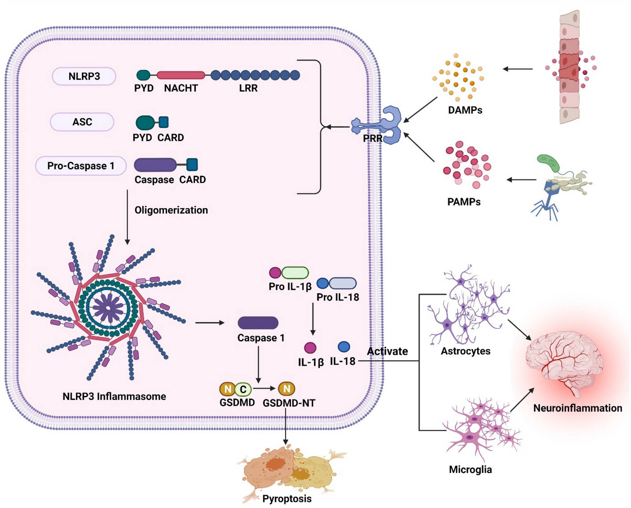
The following antibodies were used: anti-β actin (1:2,000, sc-130657, Santa Cruz Biotechnology), anti-HMGB1 (1:1,000, ab18256; Abcam), anti-RAGE antibody (1:2,000, sc8230; Santa Cruz), anti-NLRP3 antibody (1:2,000, sc-66846, Santa Cruz Biotechnology), anti-ASC antibody (1:2,000, sc-22514-R, Santa Cruz Biotechnology), pro and cleaved Caspase1 antibody (1:2,000, sc-514, Santa Cruz Biotechnology) and pro and mature IL-1β (1:1,000, AF-401-NA, R&D Systems).
Statistical AnalysisData were expressed as mean ± standard error of the mean (SEM). Using Graph-pad prism 5 statistical comparisons were carried out using one-way analysis of variance (ANOVA) followed by post-hoc Bonferroni test. The minimal level of significance was identified at P < 0.05. Correlation-coefficient analysis was performed by the Pearson's r.
留言 (0)