
記住我


This study is a retrospective cohort, single center study. The study protocol was approved by the Institutional Review Board at Stony Brook University and is conducted in accordance with the tenets of the Declaration of Helsinki. Informed consent was obtained by the subjects. Patients enrolled in the Argus II Post-Approval Study (PAS) (clinicaltrials.gov identifier, NCT01860092) at Stony Brook University Hospital were invited to enroll in the study.
Patient eligibility criteriaInclusion criteria included: patients with profound retinal degeneration and subsequent vision loss resulting in bare light perception or worse vision (> 2.9 logMAR). Eligibility was extended to those with a diagnosis of outer retinal dystrophy. Patients are to attend follow-up visits, rehabilitation measures, and continuing device fitting and programming.
Exclusion criteria included: patients with diseases that could affect successful implantation and could compromise functional optic nerves, such as optic nerve disease, trauma, severe strabismus, central retinal artery or vein occlusion, history of retinal detachment. Patients with ocular conditions that could complicate surgical implantation were not included in the study, such as axial lengths < 20.5 mm or > 26 mm and/or corneal ulcers.
Patient and public involvementPatients or the public were not involved in the design, or conduct, or reporting, or dissemination plans of our research.
Data collectionPatients were followed up at month 1 (M1), month 3 (M3), month 6 (M6), month 12 (M12), and month 24 (M24) post implantation. Fundoscopy was done at baseline prior to surgical implantation and at each subsequent follow up visit as part of the Argus II Retinal Prosthesis Post Approval Study. Images were taken using both Topcon 50 IX Retinal Camera (Topcon, Tokyo, Japan) and Optos Retinal Camera (Dunfermline, Scotland, United Kingdom).
Postoperative Fundoscopy and image analysisFundoscopy from each follow-up visit (M1, M3, M6, M12 and M24) was extracted from the IBM Merge Healthcare (IBM Corp, Armonk, New York, USA) and Optos software using unique patient identifiers. As part of our study, each patient enrolled was identified as A01, A02, A03, A04 and A05 to maintain confidentiality. Images that were selected for study purposes at each visit consisted of clear depictions of the optic disc and the retinal implant. This allowed for the ability to analyze the data using NIH ImageJ software [14].
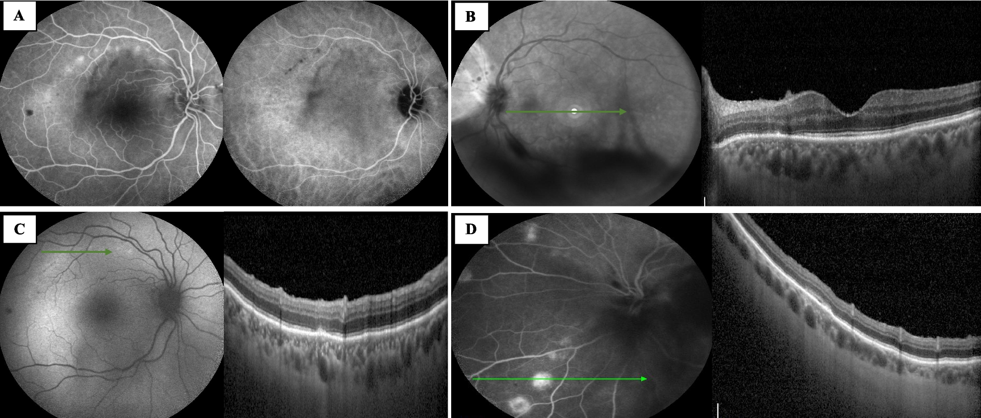
Determination of linear and angular movementFundus images from all patients taken during M1, M3, M6, M12, and M24 were imported into NIH ImageJ v1.53a to measure lengths AB, AC, BC, and angles ?, ?, ?, and ? (Fig. 1) (Schneider et al. 2012). As reported by the Argus II Retinal Prosthesis System Surgeon Manual, the diameter of each electrode is 200 μm in diameter [7]. The diameter of one electrode from each image analyzed was measured in pixels on ImageJ, and a scale was set based on the 200-μm measurement provided by the surgeon manual. Following the calibration, each length was measured in micrometers, and angle measured in degrees.
Fig. 1
Fundoscopy of patient A-03 at month 24 post implantation with markings to show distances and angles that were recorded using NIH ImageJ: A A represents the point in the center of the optic disc, B is the point at the top corner of the surgical handle on the implant, C is the point in the center of the tack. Alpha (α) represents the ∠CAB, gamma (Ɣ) represents the ∠ACB, beta (β) represents ∠ABC. B delta (?) measures the angle made by the line along the edge of the implant to the horizontal of the image frame
Length AB represents the distance from the optic disc to the surgical handle on the electrode array. This distance reflects the implant moving towards or away from the optic disc in a linear fashion. Angle ? (optic disc-tack-surgical handle), angle ? (tack-optic disc-surgical handle), and angle ? (optic disc-surgical handle-tack) are reflective of rotation around the axis of the tack. Another measure for showing the rotation around the axis is the angle to the horizontal of the image frame (angle ?). Variations in such markers between postoperative visits M1, M3, M6, M12, and M24 would be reflective of the linear movement and rotation of the implant over time (Fig. 1).
Other markers recorded in this study were length AC which is the distance from the optic disc to the tack and length BC which is the distance from the surgical handle to the tack. Both are representative of steady measures over time and are used as retinal references.
Statistical analysisRepeated ANOVA was used to assess for changes in distance of the implant over time. P values less than or equal to 0.05 were considered statistically significant. All statistical analysis was conducted on IBM SPSS Version 27.0 (IBM Corp, Armonk, New York, USA).
留言 (0)