
記住我

SVC syndrome refers to the obstruction of venous flow in the superior vena cavaIntrathoracic, malignancies are responsible for 60–85% of cases of SVC syndrome, and the occurrence of device-related SVC obstruction has increased and accounts for around 20–40% of overall cases [3]. Procedures performed on venous vasculature, resulting in a possible intimal injury or venous stenosis, provoked by transvenous leads, seem to be the most plausible explanation for the observed complication [4]. And what is striking, really remarkable, is when leads were serially evaluated with Doppler ultrasonography after initial placement, nearly 25% had associated thrombus, the majority of which occurred within 3 months [5]. In our case, although the patient had been taking anticoagulants for paroxysmal atrial fibrillation, he developed SVC syndrome after placement of a pacemaker for about 2 years. Thus it can be seen the patient's clotting was normal and no tumor was detected, but he was still at high risk for thrombosis.
Almost half of the SVC syndrome patients have relevant pulmonary embolism, which should prompt physicians to take positive therapy. Unfortunately, however, treatment options for device-related SVC obstruction are mainly supported by case series and anecdotal experience. Anticoagulation remains a common practice. The current practice is to start warfarin after 5 to 7 days of low-molecular-weight heparin. Other treatment options include lead extraction, thrombolysis, venoplasty,stenting, thrombectomy, and surgical grafts [6]. DOACs are now considered an important first-line therapy, and it has been reported that anticoagulation with Edoxaban 60 mg once daily could make complete resolution of the thrombosis after 3 months [4]. Although the etiology of pacemaker induced SVC syndrome is not identical to that of upper extremity deep vein thrombosis (UEDVT), it is important to note that the risk factors for UEDVT also include implantable pacemakers. The Swedish retrospective clinical study by Montiel et al. enrolled 55 patients treated with DOAC for 3–6 months because of UEDVT, the vast majority (84%) were treated with rivaroxaban, whereas 13% and 4% got apixaban and dabigatran, respectively., and found that DOAC can be used in the treatment of UEDVT patients with acceptable efficacy and safety [7]. The Italian multicenter retrospective study by Porfidia et al. also supports the concept that DOACs might be safe and efective for treating UEDTV [8]. Due to the similar pathophysiological mechanism, the research results and treatment experience of the two can learn from each other.
However, the choice of anticoagulant and duration of therapy remains controversial in the setting of SVC syndrome related to device leads. It is currently recommended to start 3 months of anticoagulant therapy, if symptoms persist, invasive venography should be performed to further characterize the source of occlusion. In a Canadian prospective multicenter study (CATHETER-2), 70 patients received rivaroxaban 15 mg twice daily for 3 weeks, followed by 20 mg once daily, in 51% of cases preceded by LMWH. This trial showed promise in treating central venous catheter (CVC)-UEDVT in cancer patients. However, raised concerns abput the risk of major bleeding and clinically relevant nonmajor bleeding, which occurred in 13% of patients during the 3-month follow-up [9]. This finding could be partly explained by the loading dose of rivaroxaban, since most of the events occurred in the first month. For our patient, considering the small amount of blood ooze in the left eye, we once again recommended the patient to switch to warfarin so that severe bleeding could be managed in time. However, the patient still required rivaroxaban, and we eventually increased the dose to 20 mg per day to achieve a therapeutic effect. Surprisingly, there were no significant abnormalities in coagulation function after increasing the dosage of anticoagulant drugs, no associated adverse events were observed. His symptoms of obstruction improved significantly after 4 months. Fortunately, the patient had complete resolved nine months later and has been taking adequate rivaroxaban anticoagulant.
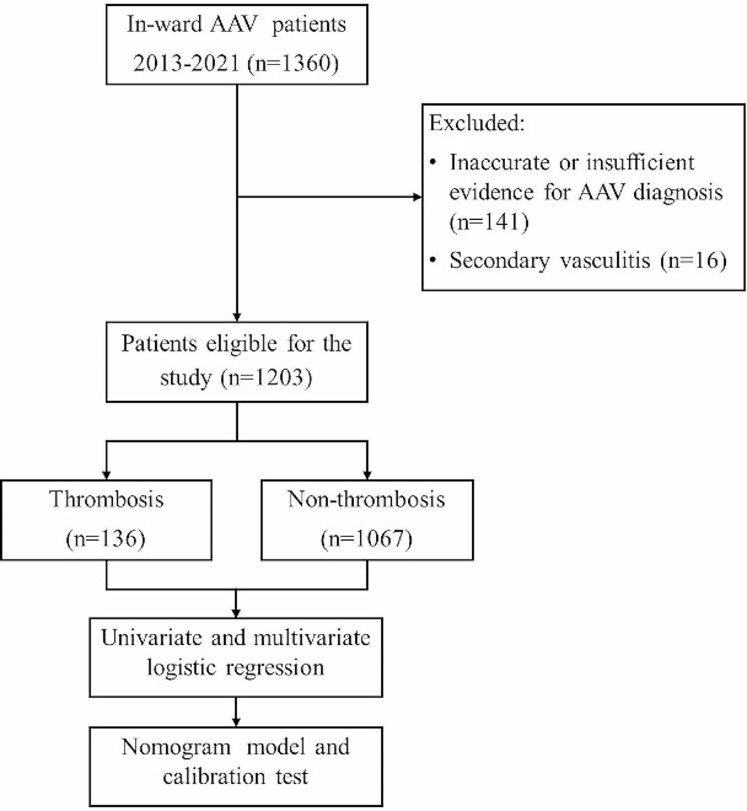
For occlusions that are relatively acute or subacute (within weeks to months) with relatively fresh thrombus, debulking strategies, including the local administration of thrombolytics or the use of venous thrombus extraction devices, should be considered. If leads are reimplanted or left in place, lifelong anticoagulation is recommended [2]. Despite the DOACs being increasingly used in real-world experiences, randomized trials for the management of the pacemaker lead induced SVC syndrome is still lacking. For the management process of Lead-Associated SVC Obstruction, we can refer to Fig. 3.
Fig. 3
This figure shows an approach to the diagnosis and management of lead-associated superior vena cava (SVC) obstruction. # If thrombus is in the first 2–4 weeks following device implantation. *If leads are left in place, consider lifelong anticoagulation. CT: computed tomography; SVC: superior vena cava
Our case highlights the medical management of SVC syndrome with its early detection and the clear effect of the use of DOACs. This is a good option for patients who do not want to undergo interventional or surgical procedures.
留言 (0)