Extraction of metabolites
After the addition of 300 μL of water, the samples were vortexed for 30 s. The samples were precooled in dry ice and subjected to three cycles of repeated freezing–thawing in liquid nitrogen. The samples were vortexed for 30 s and sonicated for 15 min in an ice-water bath. A 250 μL aliquot of the clear supernatant was transferred to a new EP tube, and 750 μL of methanol (precooled at − 40 °C) was then added. The remaining sample supernatant was used to measure the protein content. The samples were vortexed for 30 s, incubated at − 40 °C for 1 h and centrifuged at 12000 rpm (RCF = 13800(× g), R = 8.6 cm) for 15 min at 4 °C. A 900 μL aliquot of clear supernatant was collected and dried by spinning. The residue was reconstituted with 180 μL of ultrapure water, and the reconstituted samples were vortexed, filtered through the centrifuge tube filter, and subsequently transferred to inserts in injection vials for HPIC-MS/MS analysis.
Standard solution preparation
Stock solutions were individually prepared by dissolving or diluting each standard substance to obtain a final concentration of 10 mmol/L. An aliquot of each of the stock solutions was transferred to a 10-mL flask to obtain a mixed working standard solution. A series of standard solutions for calibration were then prepared by stepwise dilution of the mixed standard solution.
HPIC-MRM-MS analysis
HPIC separation was performed using an Thermo Scientific Dionex ICS-6000 HPIC System (Thermo Scientific) equipped with Dionex IonPac AS11-HC (2 × 250 mm) and AG11-HC (2 mm × 50 mm) columns. Mobile phase A was 100 mM NaOH in water, and mobile phase D was water. Another pumping system was used to supply the solvent (2 mM acetic acid in methanol), and the solvent was mixed with the effluent before entering the ESI (flow rate of 0.15 mL/min). The column temperature was set to 30 °C. The autosampler temperature was set to 4 °C, and the injection volume was 5 μL.
An AB SCIEX 6500 QTRAP + triple quadrupole mass spectrometer (AB Sciex) equipped with an electrospray ionization (ESI) interface was used for assay development. The typical ion source parameters were the following: ion spray voltage = − 4500 V, temperature = 450 °C, ion source gas 1 = 45 psi, ion source gas 2 = 45 psi, curtain gas = 30 psi.
The MRM parameters for each of the targeted analytes were optimized via flow injection analysis by injecting the standard solutions of the individual analytes into the API source of the mass spectrometer. Several of the most sensitive transitions were used in the MRM scan mode to optimize the collision energy for each Q1/Q3 pair. Among the optimized MRM transitions per analyte, the Q1/Q3 pairs that showed the highest sensitivity and selectivity were selected as the ‘quantifier’ for quantitative monitoring. The additional transitions acted as the ‘qualifier’ for the purpose of verifying the identity of the target analytes.
AB SCIEX Analyst Workstation software (1.6.3 AB SCIEX), MultiQuant 3.0.3 software and Chromeleon 7 were employed for MRM data acquisition and processing.
RNA extraction
Total RNA was isolated and purified using TRIzol reagent (Invitrogen, Carlsbad, CA, USA) following the manufacturer’s procedure. The RNA amount and purity of each sample were quantified using a NanoDrop ND-1000 instrument (NanoDrop, Wilmington, DE, USA). The RNA integrity was assessed with a Bioanalyzer 2100 instrument (Agilent, CA, USA) based on RIN > 7.0 and confirmed by electrophoresis with a denaturing agarose gel. Poly(A) RNA was purified from 1 μg of total RNA using Dynabeads Oligo (dT)25-61005 (Thermo Fisher, CA, USA) via two rounds of purification. The poly(A) RNA was then fragmented into small pieces using a Magnesium RNA Fragmentation Module (NEB, cat., e6150, USA) at 94 °C for 5–7 min. The cleaved RNA fragments were then reverse-transcribed to create cDNA using SuperScript™ II Reverse Transcriptase (Invitrogen, cat., 1896649, USA), and the resulting cDNA was used to synthesize U-labelled second-stranded DNAs with E. coli DNA polymerase I (NEB, cat., m0209, USA), RNase H (NEB, cat., m0297, USA) and dUTP solution (Thermo Fisher, cat., R0133, USA). An A base was then added to the blunt ends of each strand to prepare them for ligation to the indexed adapters. Each adapter contained a T-base overhang for ligation of the adapter to the A-tailed fragmented DNA. Single- or dual-index adapters were ligated to the fragments, and size selection was performed with AMPure XP beads. After heat-labile UDG enzyme (NEB, cat. M0280, USA) treatment of the U-labelled second-stranded DNAs, the ligated products were amplified by PCR with the following conditions: initial denaturation at 95 °C for 3 min; 8 cycles of denaturation at 98 °C for 15 s, annealing at 60 °C for 15 s, and extension at 72 °C for 30 s; and final extension at 72 °C for 5 min. The average insert size for the final cDNA library was 300 ± 50 bp. We then performed 2 × 150-bp paired-end sequencing (PE150) using an Illumina NovaSeq™ 6000 (LC-Bio Technology Co., Ltd., Hangzhou, China) following the vendor’s recommended protocol.
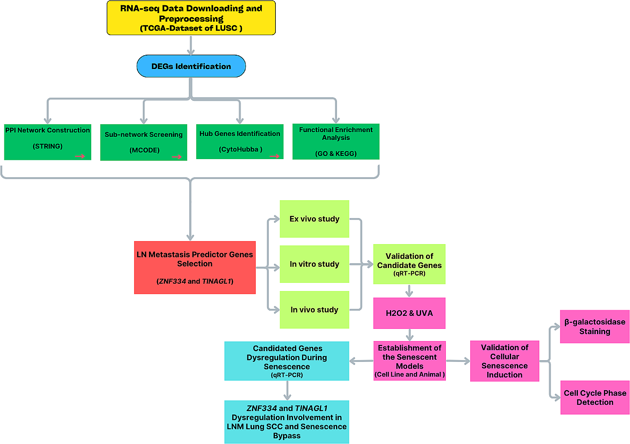
RNA-seq data analysis
Fastp software (https://github.com/OpenGene/fastp) was used to remove the reads that contained adaptor contamination, low-quality bases and undetermined bases with the default parameters. The sequence quality was then also verified using fastp. We used HISAT2 (https://ccb.jhu.edu/software/hisat2) to map reads to the Homo sapiens GRCh38 reference genome, and the mapped reads of each sample were assembled using StringTie (https://ccb.jhu.edu/software/stringtie) with default parameters. All transcriptomes from all the samples were then merged to reconstruct a comprehensive transcriptome using gffcompare (https://github.com/gpertea/gffcompare/). After the final transcriptome was generated, StringTie was used to determine the expression levels of mRNAs by calculating the FPKM values (FPKM = [total_exon_fragments/mapped_reads (millions) × exon_length (kB)]). The differentially expressed mRNAs were selected based on a fold change (FC) > 2 or < 0.5 and by performing a parametric F test comparing nested linear models (p value < 0.05) with an R package.
Integrated analysis of the metabolomic and transcriptomic data
For further integrative analysis of the transcriptomics and metabolomics data at the pathway level, all the DEGs and differentially expressed metabolites (DEMs) were used to perform pathway analysis with MetaboAnalyst 5.0 (https://www.metaboanalyst.ca/) [17, 18].
ROS detection
Briefly, HemECs were seeded in 6-well plates, and the next day, the cells were treated with propranolol and PFK15 for 24 h. The cells were then incubated with CDFH-DA, washed three times with PBS, observed using a fluorescence microscope (Olympus) and measured with a flow cytometer (Beckman).
Transmission electron microscopy (TEM) analysis
HemECs were seeded in 6-cm culture dishes, and the next day, the cells were treated with propranolol and PFK15 for 24 h. A morphological examination of the propranolol-treated HemECs was then performed with a transmission electron microscope (CM12 Philips, Amsterdam, Netherlands). The samples were stained with 2% (w/v) phosphotungstic acid and placed on copper grids for TEM observation.
Immunoprecipitation
The cells were lysed with protein lysis extract buffer and then centrifuged at 12000 × g for 10 min at 4 °C, and the supernatant was then collected. Eighty microliters of supernatant was then removed and used as input. The supernatant was incubated with the indicated antibodies and protein-A-agarose overnight at 4 °C. Isotype-matched IgG was used as a negative control. The beads were washed three times with ice-cold buffer and decoupled by boiling in sodium dodecyl sulfate (SDS) loading buffer. The samples were analyzed by western blotting.
Lentiviral vector transfection
Briefly, specific shRNA for PFKFB3 was purchased from GeneChem Co., Ltd. (Shanghai, China), and transfection was performed using Lipofectamine 2000 (Invitrogen, USA) according to the manufacturer’s protocol. Forty-eight hours after transfection, the cells were collected for various experiments.
Mouse model of IH
The in vivo study was approved by the Ethics Committee of West China Hospital of Sichuan University. To investigate the role of PFKFB3 inhibition by PFK15, the murine hemangioma model was established by subcutaneous injection of HemECs and hemangioma-derived pericytes (HemPCs). HemPCs (2 × 10⁶) and HemECs (2 × 10⁶) were mixed in 200 μL of Matrigel (BD, San Jose, CA, USA) and subcutaneously administered into the backs of BALB/C-nu mice (aged 4 weeks, male, purchased from Chinese Academy of Science, Shanghai) for 7 days. Four groups (five mice per group) were then treated with PBS (control), propranolol alone (10 mg/kg), PFK15 alone (25 mg/kg), or propranolol in combination with PFK15 every other day for 7 days by intratumoral injection. To investigate the role of PFKFB3 inhibition by shRNA, the mice were injected subcutaneously with 2 × 10⁶ PFKFB3-knockout HemECs and HemPCs (2 × 10⁶) for 14 days.
After the treatment, the experimental animals were sacrificed, and the tumors of the experimental animals were dissected.
Microvessel destiny (MVD) assay
To analyze the effect of different formations of PFK15 on the growth of microvessels, an MVD assay was performed. Paraffin-embedded sections of Matrigel explants were stained with hematoxylin and eosin (HE). For assessment of the MVD, 4 fields from mid-Matrigel HE-stained sections of each of the four animals in the group were analyzed. The microvessels were quantified by counting the luminal structures containing red blood cells [19]. Other paraffin-embedded sections of Matrigel explants were utilized for immunohistochemical staining for CD31.
Statistical analysis
The data were analyzed using SPSS version 23.0 (SPSS, Chicago, IL, USA). Continuous variables are presented as the means ± standard deviations, and Student’s t test was used to assess the differences between two groups. P values < 0.05 were considered statistically significant.


留言 (0)