Patient characteristics and placenta tissue collection
The study protocol was approved by the local ethics committee of Tongji Medical College, Huazhong University of Science and Technology. Informed consent was obtained from all 40 participants aged 22–41 years. PE was diagnosed according to the clinical criteria of the American College of Obstetricians and Gynecologists (ACOG Practice Bulletin No 2019). Exclusion criteria were as follows: (1) The healthy pregnant women with high blood pressure or proteinuria were excluded; (2) All pregnant women were excluded if they had a history of cardiovascular disease, diabetes, neurodegeneration, stroke, cancer, multiple pregnancies, placental abruption, or chorioamnionitis. Twenty healthy pregnant women were allocated to the healthy group, whereas the remaining preeclamptic women were allocated to the PE group. Their relevant clinical features are listed in the Table 1, and their placental specimens, which were approximately 2–3 cm from the edge of the umbilical cord attachment site (on the chorionic side), were collected. Each tissue sample was separated into two fractions: one for immunohistochemistry (IHC) and the other for RNA and protein extraction.
Table 1 Clinical characteristics of study populationQuantitative real-time PCR (qRT-PCR)
Total RNA was extracted from the collected placentas and cells in different groups using the TRNpure Total RNA Kit (HYCEZMBIO, China), and the concentration of RNA was quantified using Q5000 (QUAWELL, USA). A PrimeScript RT Reagent Kit (Takara, Tokyo, Japan) was used to synthesize the cDNA, and qRT-PCR was performed using a Step One Real-Time PCR System with ChamQ SYBR qPCR Master Mix (Vazyme, China), following the manufacturer’s instructions. The qRT-PCR reaction was conducted as follows: holding stage: 95 °C for 30 s, cycling stage: 40 cycles (95 °C for 10 s and 60 °C for 30 s), and melt curve stage: 95 °C for 15 s and 60 °C for 60 s. β-actin was used as an internal reference gene to quantify mRNA expression. Target mRNA expression levels were quantified according to the comparative cycle threshold (Ct; 2 − ΔΔCT) method, with β-actin as an internal reference gene. The qRT-PCR primer sequences used in this study are listed in Additional file 1: Table S1.
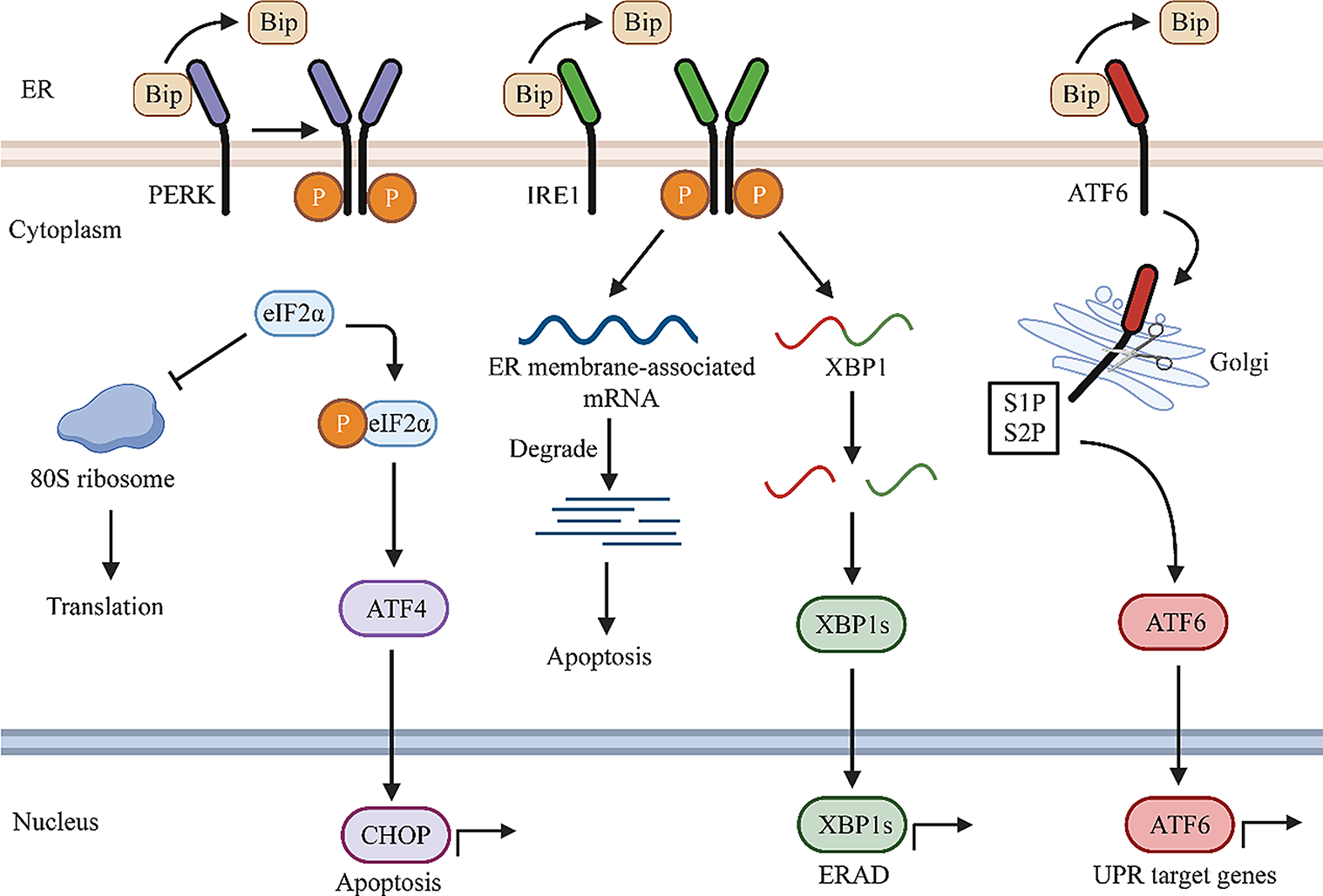
m6A colorimetric assay
The m6A modification level in total RNA from placentas and cells was determined using the EpiQuik m6A RNA Methylation Quantification Kit (P-9005; Epigentek, USA), according to the manufacturer's protocol, after RNA quality analysis using Q5000 (QUAWELL, USA). Briefly, 200 ng RNA was added to the strip wells. The capture and detection antibody solutions were then added to the assay wells separately at appropriate dilution concentrations. m6A levels were colorimetrically quantified by measuring the absorbance at a wavelength of 450 nm. Finally, the data were calculated using relative quantification according to the manufacturer’s instructions.
Immunohistochemistry
Placenta specimens were fixed with 4% paraformaldehyde. Servicebio Biotechnology Co., Ltd. (Wuhan, China) was employed for paraffin embedding, paraffin sectioning, and IHC staining. Primary antibodies against METTL3 (15073-1-AP, 1:300; Proteintech, China) and TMBIM6 (26782-1-AP, 1:300; Proteintech, China) were used. Each pair of placenta specimens from healthy and preeclamptic group was stained in the same condition. Sections of the immunohistochemical specimens were observed and imaged using a microscope (Olympus, Japan). Finally, Image Pro Plus6.0 was used to analyze the data. Specifically, the optical density (OD) of IHC images were firstly standardized and the positive areas were selected and measured to calculate the integrate OD (IOD) and mean IOD. Mean IOD was used for statistical analysis.
Western blot (WB) analysis
Total protein was extracted from the cells and tissues using the RIPA lysis buffer (Servicebio, China), mixed with protease inhibitors (Beyotime, China), and the protein concentrations were quantified using a BCA protein assay kit (CWBIO, China). The protein samples were denatured at 95 °C for 10 min and stored at –80 °C until use. Equal amounts of proteins from different groups were separated by 10% SDS-PAGE gels and transferred onto PVDF membranes (0.45 mm pore size; Millipore, USA) which were then blocked with 5% skimmed milk in Tris-buffered saline containing 0.1% Tween-20 for 1 h. Next, the membranes were incubated with the METTL3 (15073-1-AP, 1:1000; Proteintech, China), TMBIM6 (26782-1-AP, 1:1000; Proteintech, China), GRP78 (11587-1-AP, 1:1000; Proteintech, China), CHOP (2895T, 1:1000; Cell Signaling Technology, USA), METTL14 (26,158–1-AP, 1:1000; Proteintech, China), fat mass and obesity-associated protein (FTO; 27226-1-AP, 1:1000; Proteintech, China), B-cell lymphoma-2 (Bcl-2; 26593-1-AP, 1:1500; Proteintech, China), Bcl-2-associated X (Bax; 2772T, 1:1000; Cell Signaling Technology, USA), heme oxygenase-1 (HO-1; 10701-1-AP, 1:3000; Proteintech, China), nuclear factor erythroid 2-related factor 2 (NRF2; 16396-1-AP; 1:1000; Proteintech, China), YTHDF2 (24744-1-AP, 1:4000; Proteintech, China), and β-actin (66009-1-Ig, 1:10,000; Proteintech, China) antibodies overnight at 4 °C. They were then washed thrice with TBST and incubated with the appropriate HRP-conjugated anti-rabbit or anti-mouse secondary antibodies (1:5000; Proteintech, China) at room temperature for 1 h. The proteins were visualized with enhanced chemiluminescence reagents (Millipore, USA), and the protein bands were analyzed using the ImageJ software.
Cell culture and transfection
HTR-8/SVneo cell line, an extravillous trophoblastic cell line, was obtained from Dr. Charles Graham (Queen’s University, Canada) as a gift. Cells were cultured in RPMI 1640 medium (Biosharp, China) supplemented with 10% fetal bovine serum (Gibco, USA) and 100 U/mL penicillin & streptomycin (PYG0016, Boster, China) at 37 °C in an incubator containing 5% CO2. HTR-8/SVneo cells were cultured in a 6-well plate and transfected with Lipofectamine 3000 (Invitrogen, USA). A TMBIM6 overexpression model was established via TMBIM6 plasmid transfection for 24 h. Simultaneously, a negative control model was established via an empty vector transfection. Knockdown of METTL3 and YTHDF2 was achieved using three shRNAs targeting the METTL3 and YTHDF2 genes, which were synthesized by GenePharma Biotech. The target sequences are listed in Additional file 2: Table S2. Transfection efficiency was confirmed by qRT-PCR or WB. To construct a model of ER stress, HTR-8/SVneo cells were incubated with 100 nmol/L TG (T863962; Macklin Biochemical Co., Ltd, China) for 6 h after transfection with overexpression plasmids or shRNAs and incubated for the last 18 h. Finally, HTR-8/SVneo cells were collected to extract the RNAs and proteins for subsequent experiments.
Methylated RNA immunoprecipitation (MeRIP)-qPCR
The m6A modification of TMBIM6 was determined using the Magna EpiQuik CUT&RUN m6A RNA Enrichment (MeRIP) Kit (P-9018, Epigentek, USA), according to the manufacturer’s instructions. Briefly, 50 μg of total RNA was used for m6A immunoprecipitation, and 1/10 of it was saved as the input control group. First, an immunocapture solution containing the m6A antibody, non-immune IgG, affinity beads, immunocapture buffer, and RNA sample was prepared and vortexed for 90 min at room temperature to immunocapture m6A RNA. Then, the cleavage enzyme mix was used to cleave RNA, and proteinase K and RNA purification solution were added to the immunoprecipitated complex to remove the excess proteins and purify m6A-containing RNA. Finally, the immunoprecipitated m6A RNA was recovered using the elution buffer, and its level was measured using qRT-PCR.
Cell viability assay
Cell viability was assessed using the Cell Counting Kit-8 (CCK8; Proteintech, China). Approximately 0.6 × 10⁴ cells were seeded in each well of a 96-well plate and treated with TG at different concentrations (0, 25, 50, 100, 200 nmol/L) for 1, 6, 12, 24 h. Later, the CCK-8 reagent (10 μL) was added to each well, followed by 2 h incubation at 37 °C in a dark incubator. Absorbance was measured at 450 nm using a multimode reader (Infinite F50; Tecan).
Measurement of ROS
HTR-8/SVneo cells were seeded in a 6-well plate and washed thrice with PBS after various treatments. Then, 2′-7′-dichlorodihydrofluorescein diacetate (DCFH-DA, CA1410; Solarbio, China) kit, one of the most widely used commodities to directly measure the intracellular ROS levels, was added to the plates and incubated for 20 min at 37 °C in a dark atmosphere. Finally, the green fluorescence indicating the level of intracellular ROS was observed by fluorescence microscopy (Olympus, Japan) at 100 ×, and the mean fluorescence intensity was calculated using the ImageJ software to determine the level of intracellular ROS.
Flow cytometry analysis
To measure apoptosis, HTR-8/SVneo cells were harvested and stained using the Annexin V-FITC Apoptosis Detection Kit (KGA107, Keygen Biotech, China) for 10 min at room temperature in the dark. Flow cytometry was used to detect apoptosis, and the data were analyzed using the FlowJo 10.5.3 software.
Transwell assays
Cell invasion was detected using Transwell chambers (8.0 μm pore size; Corning, USA) and Matrigel mix (BD Biosciences, CA, USA). Specifically, 3 × 10⁴ cells were resuspended in serum-free RPMI 1640 medium (200 μL) in the upper chamber, while the lower chamber contained 600 μL medium supplemented with 10% fetal bovine serum. The cells were incubated for 24 h at 5% CO2 and 37 °C, fixed with 4% paraformaldehyde for 20 min, and stained with crystal violet for 15 min. Finally, cells in the lower compartment of the chamber were counted under an inverted microscope (Olympus, Japan) at a magnification of 100 ×, and five random fields were selected for cell counting in each chamber.
mRNA stability assay
After treatment with 5 μg/mL actinomycin D (7240-37-1; Macklin, China), which inhibits mRNA transcription, for 0, 2, 4, and 6 h, total RNA was extracted using a TRNpure Total RNA Kit (HYCEZMBIO, Wuhan, China). The mRNA level of TMBIM6 at the indicated time was measured via qRT-PCR, and the degradation percentage was calculated. The turnover rate and half-life of RNA were estimated according to a previously published method (Zhang et al. 2021a). The constant of RNA decay (K) was calculated using the following equation: dC/dt = − KC, where dC/dt is the change in RNA concentration at a given time and C is the RNA concentration. Subsequently, the equation ln(C/C0) = − Kt was used to estimate the K value which represents the degradation rate of RNA. RNA half-life time (t1/2), indicating that 50% of the RNA had decayed (C/C0 = 1/2), was also calculated based on the equation: t1/2 = ln2/K.
Animals and experimental groups
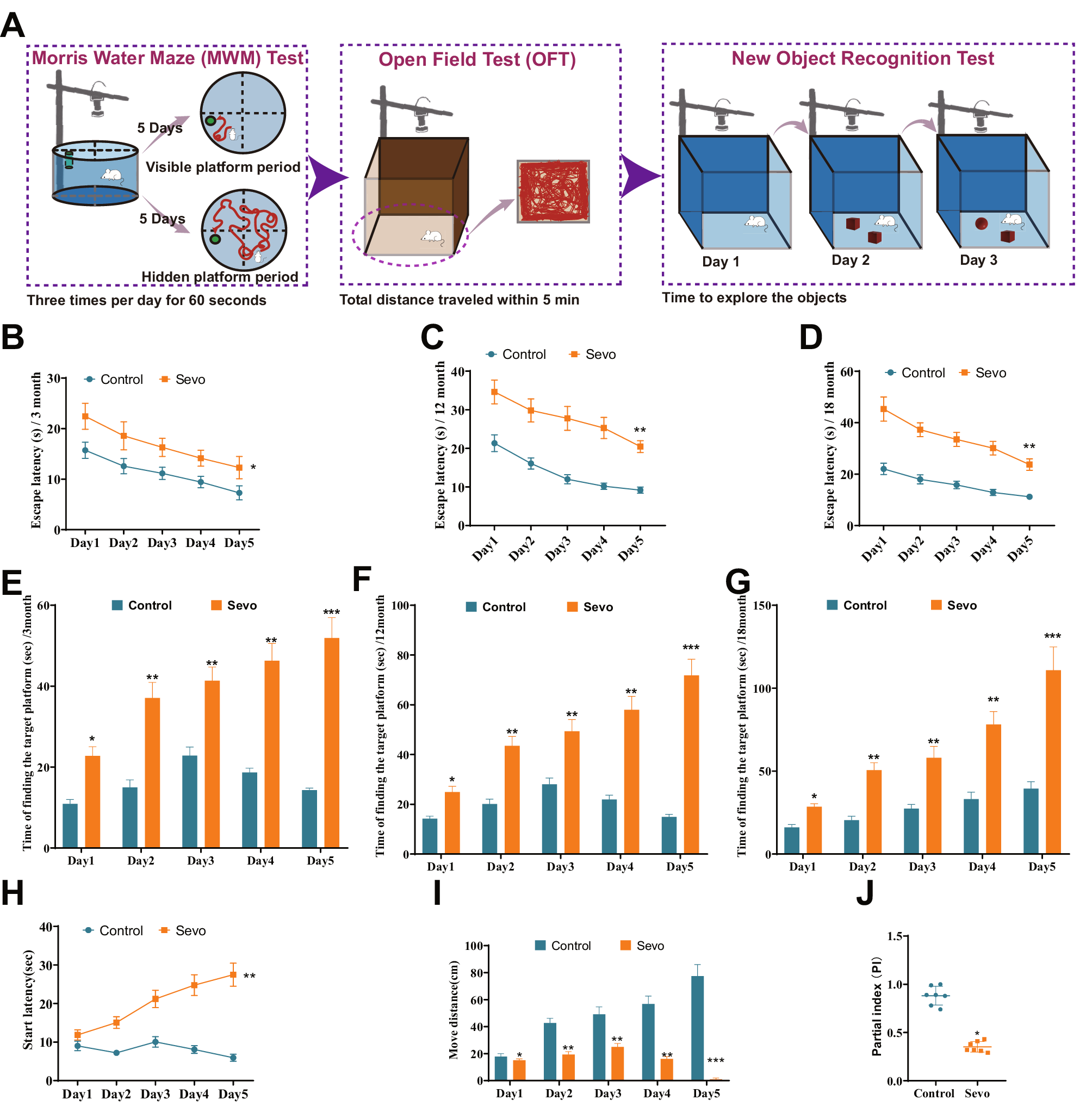
The animal study was conducted with the approval of the Animal Ethics Committee of the Huazhong University of Science and Technology, and all experimental procedures were performed according to the National Institutes of Health Guidelines and Regulations. Twenty-four SD rats aged 8–10 weeks (240–260 g) were purchased from the experimental animal center of Sanxia University (Sanxia, China) and raised in the animal laboratory of Tongji Medical College. Each rat was housed under SPF conditions with water and food ad libitum. Female rats were copulated with weight-matched male rats in a ratio of 2:1. The first gestational day (GD1) of pregnancy was defined as when the sperm or vaginal suppositories were observed under a light microscope, as shown in Fig. 8B. The time scheme of the animal experiment is shown in a schematic diagram (Fig. 8A). Pregnant female rats (n = 24) were randomly divided into healthy (n = 6) and disease (n = 18) groups, and the disease groups (n = 18) were further divided into three subgroups: PE, PE + LV-sh-Con, and PE + LV-sh-METTL3 groups. L-nitro-arginine methyl ester (L-NAME; R015327; RHAWN, China), a NOS inhibitor, was used to establish the PE model, as previously reported (Burke et al. 2016). The healthy group (NC, n = 6) received an intraperitoneal injection of sterile 0.9% NaCl, whereas the three disease subgroups were injected with L-NAME (50 mg/kg/day) between GD10 and GD15. Next, the PE + LV-sh-Con group was injected with a lentivirus containing sh-Con RNA (LV-sh-Con), whereas the PE + LV-sh-METTL3 group was injected with an equal dose of lentivirus containing METTL3 shRNA (LV-sh-METTL3) on GD15. The transfection efficiency of LV-shRNA in rats was ascertained by observing the expression of GFP in frozen placental sections under a fluorescence microscope (Fig. 8M). Then, the rats were anesthetized using 10% chloral hydrate (Macklin, China), and the embryonic, placental, and kidney samples were immediately removed by cesarean section on GD20. Finally, the fetuses and placentas were weighed, and placental samples were collected for WB and staining.
Haematoxylin‐eosin (HE) staining
The paraffin‐embedded kidney tissues were sliced to serial sections of 5-μm thickness. Subsequently, the slices were depafaffinized, debenzolized, and conventionally stained with HE. After staining was completed, the sample slides were mounted using neutral balsam and sealed with clean coverslips. Finally, the sections were photographed, and histopathological changes were observed under a microscope.
Blood pressure and urinary protein analysis
Systolic blood pressure (SBP) was monitored using a non-invasive blood pressure system (Kent Scientific, USA) on GD9, GD15, and GD20. Each rat was preheated to 38 °C for 5 min before each measurement. Urinary protein levels were determined in the urine samples using CBB kits (Jiancheng Institute of Biotechnology, China) on GD9, GD15, and GD20. Blood pressure and urinary protein levels on GD9 are shown in Additional file 3: Table S3. Increase in blood pressure to > 30 mmHg and significant increase in urinary protein levels indicated the successfully establishment of the PE rat model.
Statistical analysis
Statistical analysis was performed using the GraphPad Prism8.0 and SPSS 20.0 software. The results are presented as the mean ± SEM. Two-group comparisons were analyzed using Student’s t-test, while three or more group comparisons were carried out using one-way ANOVA. p-values less than 0.05 (p < 0.05) were considered to be statistically significant.


留言 (0)