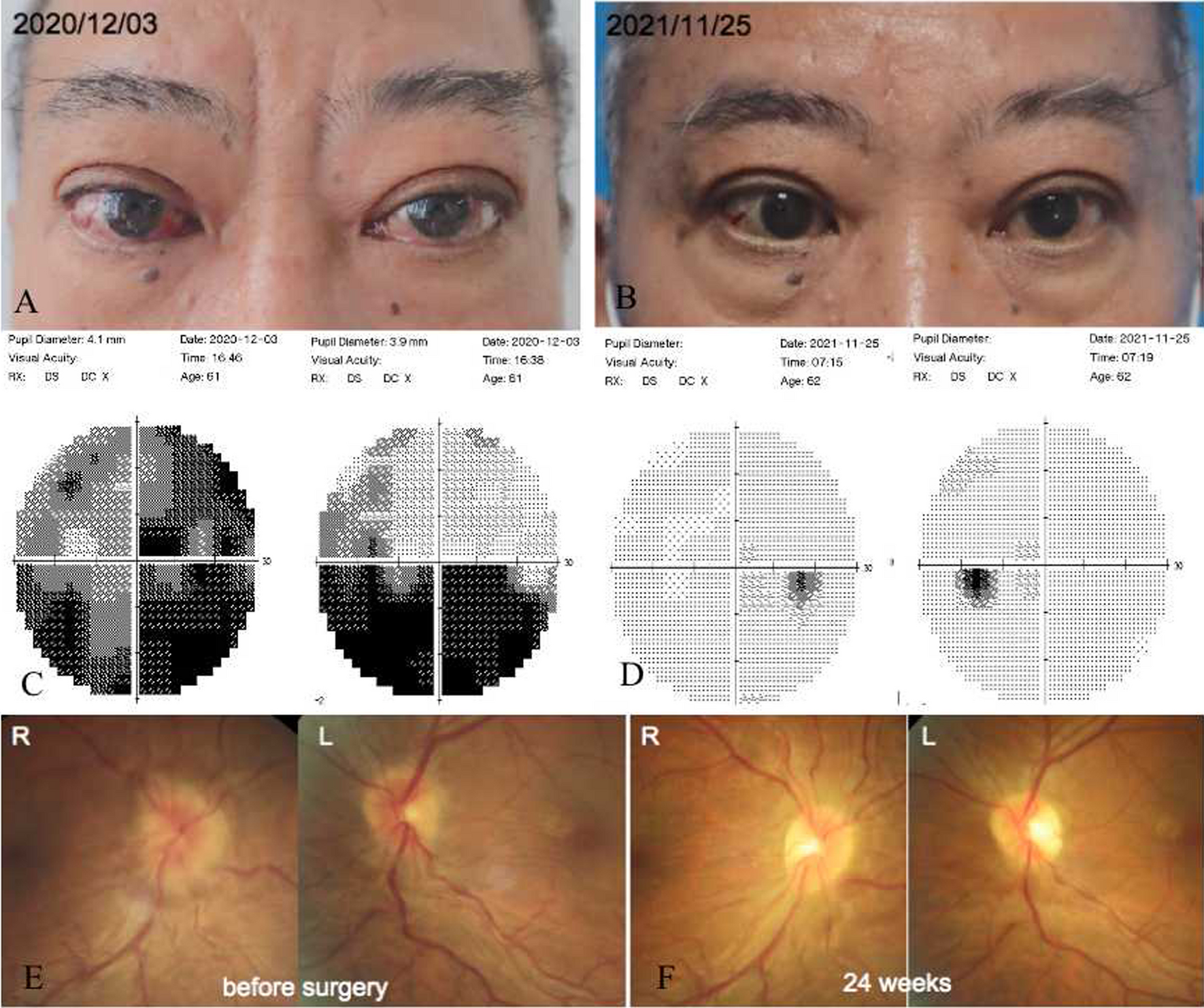
To our knowledge, the present study is the first to compare and evaluate sequential ULD temporal bone CBCT images to conventional HR CBCT images in a clinical cohort. Repetitive imaging without changes in pathology or anatomy is essential for comparing imaging modalities. There are only few clinical studies comparing the use of MSCT and CBCT to temporal bone imaging: Redfors et al. [10] were the first to evaluated the anatomical structures of the temporal bones of 20 post-stapedectomy patients (otosclerosis), and Pein et al. [9] compared images of 38 patients without pathology, but most of the compared images were from different patients. Some additional studies comparing imaging modalities have a narrow scope, such as imaging post-operative implant position or pre-operative imaging of otosclerosis.
The present study demonstrates that the IQ of ULD CBCT scans and especially that of HR CBCT scans is sufficient for several procedures in the field of ear surgery, even if off-focus scans are used. Both methods provide an anatomical map of relatively constant structures of the temporal bone, such as the external acoustic meatus, sigmoid sinus, jugular bulb, cochlea, semicircular canals and the mastoid segment of the facial nerve. It might also be possible to identify focal pathologies from ULD CBCT scans of the epitympanum, mastoid antrum or scutum.
High-resolution computed tomography (HRCT) is the gold standard for imaging the temporal bone. This method provides crisp, clear images of the bony structure with radiation doses ranging from 1.6 mSv for adults to 7.1 mSv for new-borns, an amount equivalent to 6–24 months of background radiation [8, 12]. CBCT can produce images that are as good or even better [5,6,7, 9]. However, the hypernym CBCT includes a variety of hardware, software and setting combinations, our combinations in this article are named as HR CBCT and ULD CBCT. Since the present study uses a retrospective design, we did not measure the effective dose of radiation. Furthermore, no previous studies have reported the effective radiation doses for these scanners in this imaging area. The overall mean effective doses have been published: 0.119 mSv for paranasal CBCT [13] and 0.6 mSv for paranasal MSCT [14], equivalent to two to nine weeks of background radiation. The effective dose for facial scans with the Scanora 3Dx (HR CBCT device) is reported to be 0.104 mSv (unpublished data from the manufacturer; measured in the University Hospital of Oulu, Finland, in 2013, according to the dosimetric principles of the Finnish Radiation and Nuclear Safety Authority [STUK]). For facial imaging with the Promax 3D Mid ULD (ULD CBCT device), the dose is 0.018 mSv (FOV: 200 × 170 mm) (published in a poster; ID 0920, 2015 IADR/AADR/CADR General Session, Boston Massachusetts), which equals two days of background radiation. The facial area includes radiation-sensitive organs, such as the thyroid and the submandibular glands, which were not in our FOV; but we had two 160 × 170 mm stacks instead of one 200 × 170 mm. The ULD CBCT uses six or seven times less radiation than the HR CBCT, as seen in the lower mSv and DAP values. If the ULD CBCT images were obtained in one scan focused on the ears, this reduction would double.
For our CBCT scans, we used a sinonasal focus as a reference. Although our HR CBCT is not compared to HRCT, the excellent IQ of HR CBCT is demonstrated in the samples and results. The IQ of CBCT images could be further enhanced if the patient were supine during scanning (this would reduce motion artefact) and by adjusting the primary focal area on the ear. In optimal circumstances (in vitro), superb resolution of fine structures (stapes, incudostapedial joint, tendon of the tensor tympani and the stapes footplate, to name a few) is possible with CBCT (tube voltage: 88 kV; tube current: 11 mA; voxel size: 0.1 mm; FOV: 60 × 60 mm) [5, 7]. We knew from experience that ULD CBCT with our specifications is not likely to produce clinically sufficient images of these above mentioned structures, hence majority of them were not even included in our list of structures. The results support our assumption.
It is preferable to obtain the best image possible, particularly when confirmation of a structure’s normality can rule out the need for explorative surgery or surgical intervention. For example, even when detailed images are available, tympanotomy is often performed to verify, for instance, the status and mobility of the ossicles or the footplate and, if needed, to remove the pathology and reconstruct the chain’s integrity.
Cochlear implantations are a good example of a surgical procedure conducted on ‘healthy’ ears; there is no consensus regarding the best pre-operative imaging modality [12]. Of children and adults who receive cochlear implantations, 85% undergo magnetic resonance imaging (MRI), 95% of children and 90% of adults undergo CTs and around 80% receive both [15]. We consider the MRI a baseline measurement for both children and adults as it provides a comprehensive view of the neural and inner ear structures [16,17,18,19]. Some authors recommend proceeding to surgery without imaging if there is no clinical or audiometric concerns for otosclerosis or middle ear and/or mastoid disease [15, 20]. We suggest that an ultra-low-radiation dose method of imaging, such as a ULD CBCT, could be used as the first approach to CT in children and adults with suspected healthy ears. This type of imaging can be used to evaluate the anatomical relationships of the critical structures, such as the sinus sigmoideus, facial nerve and jugular bulbus. With these said, we are still waiting our CBCT device that allows supine position, and thus enables even young children’s imaging. If obscure structures are found and the MRI does not provide enough information, reveals cochlear anomaly or some unexpected pathology, more comprehensive CT imaging (HR CBCT or MSCT) is justified.
Furthermore, there is frequently a vast reservoir of unused information in CT and CBCT images that have already been taken. In the field of otorhinolaryngology, these images focus primarily on the nasal cavity and paranasal structures, but all corners of the FOV should be utilized. The reviewer, radiologists or clinician is responsible for analysing the entire imaged area [21]. Our results show that recent imaging history might be useful. If a new image is needed for post-operative monitoring or for other non-complicated reasons, it could also be sufficient to take this image using a considerably lower radiation dose.
One strength of our study is that we used comparable images of real subjects. The images were rated in a random order, and the raters had no information about the patients or the imaging modalities. In addition, the images were viewed in same planes and with the same desktop options that clinicians normally use. However, no technical evaluation or comparison of the images was conducted, as we considered such an evaluation irrelevant to our objective. Another weaknesses of our study is that all the imaged ears were healthy. As a result, the findings cannot be generalized to ears with considerable pathology, and no surgery was planned based on our images.
Inter-rater argeement is moderate at its best regardless of the modality. Nevertheless, our findings reflect real-world circumstances, as the raters included a radiologist, an ear surgeon/neuro-otologist and a general otorhinolaryngologist. In order to remain strictly critical, every individual rating from 0 to 2 impaired the insufficient-sufficient ratio.
The ULD images in our data are combined from two stacks with an overlap of about two centimeters; this overlap is the stitching area. Its position varies according to each patient’s individual anatomy, but it usually includes the structures of interest. In an ideal situation, excess computing and geometric distortion [2] may be avoided by adjusting the FOV to include only the structures that are needed and setting the focus accordingly.
To conclude, CBCT imaging and the data at image margins are underutilized. CBCT can produce excellent structural resolution with conventional imaging parameters, even with off-focus images, and it can produce images of clinically sufficient image quality with an ultra-low dose of radiation. We encourage ear surgeons to check patients’ imaging history and to consider the use of imaging modalities that involve lower radiation doses especially for repetitive investigations and with children.


留言 (0)