
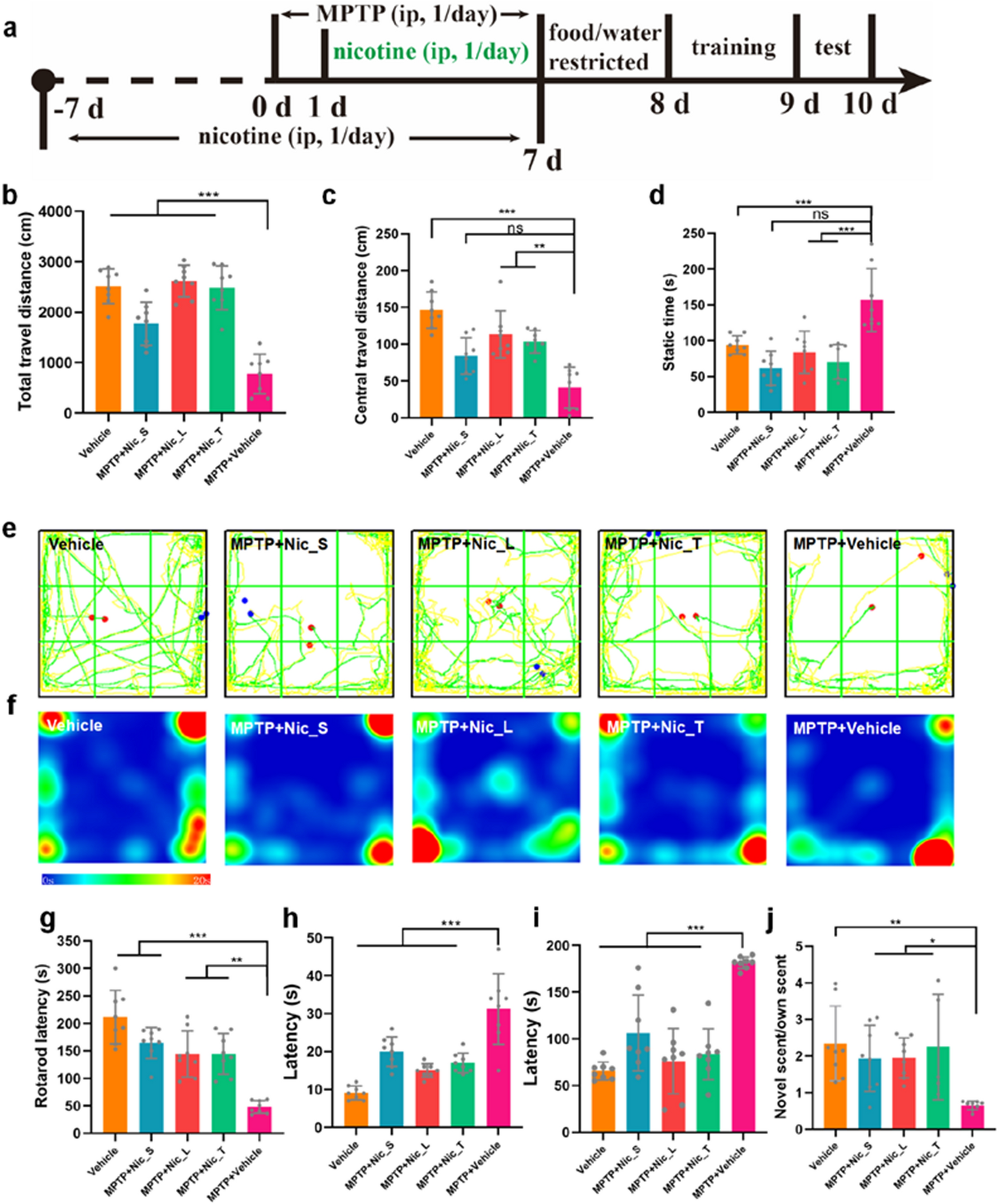
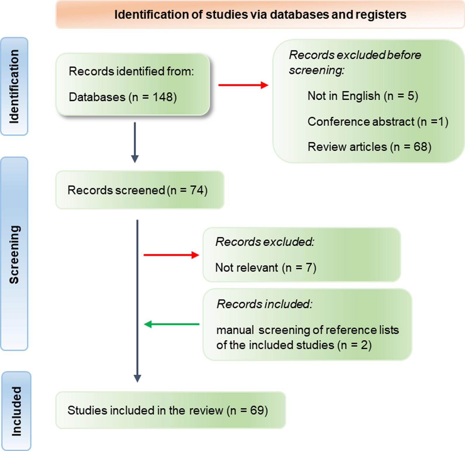
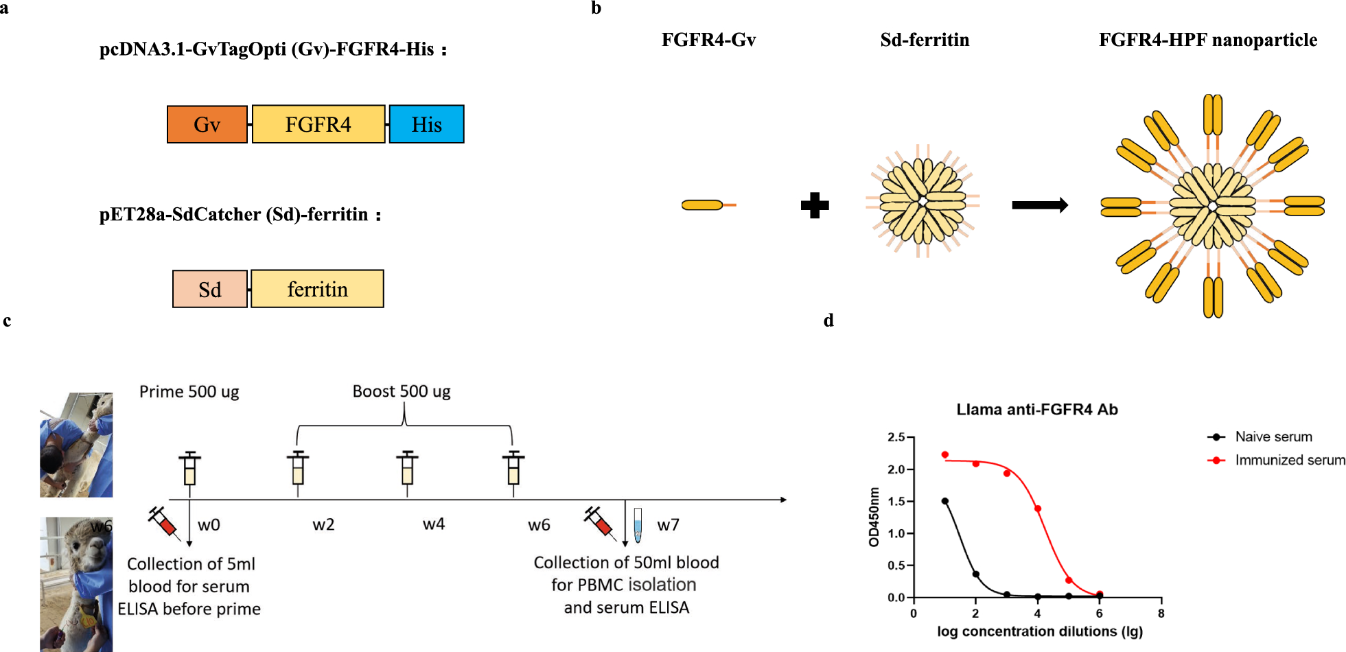
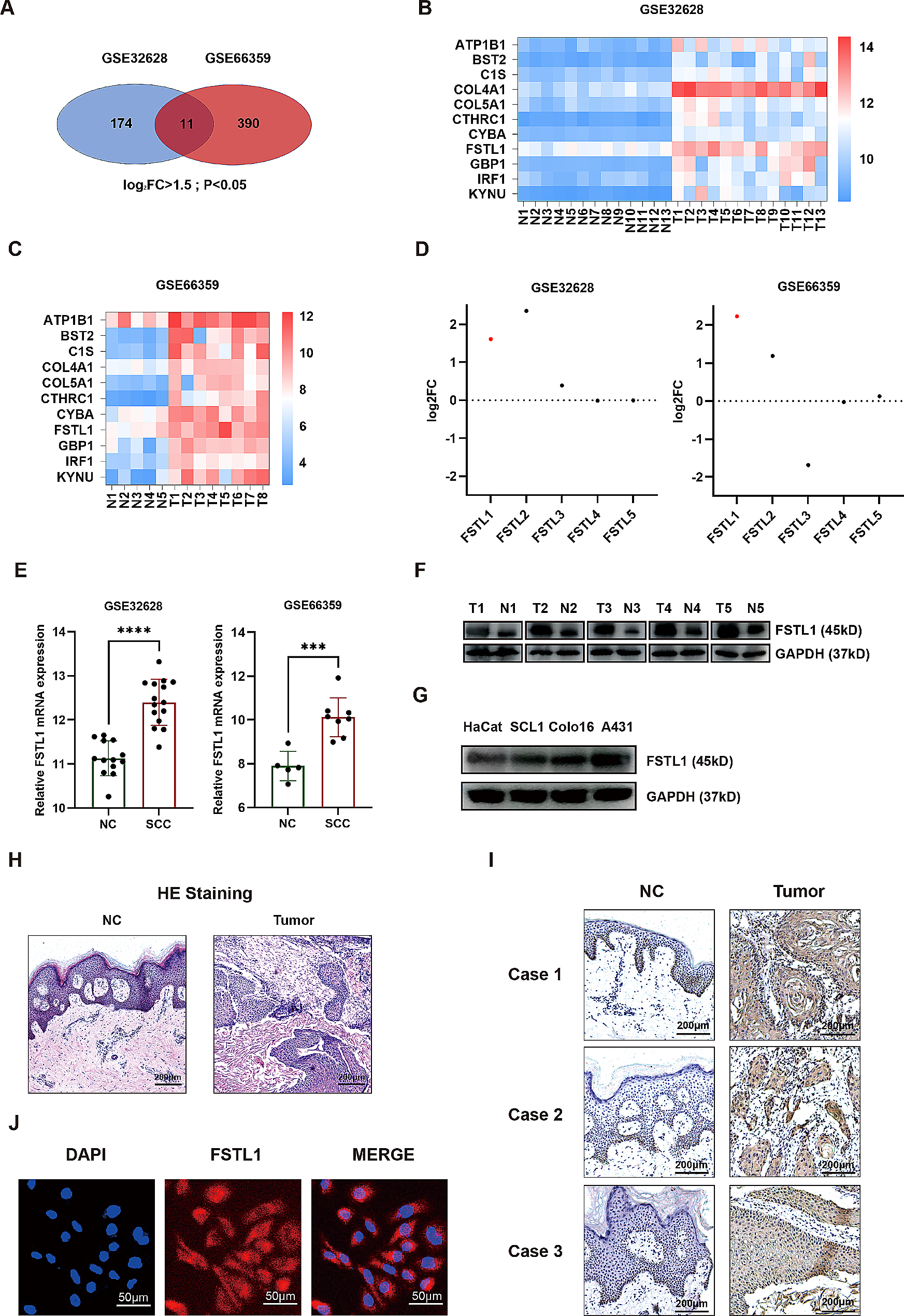
記住我
In a prior work, we found that GO patients contained CD4 CTLs that expressed more cytotoxic molecules than those from GH patients [13]. To investigate the regulators of the cytotoxicity of CD4 CTLs, transcriptome sequencing was carried out on PBMCs from GO and GH patients. There were 476 upregulated genes and 94 downregulated genes detected (Fig. 1A). After PPI analysis of the DEGs, VEGFA was identified as a potential key gene in GO (Fig. 1B). Furthermore, in the GO patients with higher VEGFA expression, the cellular response to the calcium ion process was upregulated (Fig. 1C). Additionally, VEGF-A was shown to be elevated at the protein level in the plasma of GO patients compared to that of GH patients and HCs (Fig. 1D; P = 0.0359 and 0.0284). Considering that the calcium ion response is involved in the synthesis and release of cytotoxic molecules in T cells, we hypothesized that VEGF-A contributed to the increased cytotoxic function of CD4 CTLs in GO [26]. Reanalyzing scRNA-seq data for GO patients was performed to confirm this theory, and it was discovered that GO CD4 CTLs with greater levels of GZMB, PRF1, NKG7, and GZMK expression had upregulated VEGFR1 (Fig. 1E). Furthermore, GSEA results showed that cytotoxic granule, cytolysis, and cell killing processes were enriched in GO CD4 CTLs with upregulated VEGFR1 expression (Fig. 1F). In addition, the proportions of cells expressing cytotoxic molecules such as GrmB, GrmA, and Prf were significantly increased after administrating VEGF-A to CD4+ T cells from GO patients (Fig. 1G; P < 0.0001, = 0.0286 and 0.0066, respectively). In this way, VEGF-A contributes to the enhanced cytotoxicity of CD4 CTLs in GO and can increase the production of cytotoxic molecules by these cells.
Fig. 1
VEGF-A contributes to the enhanced cytotoxicity of CD4 CTLs in GO. A Volcano plot showed differentially expressed genes between GO and GH group. Blue dots represented downregulated genes and red dots were upregulated genes. B PPI network of potential key genes. The shade of colors denoted the degree of the corresponding gene. C Gene set enrichment analysis (GSEA) showed cellular response to calcium ion process was obviously enriched in GO patients with higher expression of VEGFA compared with lower VEGFA expression. D Bar plot exhibited the O.D. value of VEGF-A in serum from HC, GH and GO patients (N = 3). E Dot plots showed the expression of GZMB, PRF1, NKG7, GZMK and VEGFR1 in CD4 CTLs and non-CD4 CTLs (other) group from GO patients respectively. Color scale represented z-score and dot size represented percentages of cells. F GSEA showed cytotoxic related processes were obviously enriched in CD4 CTLs from GO patients with higher expression of VEGFR1 compared with lower VEGFR1 expression. G Representative flow cytometry plots showed the ratios of GrmB+, GrmA+ and Prf+ cells in CD4+ T cells in the CON and VEGF-A group (N = 4). The red rectangle and number denoted in it meant GrmB+, GrmA+ and Prf+ subsets and specific ratio respectively. The quantification of the proportions of GrmB+, GrmA+ and Prf+ in CD4+ T cells was displayed on the right. Blue was CON and red for VEGF-A treated. Error bars showed SEM. The data were representative of at least three biological replicates. HC: healthy control; GH: Graves hyperthyroidism; GO: Graves orbitopathy; CON: control; Grm: granzyme; Prf: perforin. *P < 0.05, **P < 0.01, ****P < 0.0001
VEGF-A promotes the cytotoxic function of CD4 CTLsThree scRNA-seq datasets containing enriched CD4 CTLs (effector memory T cells re-expressing CD45RA, Temra) or CD4+ T cells were downloaded from the GEO database, and integrated analysis was performed with our previous scRNA-seq data (SRP226183) related to GO to further explore the role of VEGF-A in pan CD4 CTLs (Fig. 2A, Table 2). In total, 14 clusters were found (Fig. 2A). Among them, cluster 2, 6, 7, and 10 were recognized as CD4 CTLs due to the upregulated expression of cytotoxic molecules (Fig. 2B). In the HC and GO datasets (GSE106543 and SRP226183), CD4 CTLs with increased cytotoxicity (cluster 2 and 7) predominated, but these cells did not predominate in the bladder cancer or chronic rhinosinusitis with nasal polyps (CRSwNP) datasets (GSE149652 and GSE179292; Fig. 2C&E). It was assumed that CD4 CTLs in lesions tended to be exhausted with decreased cytotoxicity given that pseudotime trajectory analysis suggested that cluster 6 and 10 might differ from cluster 2 and 7 (Fig. 2D; Additional file 1: Fig. S1). Notably, CD4 CTLs with greater cytotoxicity had upregulated VEGFR1 (clusters 2 and 7; Fig. 2E). Through GSEA, it was discovered that cluster 2 and 7, which had higher VEGFR1 expression, exhibited upregulation of processes related to VEGF_VEGFR signaling and cytotoxicity (Fig. 2F). Furthermore, in contrast to cells treated with the blank control, VEGF-A-treated CD4 CTLs from HCs produced more GrmB and Prf (CON; Fig. 2G; P < 0.0001 and P = 0.0211). These findings demonstrate that VEGF-A is involved in the cytotoxicity of CD4 CTLs and promotes their cytotoxic function.
Fig. 2
VEGF-A promotes the cytotoxic function of CD4 CTLs. A The graph-based clustering and t-SNE algorithm were applied in CD4+ T cells from GSE106543 (pink), GSE149652 (green), GSE179292 (purple) and SRP226183 (blue). Clusters denoted by the same color scheme were labeled with inferred cell types (bottom). B Heatmap from scRNA-seq analysis via expression data of the top 20 genes differentially expressed for each cluster (denoted by colored bars at the top). Key genes for each cell type were shown on the left margin. Genes in red rectangle represented cytotoxicity related. C The stack plots showed the ratio of each cluster of CD4 CTLs. Each color represented different cell cluster. D Pseudo-time trajectory analysis showed the differentiation stage of each CD4 CTLs subset according to the cytotoxicity. Pink represented cluster 2, green was cluster 6, blue was cluster 7 and purple was cluster 10. E Dot plots showed the expression of GZMB, PRF1, NKG7, GZMK and VEGFR1 in each CD4 CTLs subset respectively. Color scale represented z-score and dot size represented percentages of cells. F GSEA showed (upper) cytotoxic related processes and (bottom) VEGF and cytotoxicity pathways were obviously enriched in CD4 CTLs with higher expression of VEGFR1 compared with lower VEGFR1 expression. G Representative histogram plots showed the expression of GrmB and Prf in CD4 CTLs in the CON and VEGF-A group. The quantification of the proportions of GrmB+ and Prf+ in CD4 CTLs was displayed on the right (N = 3). Blue was CON and red for VEGF-A treated. Error bars showed SEM. The data were representative of at least three biological replicates. CON: control; Grm: granzyme; Prf: perforin. *P < 0.05, ****P < 0.0001
VEGF-R1/R2 correlates with the cytotoxic function of CD4 CTLsAlthough preliminary evidence suggests that VEGF-A regulates CD4 CTLs, the specific VEGF-R involved remains unknown. To clarify the downstream receptors, qPCR was performed to measure the expression of well-known VEGF-A receptors including VEGFR1, VEGFR2, and NRP1 on CD4 CTLs with increased cytotoxicity. Because GO CD4+ T cells expressed much more cytotoxic molecules than those from HCs, GO CD4 CTLs were used as an example of CD4 CTLs with increased cytotoxicity (Additional file 2: Fig. S2; GrmB: P = 0.0083; Prf: P = 0.0301). The results revealed that VEGFR1 and VEGFR2 were upregulated in GO CD4 CTLs with increased cytotoxicity compared with HC naïve CD4+ T cells (Fig. 3A; P = 0.0286, 0.0409 and 0.4857, respectively). In contrast, there were lower proportions of VEGF-R1+ and VEGF-R2+ cells in GO CD4 CTLs with higher cytotoxicity than in HC CD4 CTLs, as measured by flow cytometry (Fig. 3B; VEGF-R1: P = 0.0001; VEGF-R2: P = 0.0016; NRP-1: P = 0.9240). We hypothesized that the decrease in the level of VEGF-R1/R2 expression on GO CD4 CTLs with increased cytotoxicity was caused by binding with overexpressed VEGF-A because VEGF-R1/R2 levels were reported to be diminished following binding to VEGF [27]. As expected, GO CD4 CTLs with higher cytotoxicity had higher levels of phosphorylated (p)-VEGF-R2 than CD4 CTLs from HCs or GH patients (Fig. 3C; P = 0.0480 and 0.0041), indicating that VEGF/VEGF-R2 signaling was upregulated in CD4 CTLs with enhanced cytotoxicity.
Fig. 3
VEGF-R1/R2 correlates with the cytotoxic function of CD4 CTLs. A Bar plots exhibited levels of VEGFR1, VEGFR2 and NRP1 in CD4 CTLs (red) from GO patients and CD4+ Naive T cells (blue) from HC (N = 4). B Representative histogram plots exhibited the expression of VEGF-R1, VEGF-R2 and NRP-1 in CD4 CTLs from GO (red) and HC (blue) (N = 3). The bar plots showed the proportions of VEGF-R1+, VEGF-R2+ and NRP-1+ cells in CD4 CTLs from GO (red) and HC (blue). C Representative histogram plots (upper) exhibited the expression of p-VEGF-R2 in CD4 CTLs from GO (red), GH (blue) and HC (gray) (N = 3). The bar plots (bottom) showed the MFI of p-VEGF-R2 in CD4 CTLs from GO (red), GH (blue) and HC (gray) group. D, E Representative histogram plots (upper) exhibited the expression of GrmB and Prf in (D) VEGF-R1/ (E) VEGF-R2 ±CD4 CTLs from GO (red and black) and HC (blue and gray) patients. The bar plots (bottom) showed the proportions of GrmB+ and Prf+ cells in (D) VEGF-R1/(E) VEGF-R2 ±CD4 CTLs from GO and HC group (N = 3). Blue denoted HC VEGF-R1/2+ CD4 CTLs, red was GO VEGF-R1/2+ CD4 CTLs, gray was HC VEGF-R1/2− CD4 CTLs and black was GO VEGF-R1/2− CD4 CTLs. Error bars showed SEM. The data were representative of at least three biological replicates. HC: healthy control; GH: Graves hyperthyroidism; GO: Graves orbitopathy; Grm: granzyme; Prf: perforin; p-VEGF-R2: phosphorylated VEGF-R2. *P < 0.05, **P < 0.01, ***P < 0.001
Furthermore, the relationships between the expression of VEGF-R1/R2 and cytotoxic molecules were examined. When VEGF-R1+ GO CD4 CTLs with higher cytotoxicity were compared to VEGF-R1+ HC CD4 CTLs, it was revealed that the proportions of GrmB+ cells were increased, while no differences were detected between GO and HC CD4 CTLs with VEGF-R1− or VEGF-R2± (Fig. 3D&E; P = 0.0184, 0.1594, 0.9745 and 0.7348). With respect to the proportion of Prf+ cells, there were no differences between VEGF-R1+ or VEGF-R2± GO and HC CD4 CTLs (Fig. 3D&E; P = 0.6380, 0.1858, and 0.9856). Interestingly, VEGF-R1− GO CD4 CTLs had higher Prf expression than VEGF-R1− HC CD4 CTLs (Fig. 3D; P = 0.0230). Hence, VEGF-R1/R2 signaling is upregulated in CD4 CTLs with increased cytotoxicity and correlated with higher cytotoxicity to some extent.
VEGF-A enhances the cytotoxic function of CD4 CTLs via VEGF-R1/R2To identify the functional downstream receptors of VEGF-A in CD4 CTLs with increased cytotoxicity, CD4+ T cells from GO patients were treated with anti-VEGF-R1/R2 neutralizing antibodies. After VEGF-R1 was blocked, the enhancement of GrmA and Prf production in CD4 CTLs induced by VEGF-A was suppressed (Fig. 4A, C, D; P = 0.0055 and 0.0128). Furthermore, only the proportion of GrmA+ cells in CD4 CTLs was diminished in the VEGF-A + anti-VEGF-R2 group compared to the VEGF-A group (Fig. 4A, C; P = 0.0450). Additionally, the expression of GrmB, GrmA and Prf in CD4 CTLs was significantly decreased in the group with VEGF-R1 and VEGF-R2 blocked simultaneously in comparison to the VEGF-A group (Fig. 4A–D; P = 0.0283, 0.0226 and 0.0468, respectively). These findings suggest that VEGF-A concurrently activates VEGF-R1 and VEGF-R2 to induce the production of cytotoxic molecules in CD4 CTLs.
Fig. 4
VEGF-A enhances the cytotoxic function of CD4 CTLs via VEGF-R1/R2. A The representative tSNE plots showed the proportions of GrmB+ (red), GrmA+ (yellow) and Prf+ (blue) in CD4 CTLs (gray). B–D The quantification of the ratio of B GrmB+, C GrmA+ and D Prf+ cells in CD4 CTLs was shown in the bar plots (N = 3). Blue was CON, red for VEGF-A treated, light gray for VEGF-A + anti-VEGF-R1 neutralizing antibody, dark gray for VEGF-A + anti-VEGF-R2 neutralizing antibody and black for VEGF-A + anti-VEGF-R1 + anti-VEGF-R2 neutralizing antibody group. Error bars showed SEM. The data were representative of at least three biological replicates. CON: control; Grm: granzyme; Prf: perforin. *P < 0.05, **P < 0.01, ***P < 0.001
AKT/mTOR signaling is upregulated in CD4 CTLs with increased cytotoxicityThe scRNA-seq data was used to further investigate downstream signaling through ssGSEA of the enriched pathways between CD4 CTLs with higher and lower cytotoxicity. In CD4 CTLs with enhanced cytotoxicity, the AKT pathway was upregulated in addition to the VEGF signaling pathway (Fig. 5A; P < 0.0001). AKT signaling might be involved in the impact of VEGF-A-VEGF-R2 on CD4 CTLs with increased cytotoxicity, as evidenced by the MFI of p-AKT being considerably increased in GO CD4 CTLs with increased cytotoxicity, particularly in p-VEGF-R2+ GO CD4 CTLs (Fig. 5B-C; P = 0.0006 and 0.0290). In addition, the MFIs of p-mTOR and p-S6K were obviously elevated in CD4 CTLs with enhanced cytotoxicity (Fig. 5D–E; P = 0.0016 and 0.0394), which indicated the activation of mTOR signaling. Therefore, the AKT/mTOR pathway may contribute to the effects of VEGF-A on CD4 CTLs with elevated cytotoxicity.
Fig. 5
AKT/mTOR signaling is up-regulated in CD4 CTLs with increased cytotoxicity. A Violin plots exhibited ssGSEA enrichment scores of VEGF signaling and AKT related pathways in CD4 CTLs from GO patients (red) and HC (blue). B Representative histogram plots (left) exhibited the expression of p-AKT in CD4 CTLs from GO (red) and HC (blue) (N = 5). The bar plots (right) showed the MFI of p-AKT in CD4 CTLs from GO (red) and HC (blue). C Representative histogram plots (left) exhibited the expression of p-AKT in p-VEGF-R2+ CD4 CTLs from GO (red) and HC (blue) (N = 4). The bar plots (right) showed the MFI of p-AKT in p-VEGF-R2.+ CD4 CTLs from GO (red) and HC (blue). D, E Representative histogram plots exhibited the expression of D p-mTOR and E p-S6K in CD4 CTLs from GO (red) and HC (blue) (N = 3). The bar plots showed the MFI of D p-mTOR and E p-S6K in CD4 CTLs from GO (red) and HC (blue). Error bars showed SEM. The data were representative of at least three biological replicates. ssGSEA: single sample gene score enrichment analysis; HC: healthy control; GO: Graves orbitopathy; mTOR: mammalian target of rapamycin; S6K: ribosomal protein S6 kinase. *P < 0.05, **P < 0.01, ***P < 0.001, ****P < 0.0001
VEGF-A promotes the cytotoxic function of CD4 CTLs via the AKT/mTOR pathwayTo determine whether the impacts of VEGF-A on CD4 CTLs were the cause of the activated AKT/mTOR signaling observed, the expression of p-AKT, p-mTOR and p-S6K was examined in CD4 CTLs treated with the blank control (CON) or VEGF-A. As anticipated, the MFIs of p-AKT, p-mTOR, and p-S6K were dramatically increased in the VEGF-A-treated CD4 CTLs (Fig. 6A; P = 0.0460, P = 0.0344 and 0.0047). In addition, at the gene expression level, there were obvious enhancements of GZMB, PRF1, and GZMK in the mTOR activator group compared with the CON group (Fig. 6D; P = 0.0048, 0.0010 and 0.0004 respectively). Moreover, after treating CD4 CTLs with an AKT inhibitor (MK-2206 2HCl) or mTOR inhibitor (rapamycin), the increases in the MFI of p-mTOR as well as the proportions of GrmB+ and Prf+ cells in CD4 CTLs induced by VEGF-A were significantly diminished, indicating that VEGF-A activates mTOR signaling via AKT and the facilitating effects of VEGF-A on the cytotoxicity of CD4 CTLs were achieved by AKT and mTOR signaling (Fig. 6E–H; AKT inhibitor: P < 0.0001, < 0.0001 and = 0.0137; mTOR inhibitor: P < 0.0001, = 0.0002 and = 0.0140). Meanwhile, when an mTOR activator (MHY1485) was administered, the decrease in the MFI of p-mTOR as well as the proportions of GrmB+ and Prf+ cells in VEGF-A treated CD4 CTLs caused by the suppressed AKT could be rescued, implying the crucial role of mTOR in the downstream of AKT pathway in CD4 CTLs (Fig. 6E–H; P = 0.0296, < 0.0001 and = 0.0003). These results suggest that VEGF-A can enhance the cytotoxicity of CD4 CTLs via the AKT/mTOR pathway.
Fig. 6
VEGF-A promotes the cytotoxic function of CD4 CTLs via AKT/mTOR pathway. A, B Representative histogram plots (upper) exhibited the expression of A p-AKT B p-mTOR and C p-S6K in CD4 CTLs from CON (blue) and VEGF-A (red) group (N = 3). The bar plots (bottom) showed the MFI of A p-AKT B p-mTOR and C p-S6K in CD4 CTLs from CON (blue) and VEGF-A (red) group. D Bar plots exhibited levels of GZMB, PRF1 and GZMK in CD4 CTLs from CON (blue), mTOR activator (red) and VEGF-A (gray) group (N = 4). E–H The levels of E p-mTOR and F–H the proportions of GrmB+ and Prf+ in CD4 CTLs (N = 4). F The representative tSNE plots showed the proportions of GrmB+ (red) and Prf+ (blue) in CD4 CTLs (gray). The quantification of E the MFI of p-mTOR and ratio of G GrmB+ and H Prf.+ cells in CD4 CTLs was shown in the bar plots. Blue was CON, red for VEGF-A treated, light gray was VEGF-A + AKT inhibitor, dark gray for VEGF-A + mTOR inhibitor, and black was VEGF-A + AKT inhibitor + mTOR activator group. Error bars showed SEM. The data were representative of at least three biological replicates. CON: control; mTOR: mammalian target of rapamycin; Grm: granzyme; Prf: perforin. *P < 0.05, **P < 0.01, ***P < 0.001, ****P < 0.0001
留言 (0)