
記住我
Although effective antitumor immunity remains multifactorial, experiences from the era of immune checkpoint inhibitors (CPIs) suggest two factors are critical in driving clinical responses. Firstly, the presence of T-cell responses targeting relevant tumor antigens, and secondly, the immune checkpoints that restrict them. Emerging biomarkers predictive of CPI efficacy largely serve as evidence of tumor immunogenicity and preformed antitumor immunity. These biomarkers include tumor mutational burden (TMB) [1], predicted neoantigens [2], INF-γ gene signature [3], PD-L1 [4], and tumor-infiltrating lymphocytes (TILs) [5]. Whereas a high-TMB score is a crude link to high-tumor immunogenicity and, thus, an increased probability of preformed T-cell responses, the predicted neoantigen score offer a more refined approach towards the same purpose. To promote tumor cell killing, antitumor T cells release effector molecules, such as IFN-γ, which in turn leads to tumor up-regulation of PD-L1 [6].
Although we have successfully inhibited the immune checkpoints releasing preexisting antitumor T-cell responses (e.g. PD-1, PD-L1, CTLA-4, and LAG-3), further enhancements of the antitumor T-cell responses are required to extend durable clinical responses to more patients. A primary limiting factor now appears to be the presence of T-cell responses specific for tumor antigens, over which the CPIs have little control. Therapeutic cancer vaccines can play an essential role by instructing the immune system to induce de novo T-cell responses, specifically targeting cancer cells, aiming to extend clinical efficacy to patients with less spontaneously immunogenic tumors. This review will present current knowledge on therapeutic vaccines targeting telomerase human telomerase reverse transcriptase (hTERT) and their advancements in the malignant melanoma field.
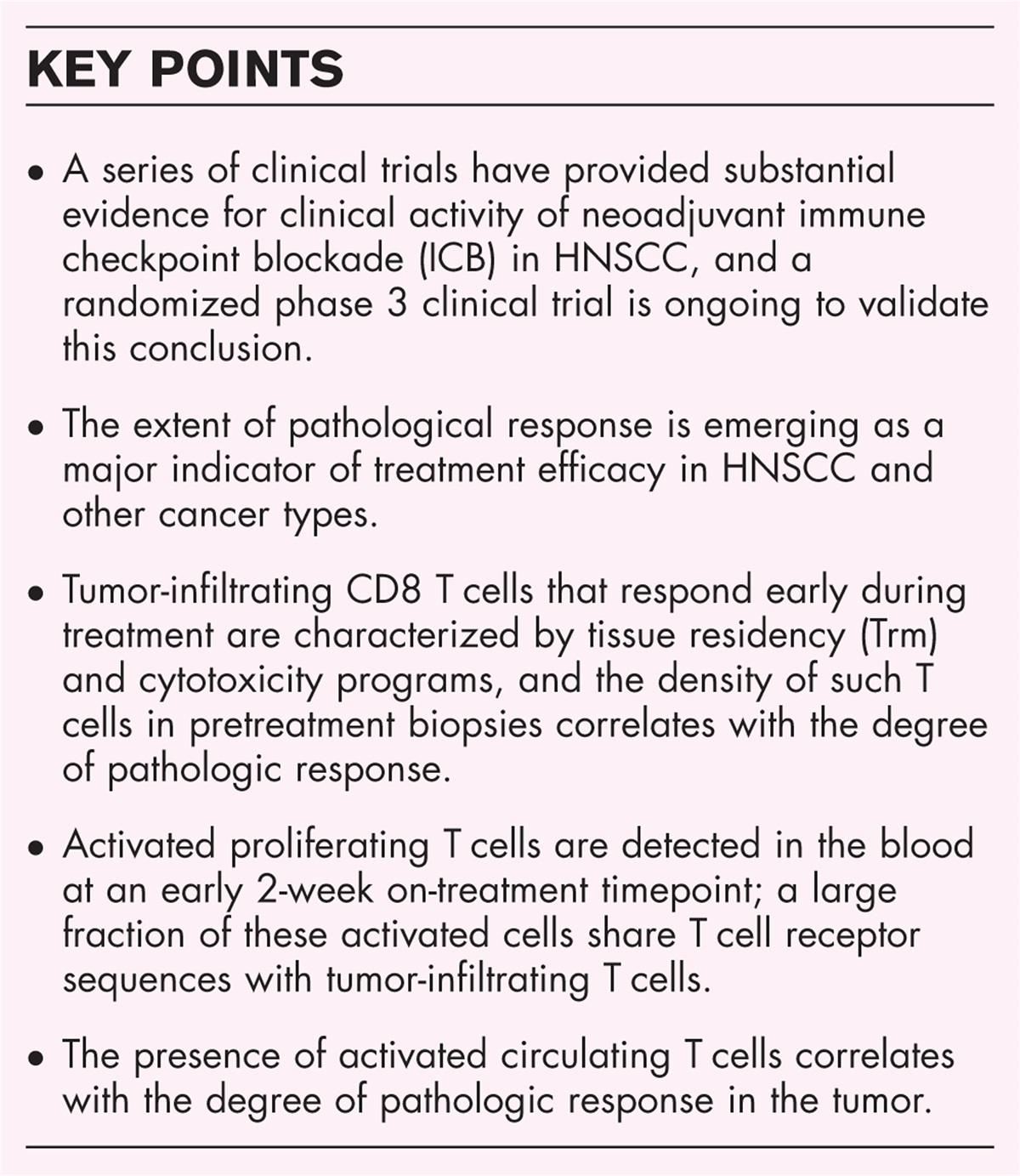
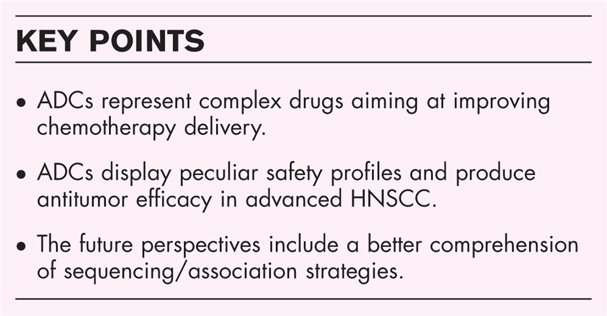
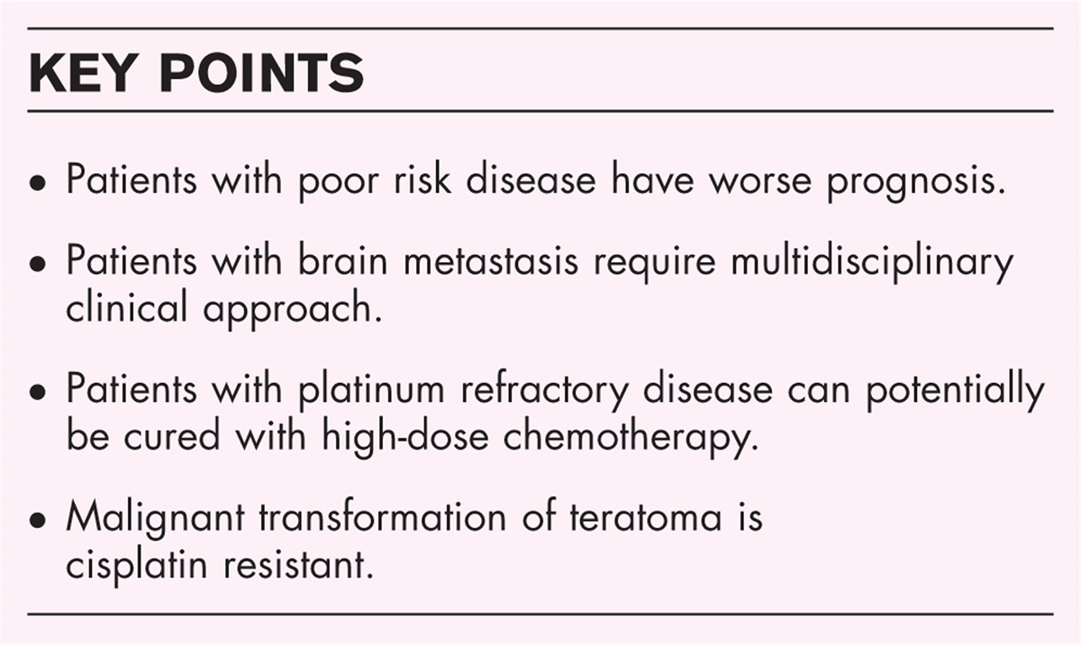
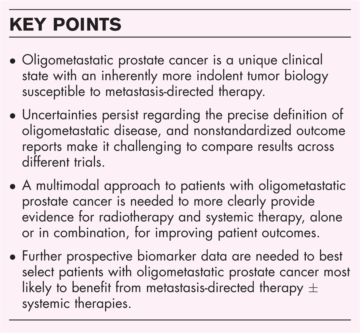
 Box 1:
 no caption available
CANCER VACCINES AND CHECKPOINT INHIBITORSTherapeutic cancer vaccination against telomerase is not a new concept. Several early vaccination attempts showed vaccine-induced tumor-antigen-specific T-cell responses but rarely resulted in an apparent clinical benefit [7–10]. In these early trials, the induced T-cell responses were likely restricted by the immune checkpoints, explaining the lack of clinical efficacy from vaccination. Despite the complicated history, therapeutic cancer vaccines are now seeing rekindled enthusiasm. This renewed excitement stems from a potential two-fold role in the immuno-oncology space. Firstly, checkpoint inhibitors (CPIs) are natural partners for vaccines, as their combination is expected to boost the induced T-cell response and release them from inhibition in the tumor microenvironment. Secondly, vaccines are designed to generate de novo antitumor T-cell responses, which is an apparent bottleneck for broader CPI efficacy. Spontaneous immune responses against a patient's tumor are the cornerstone of current cancer immunotherapy, on which the mode of action of all CPIs rests. By their nature, these responses are not under clinicians’ control but have nevertheless been shown to be required to invoke tumor regression in patients treated with the PD-1 CPI, pembrolizumab [11]. Therapeutic cancer vaccines represent a mechanistically complementary approach to CPIs by providing de novo cancer-specific T cells aiming to reinforce preexisting immune responses and pave the way for enhanced efficacy of CPIs and other cancer drugs dependent on such responses (Fig. 1).
 FIGURE 1:
 Combining a tumor-associated antigen-based vaccine and checkpoint inhibition to boost antitumor T-cell responses. Nonimmunogenic tumors may become more amenable to checkpoint inhibition through vaccination against nonmutated tumor-associated antigens, such as hTERT. Vaccination aims to increase the magnitude of T cells attacking the tumor, which can be further potentiated through checkpoint inhibitor (CPI) combinations.
TUMOR-ASSOCIATED ANTIGENS VERSUS NEOANTIGENSTwo distinct strategies for cancer vaccination have emerged; vaccination with neoantigens and vaccination with tumor-associated antigens (TAAs).
Contrary to TAA vaccines, neoantigen vaccines are personalized, one-of-a-kind, entities produced to match the individual patients’ tumor. Neoantigens are immunogenic cancer-specific antigens arising from the mutagenesis integral to tumor formation. The spectrum of neoantigens varies between tumor types, from patient to patient, and from location to location within a tumor. Neoantigen expression is also subject to temporal variation in response to evolving patterns of immune response (i.e. immuno-editing) or imposed treatment regimens [12]. Cancer-specific mutations can be regarded as potent vaccination targets as they are not subject to central immune tolerance and, thereby, likely to elicit strong immune responses. Generating neoantigen-based vaccines depends on a series of relatively complex procedures still in an exploratory phase. Identifying immunogenic tumor-specific mutations in individual patients, in general, requires tumor tissue biopsies for DNA sequencing and variant calling, together with RNA sequencing to assess expression levels. HLA typing and computational algorithms are being developed for epitope prediction. A central challenge in the design of neoantigen cancer vaccines is the selection of neoantigens that are expressed persistently and widely throughout a patient's tumor or tumors. A single personalized vaccine may have to target many neoantigens to optimize the T-cell responses and minimize the likelihood of immune evasion [13]. Recent early-phase clinical trials combining neoantigen vaccines with CPIs in melanoma have yielded encouraging efficacy signals [14–16]. Randomized phase 2 trials with neoantigen vaccines in combination with CPIs are ongoing in melanoma with anticipated read-out (NCT03897881, NCT03815058).
The alternative strategy for increasing antitumor T-cell responses, and the focus of this review, is the use of vaccines based on pan-cancer antigens that are expressed by multiple tumor types. TAAs are antigens with an expression pattern that is preferentially restricted to tumors. There are several groups of TAAs, of which many are potential targets for therapeutic vaccination (e.g. New York esophageal squamous cell carcinoma1 (NY-ESO-1) [17], melanoma-associated antigen (MAGE) [18], and human telomerase reverse transcriptase (hTERT) [19]) (Table 1). Compared with the neoantigen approach, vaccines directed towards the generation of T-cell responses to TAAs may be more relevant in cancers or individual tumors with low TMB, where the tumor expresses fewer neoantigens as a basis for vaccination. A large proportion of patients with metastatic melanoma (∼30%), for instance, have a low mutational burden at pretreatment baselines, implying expectations of few neoantigens to vaccinate against, and few spontaneously formed T-cell responses checkpoint inhibition can mobilize [20]. The use of vaccines based on highly prevalent TAAs has an additional practical benefit of allowing off-the-shelf vaccine administration and immediate use in patients whose tumors express the relevant TAAs. A recent phase I trial of a TAA mRNA vaccine [NY-ESO-1, MAGE-A3, tyrosinase, and transmembrane phosphatase with tensin homology (TPTE)] in combination with anti-PD1 therapy has shown promising results in patients with metastatic melanoma [21].
Table 1 - Expression pattern of commonly targeted tumor-associated antigens in melanoma tumors TAA Percentage positive melanoma tumors Percentage melanoma tumors with homogenous expression (>50% positive melanoma cells) hTERT [22,23] 100% ∼86–95% Tyrosinase [24] 94% ∼85% Melan-A [24] 94% ∼75–90% Gp100 ([24] 91% ∼60–75% NY-ESO-1 [24] 45% ∼10–20% MAGE-A1 [24] 36% ∼5–30% MAGE-A4 [24] 29% ∼5–25%TAA, tumor-associated antigen.
A key consideration in the utility of TAA vaccines for melanoma is the extent to which the target antigens are expressed in individual melanoma tumors. In that respect, antigens derived from telomerase reverse transcriptase (hTERT) is an attractive option. The hTERT expression pattern in cutaneous melanomas appears ubiquitous and comparatively homogenous within individual tumors (Table 1) [22–24]. Telomerase is actively expressed to preserve genome integrity in embryonic stem cells and dividing sperm cells, and it is expressed only sparsely in healthy somatic tissues [19]. In contrast, telomerase expression is essential in cancerous tissue to maintain telomere replication, unconstrained cancer cell proliferation, and metastasis [25,26]. These functions are fundamental to the oncogenesis and maintenance of a cancerous phenotype, and telomerase activation is, therefore, considered a hallmark of cancer [27]. By directing the immune response towards the active site of hTERT, tumor resistance mutations are unlikely to develop within the targeted epitopes, as these may lead to loss of hTERT function and consequent reduced tumor growth [19]. Telomerase expression has been documented in 85–90% of all tumor types [28,29], with the few remaining tumors utilizing alternative lengthening of telomeres (ALT) to achieve replicative immortality. The ALT phenotype has been documented in sarcomas, but rarely in carcinomas, and only in about 7% of melanomas [30]. Although switching to the ALT phenotype is a theoretical opportunity for the tumor to resist telomerase targeting therapies [31], such conversion has shown to significantly reduce the invasive potential of the tumor [32].
TELOMERASE EXPRESSION IN MELANOMA TUMORSMutations in the promoter region of hTERT are one of the most frequently occurring genomic aberrations in cutaneous melanoma (∼80%) [33]. These promoter mutations are a well described hTERT activation mechanism that increases transcription [34], resulting in increased hTERT expression. Gene copy number amplification is another hTERT activation mechanism, and amplification of the genomic regions containing hTERT is over-represented in melanomas [35]. Furthermore, staining of melanoma biopsies has shown a relatively homogenous hTERT expression, with a median of 72.7% positive melanoma cells [36▪,37]. High tumor hTERT activity also correlates with poor prognosis in melanoma, further supporting its relevancy as a therapeutic target [37]. Interestingly, the presence of spontaneous T-cell responses directed against hTERT appears to be advantageous for melanoma patients. A study designed to evaluate the prevalence and clinical significance of the CD4 T helper cell (Th1) response against hTERT in patients with melanoma concluded that circulating antihTERT Th1 responses were a predictive factor of response to checkpoint inhibition [38▪]. Around 55% of patients displayed circulating antihTERT Th1 immunity, and the rate of hTERT immunity was higher in patients with early-stage melanoma than in those with late-stage disease (69% in American Joint Committee on Cancer (AJCC) stage 1 versus 49% in AJCC stage ≥2), suggesting that melanoma progression was associated with a defect of antihTERT Th1 immunity. In patients subsequently treated with CPIs, a higher proportion of those demonstrating an objective response exhibited preexisting antihTERT Th1 immunity than did nonresponders – 86% (18/21) of clinical responders had preexisting anti-TERT Th1 responses, while only 32% (6/19) of clinical nonresponders did (P value 0.001). Furthermore, in a multivariate analysis, the anti-TERT Th1 response correlated with improved progression-free and overall survival, with a hazard ratio of 0.317 (95% CI, 0.14–0.72, P value 0.006) and 0.298 (95% CI, 0.09–0.94, P value 0.039), respectively.
VACCINE PLATFORMSAlthough several vaccine platforms have been utilized to mobilize an immune response against hTERT, such as DNA and mRNA vaccines, only peptide vaccines have been developed in melanoma [19]. The peptide platform is historically well established, although a few improvements have been incorporated to enhance immunogenicity and T-cell responsivity. The use of synthetic long peptides (SLPs) is now becoming the gold standard, mainly because of their ability to induce both CD4 and CD8 T-cell responses in an HLA-unrestricted manner. Previously, most peptide-based vaccines were typically tailored to fit single HLA class I alleles, necessitating prescreening of patients before administration. However, designated epitope-rich long peptides (≥15 amino acids) can be processed intracellularly to bind individual class I and II HLA alleles, thus eliciting both CD4 and CD8 T-cell responses. With the growth in available bioinformatical tools to predict immunogenicity and HLA-binding capabilities, such peptides can be readily identified [39], although their validation still requires in-vitro work. A more direct approach was utilized for selecting and validating the hTERT peptides in the UV1 vaccine, using patient-derived lymphocytes to screen overlapping peptides for immunogenicity within the active site of hTERT [40]. The resultingly identified three peptides (one 30-mer and two 15-mers), now constituting UV1, have been investigated as a vaccine product in several clinical trials in melanoma (NCT02275416, NCT03538314, NCT04382664).
ANTIhTERT IMMUNE RESPONSESThe potential clinical efficacy of therapeutic vaccination is mediated through the induced T-cell response, and the phenotype and magnitude of these responses are, therefore, important properties to consider. The immune response characterization from early clinical trials of UV1, with or without a checkpoint inhibitor combination, indicates the presence of both peripheral blood and intratumoral vaccine-specific T-cell responses. A long-term follow-up study of 52 patients treated across three trials showed that T-cell responses specific for the UV1 telomerase peptides could be measured in 78.4% of patients who received the vaccine and that the induced immune response associated with longer survival time [36▪]. The vaccine-induced T cells were of the effector memory phenotype, producing IFN-γ and TNF-α upon in-vitro stimulation. Vaccine-specific immune responses occurred earlier and in a higher proportion of patients (91%) when UV1 was administered in combination with ipilimumab, supporting the hypothesized synergy between the TAA vaccine and the CPI [36▪,41]. Translational research data from this trial confirmed telomerase expression in all baseline melanoma biopsies [22]. Based on T-cell receptor sequencing, estimated vaccine-enriched T cells accounted for up to 12% of the entire repertoire. A nonsignificant association was observed between the magnitude of peripheral T-cell responses and an increase in tumor-infiltrating CD8 lymphocytes. However, the study lacked sufficient sample size and biological material to draw conclusions.
CLINICAL EFFICACY SIGNALSThe prospects for patients with advanced melanoma have improved considerably since the introduction of regimens involving CPIs. Historically, the median survival after diagnosis was around 8 months [42]. A recent 6.5-year follow-up of the CheckMate-067 study reports median overall survival of 36.9 months for patients treated with the PD-1 inhibitor nivolumab, and 72.1 months for patients who received the combination of nivolumab and the CTLA-4 inhibitor, ipilimumab [43–45]. Five-year follow-up of the KEYNOTE-006 study reported median overall survival of 15.9 months for patients treated with ipilimumab and 32.7 months for patients treated with the PD-1 inhibitor pembrolizumab [46,47]. Further improvements may arise through judicious combinations of CPIs: The combination of relatlimab, a LAG-3–blocking antibody, with nivolumab extended median progression-free survival out to 10.1 months compared with 4.6 months for nivolumab alone [48]. The overall survival data from that study is yet to mature.
Within the next year, topline progression-free survival data are expected from INITIUM, a randomized 156-patient phase II safety and efficacy study in advanced inoperable malignant melanoma comparing treatment outcomes with ipilimumab and nivolumab with and without UV1 vaccine (NCT04382664) [49]. The study builds on promising clinical signals from earlier studies of UV1 that preceded INITIUM.
In a 12-patient phase I/IIa, single-center trial of UV1 in combination with ipilimumab in metastatic melanoma, the median progression-free survival was 6.7 months, and median overall survival was 66.3 months. Clinical responses to the combination treatment were also observed in patients with low TMB, few predicted neoantigens, and low IFN-γ gene signature, biomarkers associated with reduced CPI efficacy, thus indicating a potential added benefit of vaccination [22]. As a historical comparator, a phase 4 clinical trial (n = 151) of ipilimumab monotherapy running concurrently with the combination study used a highly similar protocol and returned a mPFS of 2.7 months and mOS of 12.1 months [50].
At the 2022 Society for Melanoma Research Conference, a second phase I study testing the combination of UV1 and pembrolizumab in 30 treatment-naive patients with unresectable advanced melanoma was presented. Patients received eight intradermal vaccinations over a 14-week period and pembrolizumab infusions according to label. The median progression-free survival was 18.9 months, and median overall survival was not reached after up to 3 years of follow-up. The objective response rate was 56.7%, with complete responses observed in 33% of treated patients. Encouragingly, similar to the UV1 ipilimumab trial, high objective response rates were observed also in subgroups with tumor biopsies characterized by PD-L1 negativity, TMB-low, few to zero predicted neoantigens, and those not enriched for the IFN-γ gene signature. There was a relative decrease in the expression of hTERT in week 14 biopsies in clinical responders, indicating eradication of hTERT-expressing cancer cells [51] (manuscript in preparation).
In both these UV1 studies, the combinations of vaccines and CPIs were considered well tolerated, and patient objective response rates were high compared with historical controls (Table 2). The safety profile of the combinations has been similar to monotherapy CPI, with vaccine-related adverse events primarily being mild injection site reactions. There have so far been no observations implicating vaccine-induced off-tumor reactivity against healthy hTERT-expressing tissues such as the bone marrow [52–54].
Table 2 - Efficacy and safety read-out of checkpoint inhibitor and hTERT vaccine trials in melanoma Nivolumab + ipilimumab Pembrolizumab UV1 + pembrolizumab Ipilimumab UV1 + ipilimumab Reference Wolchok JD et al. and Larkin J et al. [43–45] Robert et al. [46] and Carlino et al. [47] Zakharia et al. [51] Aamdal et al. [50] Aamdal et al. [41] and Ellingsen et al. [22] Phase III III I IV I N 314 556 30 151 12 mPFS (months) 11.5 8.4 18.9 2.7 6.7 mOS (months) 72.1 32.7 NRa 12.1 66.3 ORR (%) 58 42 57 9 33 ORRCR, complete response; mOS, median overall survival; mPFS, median progression-free survival; NA, not available; ORR, objective response rate; PR, partial response; TRAE, treatment-related adverse event.
aNR, not reached with up to 3 years of follow-up.
bIn first-line PD-L1 positive patients.
The current review discusses the long history of therapeutic cancer vaccines and why we may finally see their role established in the treatment of cancer. The advent of checkpoint inhibition has revolutionized the treatment of cancer, especially melanoma. Improvements are, however, still called for, and vaccines are ideally positioned to solve a central challenge in CPIs’ lack of efficacy. Accumulating data suggest telomerase reverse transcriptase is an attractive target for vaccination, and immune response data support its antitumor promise. Several phase I clinical trials have been conducted, with favorable read-outs encouraging further development. A phase II randomized clinical trial evaluating an hTERT-targeting vaccine in combination with nivolumab and ipilimumab is ongoing in frontline advanced melanoma, with read-out expected during 2023. As checkpoint inhibition is moving earlier in the treatment landscape, we expect to see cancer vaccines being developed in the (neo)adjuvant setting as well, where vaccine-induced immune responses may help clear minimal residual disease after surgery.
AcknowledgementsNone.
Financial support and sponsorshipNone.
Conflicts of interestAll authors are employees of Ultimovacs ASA. G.G. is an inventor of UV1, and a shareholder of Ultimovacs ASA.
REFERENCES AND RECOMMENDED READINGPapers of particular interest, published within the annual period of review, have been highlighted as:
▪ of special interest
▪▪ of outstanding interest
REFERENCES 1. Kim JY, Kronbichler A, Eisenhut M, et al. Tumor mutational burden and efficacy of immune checkpoint inhibitors: a systematic review and meta-analysis. Cancers (Basel) 2019; 11:1798. 2. McGranahan N, Furness AJS, Rosenthal R, et al. Clonal neoantigens elicit T cell immunoreactivity and sensitivity to immune checkpoint blockade. Science 2016; 351:1463–1469. 3. Ayers M, Lunceford J, Nebozhyn M, et al. IFN-γ-related mRNA profile predicts clinical response to PD-1 blockade. J Clin Invest 2017; 127:2930–2940. 4. Taube JM, Klein A, Brahmer JR, et al. Association of PD-1, PD-1 ligands, and other features of the tumor immune microenvironment with response to anti-PD-1 therapy. Clinical Cancer Research 2014; 20:5064–5074. 5. Uryvaev A, Passhak M, Hershkovits D, et al. The role of tumor-infiltrating lymphocytes (TILs) as a predictive biomarker of response to anti-PD1 therapy in patients with metastatic nonsmall cell lung cancer or metastatic melanoma. Med Oncol 2018; 35:25. 6. Latchman Y, Wood CR, Chernova T, et al. PD-L2 is a second ligand for PD-1 and inhibits T cell activation. Nat Immunol 2001; 2:261–268. 7. Negrini S, De Palma R, Filaci G. Anticancer immunotherapies targeting telomerase. Cancers 2020; 12:2260. 8. Middleton G, Silcocks P, Cox T, et al. Gemcitabine and capecitabine with or without telomerase peptide vaccine GV1001 in patients with locally advanced or metastatic pancreatic cancer (TeloVac): an open-label, randomised, phase 3 trial. Lancet Oncol 2014; 15:829–840. 9. Hunger RE, Kernland Lang K, Markowski CJ, et al. Vaccination of patients with cutaneous melanoma with telomerase-specific peptides. Cancer Immunol Immunother 2011; 60:1553-1564. 10. Kyte JA, Gaudernack G, Dueland S, et al. Telomerase peptide vaccination combined with temozolomide: a clinical trial in stage IV melanoma patients. Clin Cancer Res 2011; 17:4568–4580. 11. Tumeh PC, Harview CL, Yearley JH, et al. PD-1 blockade induces responses by inhibiting adaptive immune resistance. Nature 2014; 515:568–571. 12. Lo AA, Wallace A, Oreper D, et al. Indication-specific tumor evolution and its impact on neoantigen targeting and biomarkers for individualized cancer immunotherapies. J Immunother Cancer 2021; 9:e003001. 13. Blass E, Ott PA. Advances in the development of personalized neoantigen-based therapeutic cancer vaccines. Nat Rev Clin Oncol 2021; 18:215–229. 14. Ott PA, Hu-Lieskovan S, Chmielowski B, et al. A phase Ib trial of personalized neoantigen therapy plus anti-PD-1 in patients with advanced melanoma, nonsmall cell lung cancer, or bladder cancer. Cell 2020; 183:347.e24–362.e24. 15. Hu Z, Leet DE, Allesøe RL, et al. Personal neoantigen vaccines induce persistent memory T cell responses and epitope spreading in patients with melanoma. Nat Med 2021; 27:515–525. 16. Bauman J, Burris H, Clarke J, et al. 798 Safety, tolerability, and immunogenicity of mRNA-4157 in combination with pembrolizumab in subjects with unresectable solid tumors (KEYNOTE-603): an update. J Immunother Cancer 2020; 8:A477–A1477. 17. Thomas R, Al-Khadairi G, Roelands J, et al. NY-ESO-1 based immunotherapy of cancer: current perspectives. Front Immunol 2018; 9:947. 18. Jungbluth AA, Busam KJ, Kolb D, et al. Expression of MAGE-antigens in normal tissues and cancer. Int J Cancer 2000; 85:460–465. 19. Ellingsen EB, Mangsbo SM, Hovig E, Gaudernack G. Telomerase as a target for therapeutic cancer vaccines and considerations for optimizing their clinical potential. Front Immunol 2021; 12:682492. 20. Kang K, Xie F, Mao J, et al. Significance of tumor mutation burden in immune infiltration and prognosis in cutaneous melanoma. Front Oncol 2020; 10:573141. 21. Sahin U, Oehm P, Derhovanessian E, et al. An RNA vaccine drives immunity in checkpoint-inhibitor-treated melanoma. Nature 2020; 585:107–112. 22. Ellingsen EB, Bounova G, Kerzeli I, et al. Characterization of the T cell receptor repertoire and melanoma tumor microenvironment upon combined treatment with ipilimumab and hTERT vaccination. J Transl Med 2022; 20:419. 23. Bustos BDU, Torralba AS, Poveda PM, et al. Telomerase expression in a series of melanocytic neoplasms. Actas Dermosifiliogr 2019; 110:212–219. 24. Barrow C, Browning J, MacGregor D, et al. Tumor antigen expression in melanoma varies according to antigen and stage. Clin Cancer Res 2006; 12 (3 pt 1):764–771. 25. Hannen R. Bartsch JW Essential roles of telomerase reverse transcriptase hTERT in cancer stemness and metastasis. FEBS Lett 2018; 592:2023–2031. 26. Liu Z, Li Q, Li K, et al. Telomerase reverse transcriptase promotes epithelial-mesenchymal transition and stem cell-like traits in cancer cells. Oncogene 2013; 32:4203–4213. 27. Hanahan D. Weinberg RA Hallmarks of cancer: the next generation. Cell 2011; 144:646–674. 28. Shay JW, Bacchetti S. A survey of telomerase in human cancer. Eur J Cancer 1997; 33:787–791. 29. Kim N, Piatyszek M, Prowse K, et al. Specific association of human telomerase activity with immortal cells and cancer. Science 1994; 266:2011–2015. 30. Heaphy CM, Subhawong AP, Hong SM, et al. Prevalence of the alternative lengthening of telomeres telomere maintenance mechanism in human cancer subtypes. Am J Pathol 2011; 179:1608–1615. 31. Bechter OE, Zou Y, Walker W, et al. Telomeric recombination in mismatch repair deficient human colon cancer cells after telomerase inhibition. Cancer Res 2004; 64:3444–3451. 32. Chen W, Xiao BK, Liu JP, et al. Alternative lengthening of telomeres in hTERT-inhibited laryngeal cancer cells. Cancer Sci 2010; 101:1769–1776. 33. Zehir A, Benayed R, Shah RH, et al. Mutational landscape of metastatic cancer revealed from prospective clinical sequencing of 10,000 patients. Nat Med 2017; 23:703–713. 34. Huang FW, Hodis E, Xu MJ, et al. Highly recurrent TERT PROMOTER MUTATIONS IN HUMAN MELANOMA. Science 2013; 339:957–959. 35. Pirker CH, Holzmann K, Spiegl-Kreinecker S, et al. Walter chromosomal imbalances in primary and metastatic melanomas: over-representation of essential telomerase genes. Melanoma Res 2003; 13:483–492. 36▪. Ellingsen EB, Aamdal E, Guren T, et al. Durable and dynamic hTERT immune responses following vaccination with the long-peptide cancer vaccine UV1: long-term follow-up of three phase I clinical trials. J Immunother Cancer 2022; 10:e004345. 37. Hugdahl E, Kalvenes MB, Mannelqvist M, et al. Prognostic impact and concordance of TERT promoter mutation and protein expression in matched primary and metastatic cutaneous melanoma. Br J Cancer 2018; 118:98–105. 38▪. Nardin C, Laheurte C, Puzenat E, et al. Naturally occurring telomerase-specific CD4 T cell immunity in melanoma. J Invest Dermatol 2021; 142:435–444. 39. Luo H, Ye H, Ng HW, et al. Machine learning methods for predicting HLA-peptide binding activity. Bioinform Biol Insights 2015; 9s3 (BBI):S29466. 40. Inderberg-Suso EM, Trachsel S, Lislerud K, et al. Widespread CD4+ T-cell reactivity to novel hTERT epitopes following vaccination of cancer patients with a single hTERT peptide GV1001. Oncoimmunology 2012; 1:670–686. 41. Aamdal E, Inderberg EM, Ellingsen EB, et al. Combining a universal telomerase based cancer vaccine with ipilimumab in patients with metastatic melanoma - five-year follow up of a phase I/IIa trial. Front Immunol 2021; 12:663865. 42. Franken MG, Leeneman B, Aarts MJB, et al. Trends in survival and costs in metastatic melanoma in the era of novel targeted and immunotherapeutic drugs. ESMO Open 2021; 6:100320. 43. Wolchok JD, Chiarion-Sileni V, Gonzalez R, et al. Long-term outcomes with nivolumab plus ipilimumab or nivolumab alone versus ipilimumab in patients with advanced melanoma. J Clin Oncol 2022; 40:127–137. 44. Larkin J, Chiarion-Sileni V, Gonzalez R, et al. Five-year survival with combined nivolumab and ipilimumab in advanced melanoma. N Engl J Med 2019; 381:1535–1546. 45. Wolchok JD, Chiarion-Sileni V, Gonzalez R, et al. Overall survival with combined nivolumab and ipilimumab in advanced melanoma. New Engl J Med 2017; 377:1345–1356. 46. Robert C, Ribas A, Schachter J, et al. Pembrolizumab versus ipilimumab in advanced melanoma (KEYNOTE-006): posthoc 5-year results from an open-label, multicentre, randomised, controlled, phase 3 study. Lancet Oncol 2019; 20:1239–1251. 47. Carlino MS, Long GV, Schadendorf D, et al. Outcomes by line of therapy and programmed death ligand 1 expression in patients with advanced melanoma treated with pembrolizumab or ipilimumab in KEYNOTE-006: a randomised clinical trial. Eur J Cancer 2018; 101:236–243. 48. Tawbi HA, Schadendorf D, Lipson EJ, et al. RELATIVITY-047 Investigators. Relatlimab and nivolumab versus nivolumab in untreated advanced melanoma. New Engl J Med 2022; 386:24–34. 49. O’Day S, Bechter O, Lorigan P, et al. Abstract CT231: nivolumab and ipilimumab +/- UV1 vaccine as 1st line treatment in patients with malignant melanoma (INITIUM-trial). Cancer Res 2021; 81:CT231–CT1231. 50. Aamdal E, Jacobsen KD, Straume O, et al. Ipilimumab in a real-world population: a prospective phase IV trial with long-term follow-up. Int J Cancer 2021; 150:100–111. 51. Zakharia Y, Ellingsen EB, Kristedja T, et al. Clinical activity of combined telomerase vaccination and pembrolizumab in unresectable melanoma. In The 19th International Congress of the Society for Melanoma Research. Edinburgh; 2022. 52. Brunsvig PF, Aamdal S, Gjertsen MK, et al. Telomerase peptide vaccination: a phase I/II study in patients with nonsmall cell lung cancer. Cancer Immunol Immunother 2006; 55:1553–1564. 53. Danet-Desnoyers GA, Luongo JL, Bonnet DA, et al. Telomerase vaccination has no detectable effect on SCID-repopulating and colony-forming activities in the bone marrow of cancer patients. Exp Hematol 2005; 33:1275–1280. 54. Vonderheide RH, Domchek SM, Schultze JL, et al. Vaccination of cancer patients against telomerase induces functional antitumor CD8 T lymphocytes. Clin Cancer Res 2004; 10:828–839.
留言 (0)