This study investigated the baseline characteristics of ocular parameters and variables as well as longitudinal changes in ocular parameters and the TZ in myopic Chinese children over a one-year period. This is one of the first studies to evaluate AL reduction in myopic children treated with the Ortho-K lens for one year follow-up. The results of this study showed that older baseline age and smaller TZ wearing Ortho-K were associated with AL reduction. In myopic children wearing Ortho-K lenses with AL reduction, thickened CLT, decreased ACD and thinned VCD were more obvious during the one-year period, and these changes are helpful for analyzing the biological characteristics of AL reduction which provide clues for further study of the specific mechanism of myopia progression to achieve a better myopia control effect.
Possible reasons for AL reduction
Although many studies have shown that Ortho-K treatment is effective for myopia control, the mechanism by which Ortho-K might control myopia progression or even AL reduction is still controversial. AL change is a crucial index in the evaluation of myopic progression, and AL reduction due to Ortho-K treatment tends to achieve a much better myopia control effect. Some studies have reported AL reduction after Ortho-K lens wear. Zhao et al. [27] revealed that the six-month AL change was 0.04 ± 0.12 mm, and a decrease in AL was observed in 33% of the eyes treated with Ortho-K lenses. Chen et al. [21] reported that the AL in 49% of eyes treated with Ortho-K lenses for three weeks decreased. However, none of these studies extended a longer period of follow-up, and details about changes in AL components have rarely been reported.
More studies have suggested that choroidal thickness may be one of the factors contributing to AL reduction. The longitudinal orthokeratology research in children (LORIC) study by Cho et al. [28] reported AL reduction in study subjects during the first six months of Ortho-K lens treatment, where increased choroidal thickness was the speculative explanation for AL reduction. In Chen et al.’s study, the estimated mean change in the choroidal thickness was 21.8 ± 25.2 µm after three weeks of Ortho-K lens wear. In our study, the one-year AL value was reduced by 0.18 ± 0.06 mm in the AL reduction group, which was greater than 0.04 ± 0.12 mm as reported by Zhao et al. [27] and 0.03 ± 0.13 mm as reported by Cho et al. [11]. Additionally, 16.22% of the subjects had a reduction of AL of more than 0.25 mm after wearing the Ortho-K lenses even though the choroidal thickness was approximately 0.30 mm [29, 30] and the choroidal thickness change was only approximately 20 µm [21]; hence, it is difficult to explain this phenomenon by choroidal thickness change alone. In addition, the aforementioned studies measured short-term AL changes from baseline; therefore, the response may be a short-term choroidal response to Ortho-K lens treatment rather than a true AL change, and AL reduction in these patients could have led to overestimation of the effect of Ortho-K lens treatment on myopia control. To eliminate the influence of a short-term choroidal response on AL change, we studied changes in ocular parameters in subjects with AL reduction over a period of one year to better understand the long-term effects of Ortho-K lens treatment on the anterior and posterior segments and its effect on AL change. We also tried to control the influence of rhythm and factors on the change in choroidal thickness, but the choroid is a tissue that changes all the time and is affected by many other factors (environment, rhythm, visual stimuli, food, etc.). The thickening of choroidal thickness is one of the reasons for the compensation effect of AL reduction, and the definitive mechanism needs to be further studied.
The CCT change was another possible reason for the AL reduction after Ortho-K treatment that has been observed in many studies. Wang et al. observed 249 Ortho-K wearers over an average period of 2.5 years and reported that CCT was not associated with AL elongation during Ortho-K treatment [31]. Some studies reported a decrease in the CCT [20, 32] after Ortho-K lens wear. In concordance with the findings of these studies, we found CCT reduction beyond the baseline value after Ortho-K lens wear. These changes in the anterior segment of the eye partly explain AL reduction after Ortho-K lens treatment. However, in Chen et al.’s study, the perceived AL change was significantly correlated with the CCT change after Ortho-K lens treatment, but the predictive values were low (12%), indicating that there are other potentially important factors affecting AL. Epithelial change could contribute to the variation in AL, but it may not be the primary mechanism explaining these changes. Furthermore, a significant reduction in the VCD was detected in the AL reduction group. The reductions in the CCT (15.73 µm) and VCD (160 µm) were approximately the same as those in the AL reduction (180 µm), suggesting that the AL reduction was mainly due to the shortening of the VCD. We hypothesize that the reduction in AL is partly due to choroidal thickening pushing the retina forward, resulting in a shortened VCD, but there may be other factors involved. Another remarkable finding of this study was the similarity between the ACD reduction (100 µm) and the CLT increase (100 µm). This indicates that the change in the ACD may be caused by the thickening of the CLT.
As mentioned previously, we found that in children with AL reduction, their crystalline lens is thickened, corresponding to a shortened ACD. Some previous studies have reported relationships between ocular components and relative peripheral refraction, which may trigger AL elongation in myopic participants [33]. They found that a deeper ACD, a deeper vitreous chamber [34,35,36] and a thinner CLT [37] were associated with hyperopic relative peripheral refraction in myopic children, indicating a tendency for longer eyes and that a shallower ACD, thicker CLT and thinner VCD may decrease hyperopic relative peripheral refraction, slowing axial elongation. A study by Li et al. found that CLT increased during one year of Ortho-K lens treatment but did not recover after one month of lens cessation [19]. Wang et al. also observed that a greater increase in CLT was associated with a smaller increase in AL at the 12-month follow-up during Ortho-K wear in children with myopia [38]. These results may indicate that the continuous wearing of Ortho-K lenses may result in a change in the crystalline refractive power, which may remain stable and nonreversible in the short term, correlating with a smaller increase in AL. Another study found a significant ACD reduction during Ortho-K treatment, and the ACD reduction at the center of the cornea was similar to the significant AL reduction that accompanied it [39]. Consistent with these studies, we also observed a reduced ACD and VCD and an increased CLT in the AL reduction group compared with those in the AL elongation group. However, increased CLT might also be considered a result of AL reduction as more accommodation could be introduced as compensation for overcorrection caused by shortening of AL.
Notably, in this study, 16.22% of subjects had an AL reduction of more than 0.25 mm, which is difficult to explain by changes in corneal thickness and choroidal thickness, as the magnitude of the changes exceeds that for which the cornea and choroid can compensate. Therefore, we speculate that the sclera may have remodeled during the process of AL reduction. Previous studies have confirmed that the choroid, a vascular structure between the retina and the sclera, supplies RPE and the outer retina. In addition, it contains secretory cells, which may be involved in mediating scleral growth. Thus, the choroid has a crucial role in relaying signals derived from the retina to the sclera, further altering the synthesis of scleral extracellular matrix molecules and changing the size of the eye. Therefore, we hypothesized that choroidal thickening after wearing Ortho-K lenses might modulate the sclera and thus further affect AL. The exact mechanism underlying the results observed in this study remains to be explored.
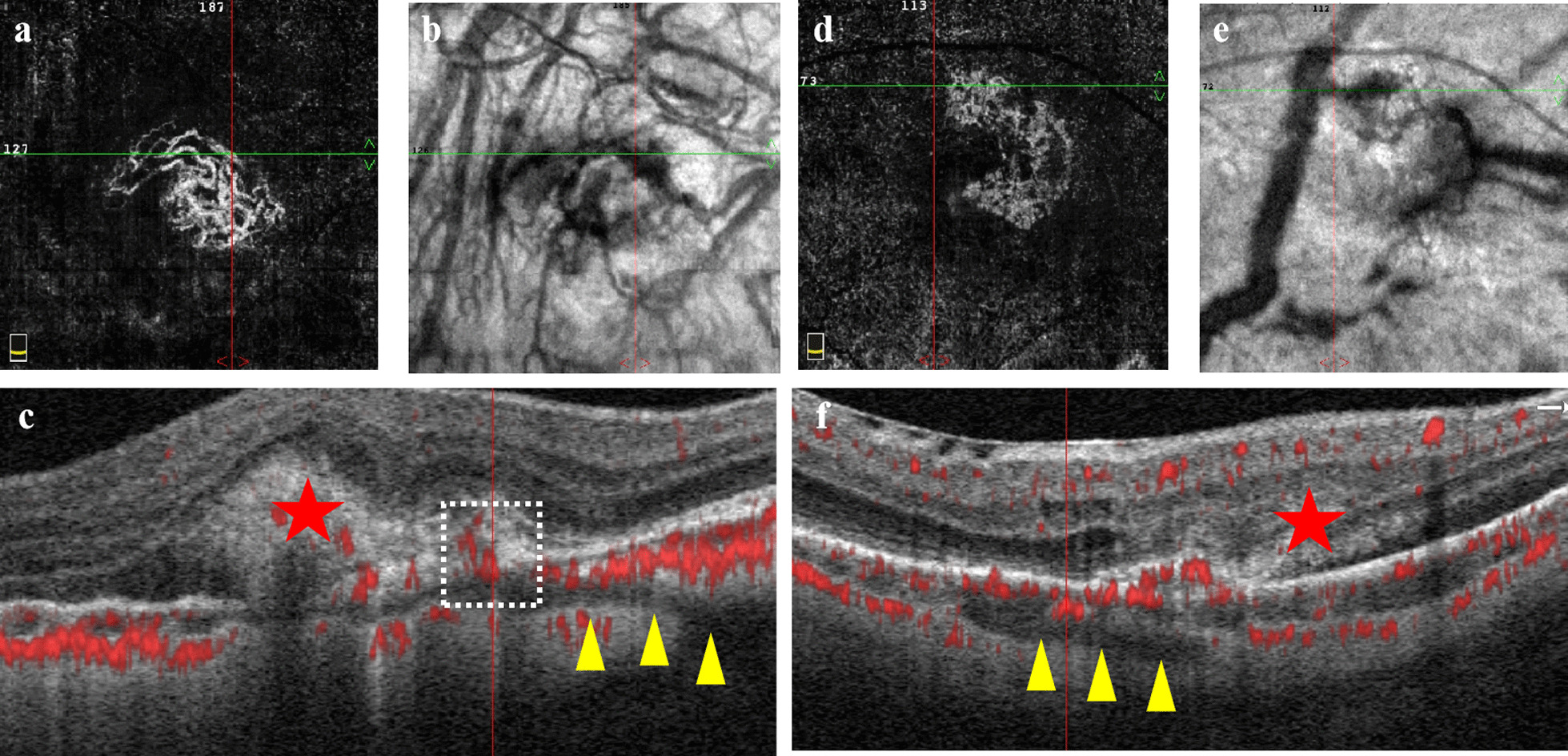
Factors affecting the Ortho-K control effect
Some studies have reported that the effect of Ortho-K on the control of myopia has great individual variability, which may be affected by age at the initiation of Ortho-K wear [25, 40]. The same conclusion was also reached in this study. In addition, the TZ is also a factor affecting the progression of myopia. As mentioned previously, changes in peripheral defocus and aberration have been hypothesized as explanations for the mechanism of Ortho-K in myopia control [41, 42]. Lau et al. [43] analyzed 103 myopic children treated with Ortho-K lenses over a two-year follow-up and found that ocular higher-order aberrations, particularly spherical aberrations, had a negative correlation with axial elongation. Increasing higher-order aberration may slow axial elongation in Ortho-K treatment. In addition, myopia control of Ortho-K is associated with relative peripheral refraction. According to studies by Carracedo et al. [44] and Guo et al. [25], a smaller TZ tends to cause more myopic relative peripheral refraction and a larger change in higher-order aberrations, achieving an improvement in myopia control. Consistent with their findings, our study found that children in the AL reduction group had a smaller TZ than those in the AL elongation group in the absence of a statistically significant difference in pupil size between groups, and AL reduction was significantly positively correlated with the TZ. This result suggests that a smaller TZ plays a potential role in controlling AL elongation using Ortho-K and provides a new perspective on the personalized design of Ortho-K lenses for myopic children.
Limitations
Our study had some limitations. First, it would have been ideal to accurately measure SER after Ortho-K treatment to further confirm the degree of myopia progression in this study. In addition, some studies have revealed that AL changes are significantly correlated with choroidal thickness changes after Ortho-K treatment; however, we did not evaluate the change in choroidal thickness after Ortho-K lens wear. Therefore, further studies should assess changes in the anterior and posterior segments to predict AL changes in Ortho-K patients. In addition, the one-year observation period chosen for this study is because most myopic children treated with the Ortho-K lens change their lenses after approximately one year of wear in view of safety concerns. For cases of AL reduction, a longer follow-up period is still needed to observe the effect of AL control. In addition, the relatively small number of children with AL reduction limited the sample size of this study, and larger sample sizes should be undertaken in future studies.


留言 (0)