
記住我
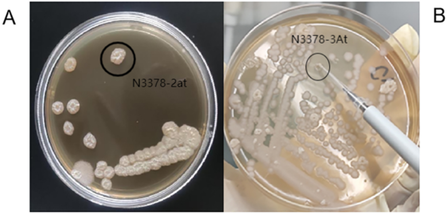


A total of 77 strains (Table 1) of endophytic fungi were isolated from P. ternata and P. pedatisecta for the first time. There were 53 and 24 strains of fungi that were isolated from all tissues of P. ternata and P. pedatisecta, respectively. The ITS1-ITS4 region of 77 strains was sequenced and compared with available GenBank reference sequences. The obtained 5.8S rDNA sequences were uploaded to NCBI under the accession numbers ON677855-ON677931. The results of sequence analysis showed that 77 fungi were attached to the phyla Ascomycota and Basidiomycota (Fig. 1). The 68 strains were grouped into four classes [Dothideomycetes (14.3%), Eurotiomycetes (32.5%), Saccharomycetes (2.6%) and Sordariomycetes (39.0%)] within the phylum Ascomycota. Nine other strains (11.7%) were distributed in the Agaricomycetes within the phylum Basidiomycota. The fungi of Sordariomycetes were the dominant species of cultivable fungi in phylogenetic diversity from P. ternata and P. pedatisecta. The largest number (Sordariomycetes, 30) of isolates was distributed in 5 orders, including the Glomerellales, Hypocreales, Pleurotheciales, Sordariales and Xylariales.
Table 1 Phylogenetic analysis of cultivable fungi associated with P. ternata and P. pedatisectaFig. 1
Neighbor-joining phylogenetic tree of 77 fungi isolates from P. ternata and P. pedatisecta. The phylogenetic tree based on ITS gene sequences. The values at each node represent the bootstrap values from 1000 replicates
There were 10, 17, 16 and 34 strains, which belonged to 7, 9, 7 and 13 genera, from roots, stems, leaves, and tubers, respectively. Among them, Alternaria angustiovoidea, Macrophomina phaseolina and Schizophyllum commune were isolated from their roots only. Similarly, Clonostachys rosea, Lecanicillium dimorphum, Nigrospora pyreformis, Pseudoechria longicollis, Trichoderma atroviride and Zygosporium masonii were isolated from tubers only. Therefore, the results of phylogenetic diversity showed that the endophytic fungi were different in different plant tissues.
Diversity analyses of endophytic fungi from P. ternata and P. pedatisectaThe diversity analyses of endophytic fungi genera isolated from P. ternata and P. pedatisecta were summarized and shown in Table 2. The Species richness (S) and Margalef index (D') values showed positive correlation with endophytic fungal genera [21]. The higher the Shannon–Wiener index (H') and the Simpson diversity index (Ds), the more diverse the microbial community [22]. Therefore, the endophytic fungi of tubers from P. ternata showed high species richness and diversity, with the values of S (12), D' (3.3011), H' (2.2299), Ds (0.8673), and PIE (0.8995). However, the endophytic fungi of the leaf from P. pedatisecta had high species richness and diversity with the values of S (6), D' (2.4045), H' (1.7329), Ds (0.8125), and PIE (0.9286). The Pielou indexes (J) could reflect the level of uniformity in the distribution of the number of individuals of the species in the community [23]. In this study, the endophytic fungi of stem from P. ternata and tuber from P. pedatisecta showed high Pielou indexes (J) with values of 0.9732 and 0.9697, respectively, which indicated a convergence in the number of individuals of each species.
Table 2 Diversity analyses of endophytic fungi from P. ternata and P. pedatisectaAntibacterial activities of the crude extracts of fungiThe filter paper disk method was used to evaluate the antibacterial activity of 77 fungal extracts from P. ternata and P. pedatisecta. The results showed that 21 extracts (27.3%) showed antibacterial activities against at least one pathogenic bacterium (Supplementary Table S1). Five of them (PT02, PP35, PP37, PP39, and PT58) had inhibitory activities against all four pathogens (Escherichia coli, Micrococcus tetragenus, Staphylococcus aureus, and Pseudomonas syringae pv. actinidiae). Among them, PP39 exhibited strong antibacterial activity against S. aureus with an IZD of 20.0 mm, which was equivalent to that of positive gentamicin sulfate with an IZD of 21.7 mm. PP39 also showed potent inhibition activities against M. tetragenus, E. coli and P. syringae pv. actinidiae with the IZD of 14.2, 15.2, and 14.0 mm, which were weaker than those of positive gentamicin sulfate with the IZD of 25.7, 26.7, and 24.3 mm, respectively. Besides, the strain PT83 showed strong inhibition activity against P. syringae pv. actinidiae with an IZD of 20.0 mm, which was comparable to that of positive gentamicin sulfate. In addition, the crude extracts of strains PT56 and PT82 also exhibited potent antibacterial activities against P. syringae pv. actinidiae with the IZD of 15.2 mm and 15.7 mm, respectively.
Phytotoxic assayAs the results shown in Table 3, 52 fermentation broth of endophytic fungi exhibited significant phytotoxic activity against the radicle of Echinochloa crusgalli with the inhibition rate of more than 50%. Among them, 22 strains showed strong phytotoxic activity against E. crusgalli with the inhibition rate of 100%. Interestingly, these fungal strains were mainly assigned to three genera (Aspergillus, Fusarium, and Talaromyces) and most of them (20 strains) were isolated from P. ternata. Besides, 16 strains showed outstanding phytotoxic activity against E. crusgalli with the inhibition rate of 80%-99%. Moreover, nine strains showed potent phytotoxic activity against E. crusgalli with the inhibition rate of 60%-79%. In addition, 25 strains showed relatively weak phytotoxic activity against E. crusgalli with the inhibition rate of 10%-60%.
Table 3 Inhibitory activity of the fermentation broth of 77 endophytic fungi of P. ternata and P. pedatisecta on the radicle growth of E. crusgalliIdentification of the secondary metabolites isolated from PT09 and PP39The fermentation broth PP39 from P. pedatisecta had the best antibacterial and phytotoxic activities among all tested endophytic fungi. PT09 from P. ternata also had strong phytotoxic activity against E. crusgalli with the inhibition rate of 91.1% and moderate antibacterial activity against S. aureus and P. syringae pv. actinidiae. Therefore, both PT09 and PP39 were selected as further research objects of active secondary metabolites.
Four monomer compounds (Fig. 2) were isolated from the liquid fermentation product of strain PT09. They were further identified as alternariol monomethyl ether (1), alternariol (2), dehydroaltenusin (3) and altertoxin II (4) by spectroscopic analyses, including HR–ESI–MS, NMR, and compared with data described in previous literature.
Fig. 2
The structure of compounds 1–7
Alternariol monomethyl ether (1) (Figures S1-S2): colorless crystal; HR-ESI–MS: m/z: 271.0599 [M—H]−, calculated for C15H11O5 271.0621; 1H NMR (600 MHz, DMSO-d6) δ: 11.81 (s, OH-7), 10.35 (br s, OH-3), 7.20 (s, H-10), 6.72 (s, H-4), 6.63 (s, H-2), 6.60 (s, H-8), 3.90 (s, 3H, OCH3-9), 2.72 (s, 3H, H-11). Due to the chemical shifts and relative molecular masses in agreement with reported in the literature [24], the structure of the compound was determined to be alternariol monomethyl ether.
Alternariol (2) (Figures S3-S4) [25]: white crystal; HR-ESI–MS: m/z: 257.0453 [M—H]−, calculated for C14H9O5 257.0464; 1H NMR (600 MHz, Acetone-d6) δ: 11.93 (s, OH-3), 9.69 (br s, OH-5), 9.21 (br s, OH-4’), 7.34 (d, J = 2.4 Hz, H-6), 6.78 (d, J = 2.7 Hz, H-5’), 6.69 (d, J = 2.7 Hz, H-3’), 6.44 (d, J = 2.4 Hz, H-4), 2.76 (s, 3H, CH3-6’).
Dehydroaltenusin (3) (Figures S5-S7) [26]: yellow green solid; HR-ESI–MS: m/z: 287.0558 [M—H]−, calculated for C15H11O6 287.0570; 1H NMR (600 MHz, CDCl3) δ: 11.29 (s, OH-7), 6.73 (d, J = 2.3 Hz, H-10), 6.69 (s, H-1), 6.63 (d, J = 2.3 Hz, H-8), 6.41 (s, H-3), 6.28 (s, H-4), 3.91 (s, 3H, OCH3-9), 1.73 (s, 3H, CH3-4a); 13C NMR (150 MHz, CDCl3) δ: 180.9 (C-2), 167.5 (C-6), 166.5 (C-9), 164.9 (C-7), 153.3 (C-10b), 146.3 (C-3), 135.2 (C-10a), 121.0 (C-1), 116.3 (C-4), 104.5 (C-10), 103.9 (C-8), 100.0 (C-6a), 79.3 (C-4a), 56.1 (OCH3-9), 29.6 (CH3-4a).
Altertoxin II (4) (Figures S8-S10) [27]: white crystal; HR-ESI–MS: m/z: 351.0849 [M + H]+, calculated for C20H15O6 351.0854; 1H NMR (600 MHz, CDCl3) δ: 12.71 (s, OH-18), 12.12 (s, OH-13), 7.91 (d, J = 8.8 Hz, H-20), 7.86 (d, J = 8.8 Hz, H-11), 7.11 (d, J = 8.8 Hz, H-19), 7.06 (d, J = 8.7 Hz, H-12), 4.23 (d, J = 3.7 Hz, H-10), 3.71 (d, J = 3.5 Hz, H-9), 3.54 (s, H-6), 3.30 – 3.21 (m, H-15), 2.89 (m, H-14),2.83 (m, H-15), 2.41 (td, J = 13.6, 4.0 Hz, H-14); 13C NMR (150 MHz, CDCl3) δ: 204.3 (C-16), 196.8 (C-8), 163.5 (C-18), 162.9 (C-13), 139.0 (C-5), 133.7 (C-2), 133.0 (C-20), 132.7 (C-11), 124.1 (C-3), 122.6 (C-4), 120.0 (C-19), 118.2 (C-12), 114.8 (C-17), 113.7 (C-7), 68.5 (C-1), 55.9 (C-10), 52.9 (C-9), 45.3 (C-6), 33.4 (C-14), 32.3 (C-15).
Three monomeric compounds (Fig. 2) were isolated from the liquid fermentation product of strain PP39. The compounds were further analyzed and identified as terreic acid (5), terremutin (6), citrinin (7) by spectroscopic analysis, including HR–ESI–MS, NMR, and compared with data described in previous literature.
Terreic acid (5) (Figures S11-S13) [28]: white crystal; HR-ESI–MS: m/z: 153.0193 [M—H]−, calculated for C7H5O4 153.0202; 1H NMR (600 MHz, CDCl3) δ: 1.93 (s, 3H, 3-Me), 3.87 (d, J = 3.7 Hz, H-6), 3.90 (d, J = 3.5 Hz, H-5), 6.83 (s, 2-OH); 13C-NMR (150 MHz, CDCl3) δ: 190.78 (C-4), 187.66 (C-1), 152.03 (C-2), 120.57 (C-3), 53.97 (C-5), 51.74 (C-6), 8.88 (3-Me).
Terremutin (6) (Figures S14-S16) [29]: white crystal; HR-ESI–MS: m/z: 157.0495 [M + H]+, calculated for C7H9O4 157.0487; 1H NMR (600 MHz, Acetone-d6) δ: 1.65 (s, 3H, H-7), 3.35 (s, H-6), 3.64 (s, H-5), 4.60 (s, H-4); 13C-NMR (150 MHz, Acetone-d6) δ: 109.0 (C-2), 66.3 (C-4), 55.3 (C-6), 52.3 (C-5), 7.5 (C-7).
Citrinin (7) (Figures S17-S19) [30]: yellow crystal; HR-ESI–MS: m/z: 251.0913 [M + H]+, calculated for C13H15O5 251.0905; 1H NMR (600 MHz, CDCl3) δ: 1.23 (d, J = 7.3 Hz, 3H, H-10), 1.34 (d, J = 6.8 Hz, 3H, H-9), 2.02 (s, 3H, H-11), 2.98 (q, J = 7.3 Hz, H-4), 4.77 (q, J = 6.5 Hz, H-3), 8.23 (s, H-1); 13C-NMR (150 MHz, CDCl3) δ: 184.0 (C-6), 177.4 (C-8), 174.7 (C-12), 162.7 (C-1), 139.1 (C-4a), 123.3 (C-5), 107.7 (C-8a), 100.6 (C-7), 81.8 (C-3), 34.8 (C-4), 18.6 (C-9), 18.4 (C-10), 9.6 (C-11).
Antibacterial activities of secondary metabolites isolated from PT09 and PP39The antibacterial activities of seven compounds isolated from strains PT09 and PP39 were tested as shown in Table 4. The results showed that compound 5 exhibited strong antibacterial activities against E. coli, M. tetragenus, S. aureus, and P. syringae pv. actinidiae with the IZD of 36.0, 31.0, 33.7, 40.2 mm and MIC values of 1.56, 3.13, 1.56, 1.56 μg/mL, which were better than or equal to those of positive gentamicin sulfate with the IZD of 26.7, 25.7, 21.7, 26.0 mm and MIC values of 3.13, 3.13, 1.56, 6.25 μg/mL, respectively. The metabolite 7 also exhibited strong antibacterial activity against P. syringae pv. actinidiae with the IZD of 26.0 mm and MIC value of 6.25 μg/mL, and moderate activity against S. aureus with the IZD of 10.0 mm and MIC value of 100 μg/mL. Moreover, compound 4 showed potent or moderate antibacterial activity against P. syringae pv. actinidiae and S. aureus with the IZD of 16.2, 13.3 mm, and MIC values of 25, 100 μg/mL, respectively. In addition, compounds 2 and 3 showed moderate antibacterial activities against P. syringae pv. actinidiae and S. aureus with the IZD of 15.3 mm and 14.2 mm, respectively. However, they were found to have MIC values of more than 100 μg/mL on both pathogenic bacteria. Besides, compounds 1 and 6 were found to have no effect on four tested pathogenic bacteria.
Table 4 IZD (mm) and MIC(μg/mL) of compounds 1–7 against the tested bacteriaPhytotoxic assay of secondary metabolites isolated from PT09 and PP39Metabolites 1–7 were assayed for their ability to inhibit radicle growth of E. crusgalli and Abutilon theophrasti using a Petri dish bioassay (Table 5). The results showed that metabolite 7 had potent phytotoxic activity against E. crusgalli and A. theophrasti with the inhibition rate of 73.4% and 41.7%, respectively, which was weaker than those of the positive 2,4-D with an inhibition rate of 100% at a concentration of 100 μg/mL. Compound 5 had moderate phytotoxic activity against E. crusgalli and A. theophrasti with the inhibition rates of 38.4% and 38.0%, respectively. However, metabolites 1–4 and 6 showed weak inhibitory activity in this bioassay with the inhibition rate of less than 31%.
Table 5 Inhibition rate (%) of compounds 1–7 on the radicle growth of E. crusgalli and A. theophrasti
留言 (0)