
記住我
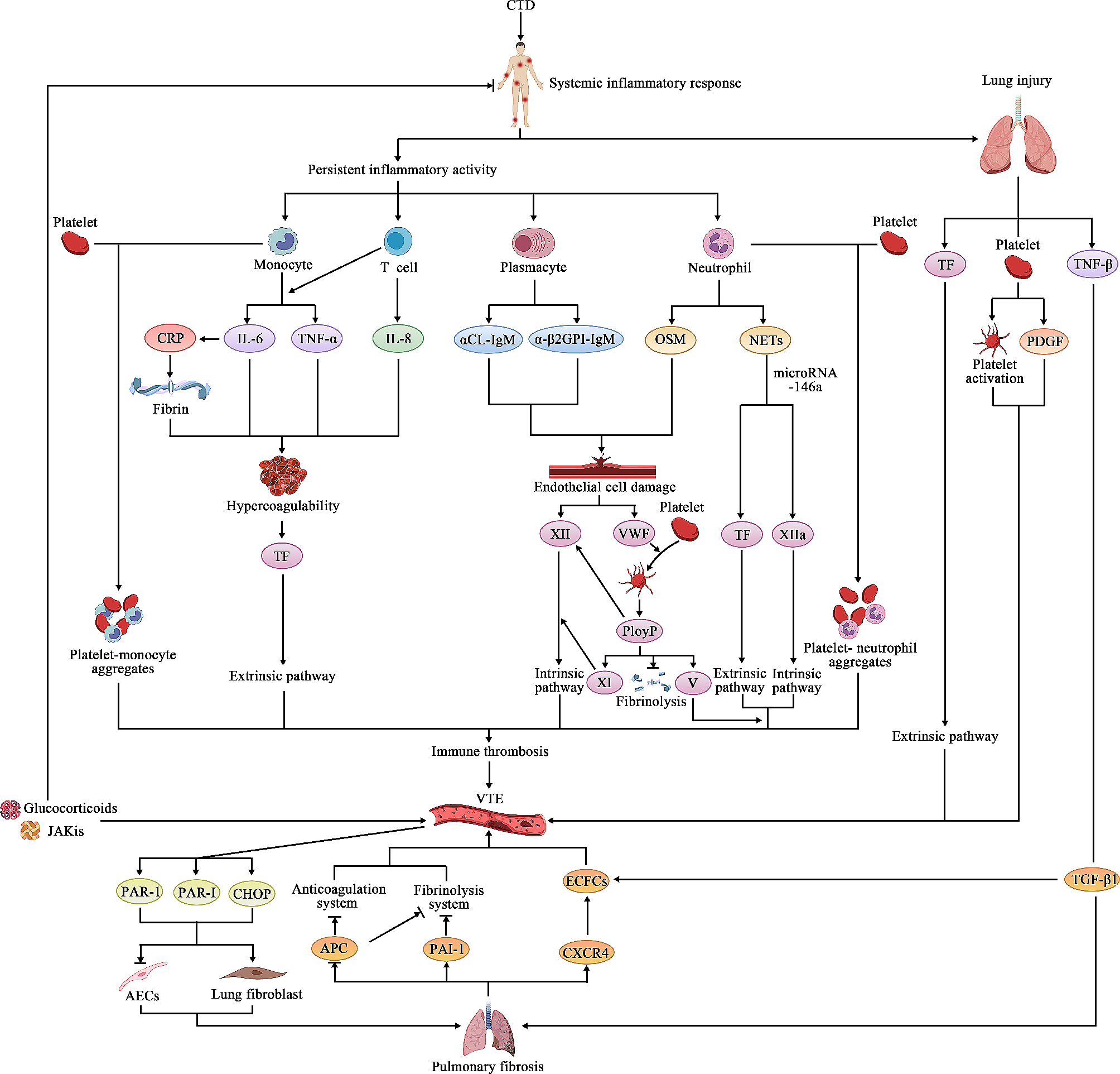

Extracorporeal membrane oxygenation (ECMO) is a treatment option for patients with advanced cardiorespiratory failure and can be utilized in both children and adults [1]. ECMO is a life-saving heart–lung machine that delivers oxygen to patients with refractory severe respiratory or cardiac failure as they await a transplant or recover from a serious illness for a few days or weeks [2]. In an ECMO circuit, the deoxygenated blood drained from the venous circulation is pumped to the oxygenator via a pump to exchange carbon dioxide with oxygen. The blood is then returned to either the venous (VV-ECMO) or arterial (VA-ECMO) circulation [3]. The ECMO circuit components (Fig. 1) include a pump, membrane oxygenator, heat exchanger, venous cannula, arterial or venous infusion canula, tubing, and connectors [4].
Fig. 1
Schematic of ECMO setup indicating the most likely clot formation sites
The most frequent complications and one of the common causes of death for patients on ECMO are thrombus formation and bleeding [5, 6]. The risk of bleeding is related to patient-specific and treatment-related factors associated with ECMO. According to Virchow's triad, which explains the thrombosis etiologies as hypercoagulability, vessel wall injury, and blood flow stasis, the ECMO circuit contains non-biological surfaces, regions of very high shear stress and regions of prolonged blood residence time, all of which act to promote thrombus formation at a level requiring systemic anticoagulation, which in turn increases bleeding complications [7].
ECMO patients are frequently critically ill, increasing the risk of bleeding complications [8]. The bleeding rate during ECMO is 20.8–39.6% [6, 9] with the cannula site (13.2%), gastrointestinal tract (5.5%), lungs (6.1%), and central nervous system (3.9%) being the most prevalent sites [10]. ECMO patients are also at risk of thrombosis complications, including ischemic stroke, right ventricular thrombus [11], left ventricular thrombus [12], and pulmonary embolism [13]. The rate of thrombosis formation in patients undergoing ECMO is 10–46.1% of patients depending on the circuit type and age of the patient in various centres [14]. Finding the balance between bleeding and thrombosis necessitates continuous monitoring of various parameters including coagulation factors, fibrinogen, and platelets. In this review, conventional and recently developed anticoagulants used in ECMO are presented, including their mechanism of action, monitoring methods, strengths and limitations. It expands on existing anticoagulant monitoring systems, indicating their target range, advantages, and disadvantages. It also introduces various unique real-time coagulation monitoring techniques, as well as their operating principles and future research prospects.
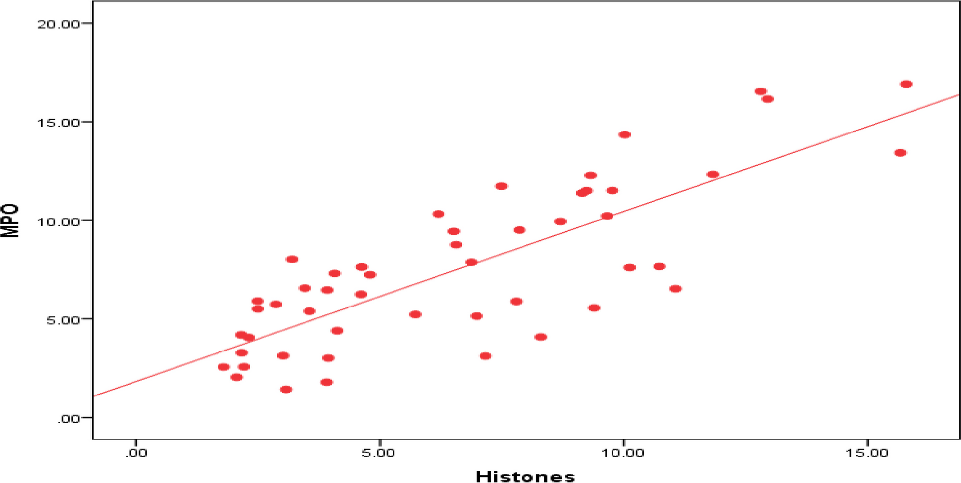
Anticoagulation during extracorporeal circulationFirstly, anticoagulants utilized in ECMO are summarized, including their benefits and limitations to provide a comprehensive viewpoint for future research. In addition, this review takes a fresh view on anticoagulant monitoring strategies by classifying them according to their purpose (Fig. 2).
Fig. 2
Various anticoagulants used in ECMO devices and their monitoring methods (AT: Antithrombin; DTI: Direct Thrombin Inhibitor; DOAC: Direct Oral Anticoagulant; NM: Nafamostat mesylate; FBC: Full Blood Count; aPTT: Partial Thromboplastin Time; PT/INR: Prothrombin time/international normalized ratio; ECT: Ecarin Clotting Time; POC: Point-of-Care; ACT: Activated Clotting Time; ROTEM: Rotational Thromboelastometry; TEG: Thromboelastography)
Anticoagulant agentsAnticoagulants commonly used for anticoagulation in ECMO patients include heparin, direct thrombin inhibitors (DTIs) (bivalirudin, argatroban, lepirudin), factor Xa inhibitors (danaparoid and fondaparinux), direct oral anticoagulants (DOAC) (DTI (dabigatran), Xabans (rivaroxaban, apixaban, darexaban, edoxaban, and betrixaban)), factor-XIIa inhibitors, nafamostat mesylate (NM), and warfarin so that heparin and DTIs are the most prevalent anticoagulants used in ECMO patients. Although heparin is the most commonly used in clinical applications, heparin resistance is a major concern in ECMO. It is defined as a situation where the ability of heparin to inhibit thrombin (factor IIa) and fibrin formation is reduced such that the correlation between dose and response is lost and increasing the heparin dosage will not result in the desired anticoagulation effect [7, 15]. Moreover, its usage is associated with the rare but life threatening immune-mediated disorder Heparin-induced thrombocytopenia (HIT), specified by thrombocytopenia and a paradoxical prothrombotic state in heparin treatment [16]. Alternative anticoagulants, on the other hand, offer excellent potential for usage in HIT patients, but present some other challenges. The working mechanism, advantages, disadvantages, and other aspects of different anticoagulants used during ECMO support including heparin and its alternatives are provided in Table 1.
Table 1 Anticoagulant AgentsHeparinUFH is the most frequently used anticoagulant in patients undergoing ECMO due to its advantages including its low cost, titratability, and easy reversibility by protamine. Heparin inhibits thrombin by binding to antithrombin (AT). AT has low anticoagulation activity but when conjugated with heparin, its anticoagulation activity increases 1000–2000-fold [17]. Heparin resistance is the main concern in ECMO, which is defined as a situation where the heparin ability to inhibit thrombin and fibrin formation is reduced so that heparin dosage response is not correlated with the injected amount of heparin and increasing the heparin dosage will not result in the desired anticoagulation effect [7]. More heparin injections are required under these conditions to achieve desired activated clotting time (ACT) values, which may result in bleeding. The situation is significantly worse in newborns as their AT level is lower than that of adults [18].
Heparin use is associated with immune-mediated side effects known HIT and specified by thrombocytopenia and a paradoxical prothrombotic state in heparin treatment. While it has been reported that HIT is a rare phenomenon in neonates [19, 20], it occurs in 0.8–7% of the adult ECMO patients [21, 22]. It has been demonstrated that 30–60% of HIT patients experience thrombotic complications [23, 24]. Therefore, in order to address the aforementioned issues, it is necessary to provide alternative anticoagulants to heparin, such as bivalirudin [20].
BivalirudinDTIs, unlike UFH, do not rely on AT to act as an anticoagulant, but instead they directly inhibit both free circulating and fibrin-bound thrombin. Bivalirudin is a reversible thrombin-binding synthetic bivalent analogue of hirudin with excellent pharmacological profiles [25]. Since bivalirudin can inhibit plasma thrombin, clot-bound thrombin, and collagen-induced platelet activation without forming a complex with the cofactor AT III, it has a much higher bioavailability than heparin [26, 27]. It has a short half-life of approximately 25 min in patients with normal renal function, making it suitable for rapid titration [28, 29]. It is mostly metabolized by the liver through proteolytic cleavage, but it also partially cleared by the kidney (20%), so the dose should be adjusted during renal dysfunction as it prolongs its half-life [30, 31]. Bivalirudin has been used to prevent clotting during ECMO in both HIT patients and non-HIT patients [32,33,34]. It is administered intravenously in doses ranging from 0.025 to 0.48 mg/kg/hour, with an action time of 2–4 min [35]. Bivalirudin efficacy has been shown to correlate well with both ACT and aPTT results [36, 37]. The researchers also compared the bivalirudin and UHF aPTT results and discovered that using bivalirudin yields more stable aPTT results [34, 38]. There is no unified approach for bivalirudin infusion, so it can be used with or without an initial bolus of bivalirudin with initial loading ranging from 0.04 to 2.5 mg/kg followed by continuous infusion [34, 39]. In particular, Koster et al. used bivalirudin for HIT patients with a bolus of 0.5 mg/kg followed by a continuous infusion of 0.5 mg/kg/h to maintain an ACT of 200–220 s [33]. In another study, Jyoti et al. [39], were able to achieve a target ACT of 200–220 s with an injection rate of 0.1–0.2 mg/kg/h and no bolus dosage of bivalirudin. The dose of bivalirudin is maintained at 0.03–0.2 mg/kg/h to maintain therapeutic targets, with [19, 32, 33] or without [34] an additional initial amount of 0.5 mg/kg. In a meta-analysis, it has been reported that in-hospital mortality, major bleeding events and pump-related thrombosis were less frequent in DTI compared to heparin [40].
There are some considerations before bivalirudin usage in ECMO. First, it affects the renal clearance process in patients with impaired renal function, resulting in drug accumulation. Therefore, lower dosage of bivalirudin is required for patients with renal dysfunction [41]. Moreover, since bivalirudin is rapidly metabolized where the blood is in stasis, it is not a viable option in venoarterial ECMO [42]. Another limitation of the bivalirudin is that there is no antidote in case of overdose or bleeding, which makes bleeding management challenging. Bivalirudin resistance may exist in the absence of a clear etiology [43]. APTT, ECT, and plasma-diluted thrombin time tests are typically used for monitoring anticoagulant effect of bivalirudin [44, 45].
ArgatrobanArgatroban is a synthetic direct thrombin inhibitor with a half-life of 39–51 min [46] and is not recommended for patients with severe hepatic dysfunction since it is metabolized in the liver, therefore, renal failure is not a concern. One of the primary issues in ECLS that limits DTIs adoption is a lack of pharmacologic antidote. However, due to their short half-lives, if bleeding occurs, the injection of DTIs can be stopped or reduced to stop the bleeding. Argatroban has been utilised as an alternative to UHF in cases of suspected HIT in adults, pediatrics, and neonates on ECMO. Its maintenance dose is 0.1–0.65 \(\mathrm/\mathrm/\mathrm\) [47, 48], and centres use a 100–200 \(\mathrm/\mathrm\) initial bolus dose [35].
FondaparinuxFondaparinux is a factor Xa inhibitor that has been indicated to be effective as an anticoagulant agent in severe acute HIT [49]. Parlar et al. [50] used fondaparinux daily subcutaneous injections (2.5 mg per day) in ECMO loop for a patient with HIT and found no adverse effects. Compared with DTIs which require the aPTT or the ECT monitoring methods, anti-FXa assays are more reliable for fondaparinux monitoring since anti-FXa assays do not rely on patient factors. To delineate, the aPTT and the ECT are influenced by the prothrombin level of the HIT patients which is often low and results in falsely long aPTT and, consequently, in inappropriate dose of the anticoagulant [51].
Nafamostat mesylateNafamostat mesylate is a synthetic serine‐protease inhibitor that inhibits many procoagulant factors, including thrombin, plasmin, trypsin, kallikrein, factors XIIa and Xa [52]. In the literature, there are conflicting findings for the use of NM in ECMO. Lim et al. [53] investigated thromboembolic or bleeding complications during ECMO using heparin and NM. According to their findings, bleeding complications were more common in patients receiving NM, while thromboembolic problems were comparable in both cases. Other studies, on the other hand, claim that it is an appropriate alternative to heparin that reduces the risk of bleeding in ECMO patients [54, 55]. Like other anticoagulants, there is no unified approach for the dosage rate but typically it falls into the range of 0.26–0.93 mg/kg/hr [54,55,56].
WarfarinWarfarin is an oral anticoagulant that inhibits the utilisation of vitamin K (factors II, VII, IX, and X). The main advantage of this method is its ease of administration and reversibility [57]. When patients have been adequately anticoagulated with DTIs and require long-term anticoagulation after the acute period of HIT, they are typically switched to vitamin K antagonists (VKA) such as warfarin and phenprocoumon [58]. Warfarin dosage should be determined based on the patient's response to the drug, and it can be monitored using international normalised ratio (INR) analysis to keep its results in therapeutic range [2, 3, 59]. Lee et al. describe the successful use of ECMO as a bridge-to-recovery therapy in a patient suffering from fatal warfarin-exacerbated DAH [60].
Anticoagulation monitoring methodsGiven the importance of anticoagulant monitoring and dose adjustment, it is vital to determine the appropriate approach for each anticoagulant and its dosage. Partial thromboplastin time (aPTT), anti-factor Xa assay, D-dimer, PT/INR, Full blood counts (FBC), Fibrinogen, ECT, activated clotting time (ACT), and viscoelastic tests (ROTEM/TEG) are discussed in this section. Also, recently developed novel real-time monitoring methods including sound, optical, fluorescent, and electrical measurements methods are presented. Table 2 summarises the sample type, purpose, target range, advantages, and disadvantages of different techniques.
Table 2 Conventional anticoagulation monitoring methodsActivated partial thromboplastin timeAPTT is an anticoagulant monitoring technique that is most commonly used to assess the effect of heparin and bivalirudin [76]. It is defined as the time required for calcium-free plasma to generate clots after being exposed to fibrin-activating reagents and calcium. Clot formation can be detected using a variety of analytical methods, including optical, mechanical, and electrochemical techniques [77]. This method involves combining citrated plasma, a phospholipid, calcium, and a contact pathway activator (silica, celite, kaolin, ellagic acid, polyphenolic acid) to trigger clot formation [78]. The normal range is defined in most laboratories as 25–90 s (an aPTT level of 1.5–2.5 times baseline is recommended for anticoagulation monitoring) but it varies from clinic to clinic and is determined by the instrument and reagents used and it is critical not to extrapolate data from one ECMO centre to another without knowing the method and assay used [79, 80]. Bates et al. investigated the relationship between aPTT and anti-factor Xa assay heparin level using four different automated coagulometers and six commercial aPTT reagents. Their findings revealed that, while there is a good correlation (r = 0.64 to 0.95) between aPTT anti-factor Xa assay results, the aPTT values at 0.3 IU/mL plasma heparin concentration determined by anti-factor Xa assay will range from 48 to 108 s, depending on the instrument and reagent utilised [81].
Another concern with this method is that it can be influenced by parameters, including drugs, hematocrit, acute phase reactants, abnormalities in coagulation factors, high C-reactive protein, hyperbilirubinemia, hyperlipidemia and lupus anticoagulant [82] so that deficiencies in common pathway factors I, II, V, and X, as well as contact pathway components such as high-molecular-weight kininogen, prekallikrein, and factors VIII, IX, XI, and XII, and lupus anticoagulants can prolong the aPTT results [78].
Anti-factor Xa assayAnti-factor Xa is a functional chromogenic assay for coagulation monitoring and evaluating the effective anticoagulant concentration. The anti-Xa assay is specific to heparinoid’s action and is unaffected by deficits in other coagulation factors and can be used with or without exogenous AT. In the former method, the sample is treated with sufficient AT, so that the rate-limiting reagent, which is heparin, can inhibit Xa and produce a precise measurement of heparin in the patient sample. In this method, a specific amount of coagulation factor Xa conjugated with chromophore and AT is added to patient plasma containing heparin [83]. Following that, as a result of the chromogenic reaction, AT and heparin form an inhibitory complex that inactivates factor Xa. The activated factor X is then introduced to the sample, which cleaves the chromophore compound, and the amount of released chromophore is measured using spectroscopy. As the amount of remaining factor Xa in the sample is inversely proportional to the original amount of heparin, the colour change will be greater as less heparin/AT complex interacts with factor Xa, indicating a lower drug level. The relationship between AT and heparin is critical because, even with a high level of heparin, a deficiency in AT causes more unbound factor Xa, implying lower heparin levels. On the other hand, kits without AT do not add extra AT and give a more precise measurement of in-vivo anticoagulation because the patient's AT and heparin levels are both rate-limiting reagents. However, this approach has the problem of being unable to differentiate between AT deficiency and inadequate heparin [84, 85].
The widely accepted target range for anti-factor Xa levels during ECMO is 0.3–0.7 IU/mL [86, 87]. Unlike the ACT and aPTT methods, this method is unaffected by coagulopathy, thrombocytopenia, or dilution and best represents the overall heparin anticoagulation level. However, some parameters which can occur in patients on ECMO, such as hyperbilirubinemia, haemolysis, lipaemia, hyperlipidemia, and plasma-free haemoglobin affect anti-Xa assay results [88, 89]. Anti-Xa activi
留言 (0)