We comprehensively analyzed the long-term clinical outcomes of patients with PVT, identified predictors of the three clinical outcomes, and established three simple, intuitive, and rapid prognostic prediction models. Additionally, we evaluated the effects of anticoagulation therapy on PVT. The results indicated that anticoagulation therapy may increase the rate of recanalization without increasing the risk of GIB and death.
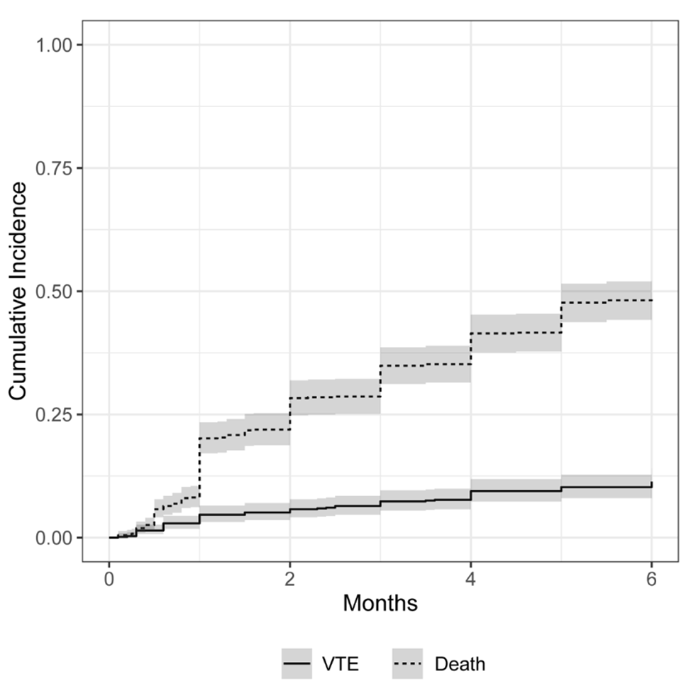
Recently, the incidence of PVT, which is estimated to range from 1.6 to 15.8% in patients with cirrhosis or portal hypertension, has gradually increased due to improvements in diagnostics brought about by advancements in imaging modalities [4]. PVT increases the risk of portal vein hypertension and related complications, such as bleeding, thrombus progression, and death [8, 14, 15]. Therefore, monitoring the prognosis of patients with PVT is crucial for clinical decision-making. According to recent studies, the incidence rates of bleeding, PVT resolution, PVT progression, and death after a diagnosis of PVT ranges from 12 to 30.8%, 31.6 to 71%, 5.7 to 15.8%, and 13 to 24.2%, respectively [14,15,16,17]. In the present study, the overall incidence rates of GIB and death were 9.9, and 15.6%, respectively, while 45.5 and 20.9% of patients who have been evaluated for morphological change of PVT were observed to have PVT resolution and progression, respectively, which are similar to the previously reported rates.
Previous studies have confirmed that anticoagulation therapy, an interval of less than 6 months between a diagnosis of thrombosis and initiation of anticoagulation therapy, and splenic thickness may be positively associated with portal vein recanalization [7, 18, 19]. Anticoagulation therapy is a crucial treatment option for patients with PVT; however, it was rarely implemented previously as clinicians and patients were concerned about the risk of complications such as GIB [16]. Moreover, among the patients who were evaluated for morphological PVT changes in the present study, the PVT recanalization rate (45.5%) was higher than the proportion of patients who received thrombolytic therapy (38.0%), suggesting that spontaneous recanalization occurred in a small number of patients with PVT, similar to the findings of previous studies [6, 17]. Notably, portal vein hypertension is a predictor of nonresponse to anticoagulation therapy [9, 20]. In our study, 63.3% of patients treated with anticoagulation therapy achieved thrombus resolution; this result is consistent with those of previous studies (30–80%) [8, 21] and also confirmed that anticoagulation therapy was a significant predictor of PVT resolution. Therefore, we suggest that most patients with PVT should receive anticoagulation therapy unless there is a high risk of bleeding.
Hemoglobin is essential for maintaining cellular bioenergetic homeostasis and modulating cell functions (inflammation and redox status of cells) through its ability to bind and transport oxygen to tissues, which may decrease the incidence of thrombosis [22]. However, excessively high levels of hemoglobin can lead to local inflammation and even tissue damage among patients with hemoglobinemia, which further accelerates the formation of thromboses [23]. The C-reactive protein level is a common indicator of inflammation and infection and is often used to assess their severity [24]. Darzi et al. demonstrated that a C-reactive protein level > 10 mg/L was positively associated with venous thromboembolism and that it could lead to a transient hypercoagulable state [25]. Additionally, the 2020 Chinese consensus regarding PVT indicated that inflammation or infection of the abdominal cavity may be an important risk factor for PVT in patients with cirrhosis [3]. PVT may be a potential consequence of any inflammatory intra-abdominal process (including cholecystitis, pancreatitis, or inflammatory bowel disease), and its risk will increase in a setting of acute infection and recurrent infections [26]. Therefore, patients with a higher C-reactive protein level and a history of abdominal infection may have higher levels of inflammation, which lowers the probability of PVT resolution.
Portal vein hypertension is the main determinant of esophagogastric variceal bleeding [11]. Previous studies have demonstrated that esophageal varices, the red color sign observed during endoscopy, advanced stage of liver disease (Child–Turcotte–Pugh class C patients), and ascites were possible predictors of bleeding events [16, 27]. Although the same conclusions were reached in the present study, we also demonstrated that a history of GIB and thrombus extension into the mesenteric veins were significant predictors of GIB. Portal vein hypertension results in redistribution and increased blood flow through the short gastric and coronary veins, causing esophagogastric varices. Esophagogastric varices begin to form at a pressure gradient of 8–10 mmHg, and bleeding risk increases at a gradient of at least 12 mmHg [27]. Certain endoscopic variceal stigma, collectively referred to as “red color sign” (red-whale markings, nipple symptoms, cherry-red spots), correlated with a significantly higher risk of acute variceal bleeding and re-bleeding [28]; hence, early preventive endoscopic treatment and shortening of the prothrombin time may decrease the occurrence of GIB [3]. Additionally, when hepatic encephalopathy occurs in patients with advanced liver disease due to liver failure, and imaging also shows PVT extension into the superior mesenteric vein, further reductions in the flow velocity and increases in the portal vein pressure, even GIB, may occur. In the long-term, approximately 70% of patients with GIB may experience further variceal bleeding because of superficial varices and a thinner vessel wall [29]. Therefore, a history of GIB may increase the risk of re-bleeding. These new findings may help clinicians identify patients at high risk for GIB, and they may also facilitate the timely initiation of anticoagulation therapy.
Patients with cirrhosis or hepatocellular carcinoma are in a state of imbalanced coagulation function that can promote the propensity for bleeding or thrombosis [4, 30], thus making it challenging for clinicians to initiate anticoagulation therapy for PVT. Mohan et al. reported that the incidence of GIB for patients receiving anticoagulation therapy was 7.8% [31], which is very close to the rate (8.8%) observed in the present study. Furthermore, the univariate analysis performed in the present study showed that anticoagulation therapy did not promote GIB events, thus demonstrating the safety and efficacy of anticoagulation therapy for PVT. Therefore, our conclusion is consistent with those of existing studies. Qi et al. confirmed that prophylactic anticoagulation therapy for deep venous thrombosis in hospitalized patients with cirrhosis and without active bleeding was safe and did not increase the incidence of GIB or death [18]. Furthermore, Ageno et al. found that the duration of anticoagulation therapy was associated with a reduced risk of bleeding [21]. However, this variable was not statistically significant in the univariate analysis for GIB events in the present study, which may be due to its dual effect on GIB. Prolonging anticoagulation therapy will increase the risk of bleeding by preventing coagulation. Simultaneously, anticoagulation therapy may reduce the severity of esophagogastric varices due to PVT resolution, which decreases the incidence of GIB.
Regarding the analysis of the overall death of patients in our study, most of the fatal events were related to the underlying disease. In one case, the progression of PVT into the superior mesenteric vein caused intestinal obstruction, resulting in death. In a large prospective study of 178 patients with PVT, few deaths occurred during follow-up, and the 5-year survival rate was 96% [11]. Moreover, overall 5-year survival rates ranging from 70 to 78% have been reported by a previous study [10]. In the present study, 23.1% of all deaths were caused by fatal GIB events; however, GIB caused only 3% of the deaths in a study of 120 non-cirrhotic patients with PVT [32]. Most patients in our study had cirrhosis, hepatocellular carcinomas, and coagulation disorders, which led to a higher mortality rate. Significantly relevant factors for PVT-related mortality events include the Child–Turcotte–Pugh score, age, and ascites [32]. In partial agreement with the results of previous studies, the univariate analysis performed during the present study confirmed that the Child–Turcotte–Pugh score was related to survival (P = 0.0002). Because multiple variables of the Child–Turcotte–Pugh score may interact with each other during statistical analyses, these variables were analyzed separately in our study. We found that hepatic encephalopathy and ascites were independent predictors of PVT progression in the Cox regression analysis of death. Both hepatic encephalopathy and ascites are important signs of liver failure, which has a high risk for mortality and allows for a more accurate assessment of a patient’s prognosis. In addition, abdominal infections, history of abdominal surgery, and aspartate aminotransferase level > 35 U/L were positively associated with death in the present study. Intra-abdominal infections and surgery pose serious clinical challenges, and may result in wide variety of conditions ranging from uncomplicated cases to fulminant septic shock and multi-organ dysfunction, further increasing the risk of death [26].
This study had some potential limitations. First, the effectiveness of the data analysis was limited because of the retrospective nature of the study. Additionally, the etiology ratio of PVT was not compared with those reported by other studies to confirm the external implementation performance of the nomograms. Second, some data related to PVT (e.g., portal vein velocity and morphological changes in the thrombus) were not included in this study. Moreover, many patients underwent only one blood or endoscopic examination. Therefore, a dynamic follow-up could not be performed. Future prospective, multicenter, randomized clinical trials with larger sample sizes are needed to corroborate the findings of this study.
In conclusion, we developed three easy-to-use nomogram prediction models to evaluate the prognosis and assist with the initiation of early intervention for patients with PVT. Additionally, the results of this study suggested that most patients with PVT should undergo anticoagulation therapy. Furthermore, these findings provided evidence of the benefits and risks of anticoagulation therapy for patients with PVT, which will help clinicians balance the benefit-to-risk ratio of anticoagulation therapy.


留言 (0)