
記住我


Pigment cells are called melanocytes, which are dendritic cells (pseudopod extensions facilitating movement). The inclusion bodies for pigments are called melanosomes, and they can move within the cell. Baseline skin pigmentation is determined by the amount of melanin in the melanosomes; that is, black skin has larger melanosomes with more melanin than lighter skin. UV light exposure causes melanosomes to migrate peripherally, leading to darker pigmentation colloquially known as a “sun tan.”
The Fitzpatrick classification categorizes skin types according to melanin pigment concentration, ranging from type 1 (persons with very light white skin) to type 6 (persons with dark black skin; Figure 1).1 To provide appropriate care for all patients, podiatrists and healthcare professionals who perform skin assessments must competently assess all Fitzpatrick skin types.
 Figure 1: FITZPATRICK CLASSIFICATION 1
Melanoma development is related to many factors, and skin pigmentation is crucial. In a two-part model, some individuals have atypical melanocytes that act as a genetic initiator of malignant transformation, often with the sun serving as a promoter. In addition, persons with more than 100 normal nevi (round and regular) or irregular dysplastic nevi (often larger, with a “fried egg” appearance, darker central areas, and increased pigment splay around the periphery) have an increased melanoma risk. When melanocytes become damaged by sunlight or other carcinogens, aberrant changes may lead to melanoma in situ. Once the melanoma invades past the basement membrane of the epidermis, it is more likely to spread to other parts of the body, leading to poor outcomes.2–4 Melanoma is more common in lighter-skinned individuals (Fitzpatrick types 1 and 2) because they are typically prone to sun damage and potential melanocytic mutations.
Early detection of melanoma leads to better survival outcomes for all skin types: Successfully removing lesions with excisional surgery with adequate peripheral margins prior to dendritic melanocytic invasion2,3 prevents malignant spread through the body. Early surgical removal is often curative, given an adequate histologic peripheral margin from the melanoma cells along with a clear zone free of melanoma cells in the deeper portion of the biopsy. On biopsy, the deeper the melanoma cells have migrated (ie, from the stratum corneum toward the dermis), the greater the concern for distant metastases via the bloodstream or lymph nodes.
Because early detection of melanomas relies on visual inspection, these lesions are often underdiagnosed in individuals with darker skin (Fitzpatrick types 5 and 6). According to Wu and colleagues,2 early melanoma diagnosis is successful in 80% of White individuals, compared with a 56% success rate in Black individuals. Melanoma can be detected more readily on the feet of individuals with darker skin complexions because the feet often have a lighter color tone. This is especially true in the two foot sites where melanoma most commonly occurs: the toenail bed (under the nail plate) and the plantar aspect of the feet.3
Podiatrists, chiropodists, and other foot specialists need to be familiar with the appearance of pigmented lesions in all skin tones and both sexes to successfully identify and biopsy suspicious lesions early in their melanoma evolution. At particular risk are women of all skin types; their lower legs and feet are more prone to melanoma. This group warrants additional clinical inspection and attention. Serra-García et al5 documented that the identification of pigmented lesions as part of podiatry training increased diagnostic accuracy for lower extremity melanoma by 33%.
Both health providers and patients can benefit from knowing that early melanomas may express clinical components that follow an ABCDE mnemonic:6
• Asymmetry: One side of the lesion is not a mirror image of the other side.
• Border: Edges are irregular and may go up and down like a ski slope.
• Color: Black indicates a concentration of melanin that may be darkly pigmented in an irregular pattern for persons with brown or black skin (more easily detected with a dermatoscope).
- Blue via the Tyndall effect (ie, deeper black pigment that is not covered with overlying epidermal pigment appears blue) - White for areas of regression - Red for inflammation on the skin surface• Diameter: Larger than 6 mm (the size of an eraser at the end of a pencil)
• Evolving: Lesions that change more rapidly than other nevi (Figure 2)
 Figure 2: ABCDE OF MELANOMAA, A nevus in a relatively young person; these usually develop in late childhood or teen years. B, Congenital nevus in a person of color. Congenital nevus is present at birth and enlarges with aging. Note the dark but even color. C, Nevi often become elevated with pigment in adult life. D, Neuroid nevi lose pigment and may disappear with aging. E, Dysplastic nevus: Note the “fried egg” appearance with subtle irregular margins; slightly larger than normal nevi. F, ABCDs of malignant melanoma (modified from the National Cancer Institute. Common Moles, Dysplastic Nevi, and Risk of Melanoma. 2018. www.cancer.gov/types/skin/moles-fact-sheet).Skin and the Fitzpatrick Skin Type Classification
The basal layer of skin contains keratinocyte precursors, Merkel cells, Langerhans cells, and melanocytes. The melanocytic cells produce melanin, which is contained in melanosomes and dispersed in the dendrites of the melanocytes. Melanosome size is larger in black skin than in white skin, and brown skin melanosomes are of intermediate size. The pigment is taken up by keratinocytes as they develop and migrate toward the superficial layer of the epidermis.4 The amount of pigment produced by melanocytes serves as protection against the harmful UV portion of the sun’s rays. Melanin taken up by the keratinocytes determines the color of a person’s skin (in part) and is additionally triggered by the skin’s ability to react to sun exposure with tanning.
When the skin is overexposed to UV radiation, sunburns can occur. The Fitzpatrick skin type classification1 organizes skin types into six different categories based on skin color and skin reaction to sun exposure (Figure 1). However, habituation does play a part in skin damage; persons with skin types 4 through 6 skin who live in the northern hemisphere in cold or temperate climates may suffer sunburns without adequate protection when on holiday in the tropics or during a southern hemisphere summer period.
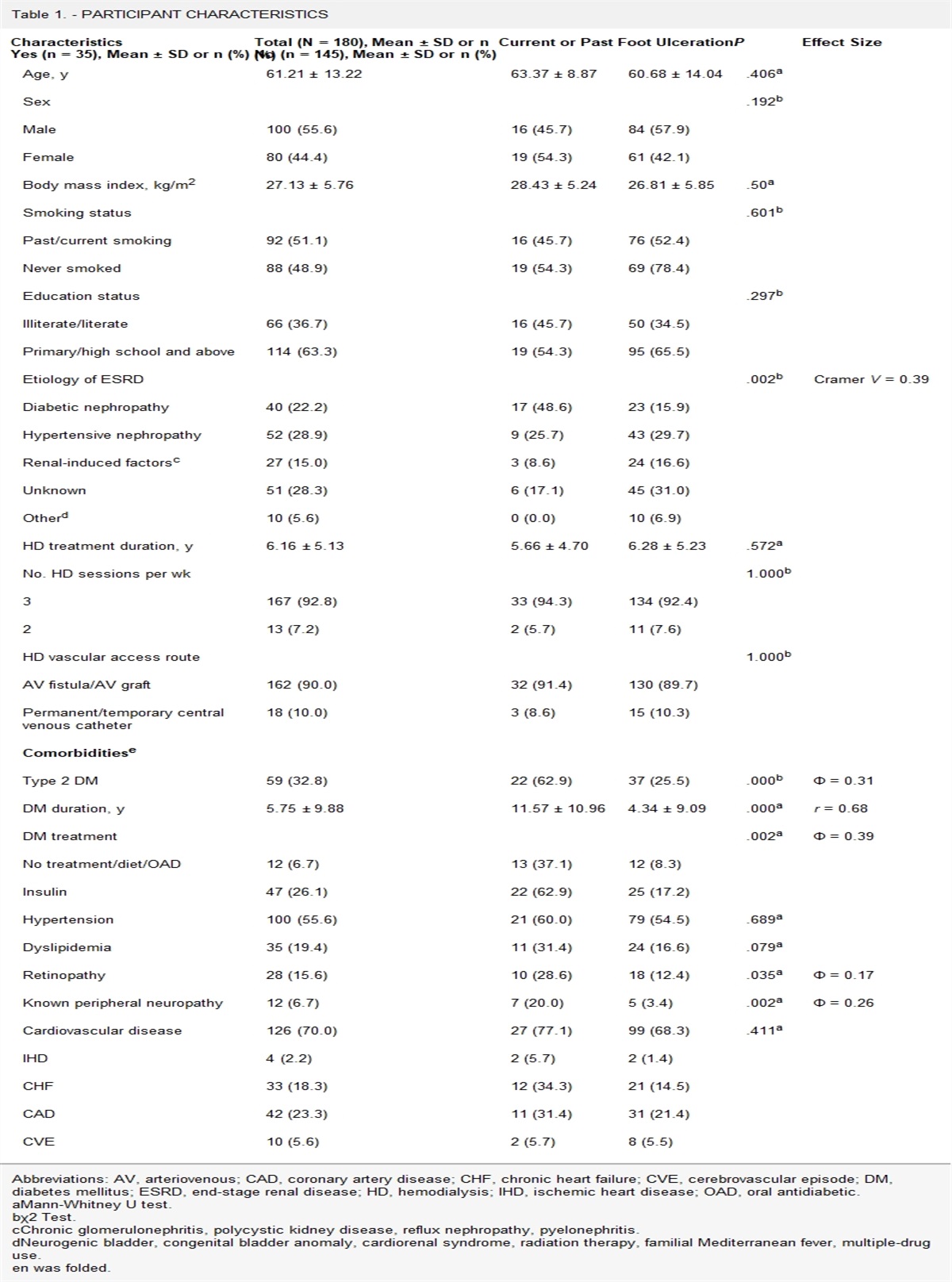
Whereas most versions of the Fitzpatrick skin type classifications use faces as illustrations, the authors have developed an educational enabler depicting two views of feet showing skin types 1 through 6 for podiatrists, chiropodists, and other foot specialists (Table 1). This serves as a useful tool when examining feet across the entire skin pigmentation spectrum.
 Table 1:
 FITZPATRICK SKIN TYPES 1 THROUGH 6 SHOWN IN FEET
Characteristics of Cutaneous Malignant Melanoma Pathology overviewAlthough melanocytes protect the skin, they can also be the genesis of malignant melanoma. Abnormal melanocytes may initiate melanoma with sun exposure, especially when blistering burns or long-term skin damage occurs. When enough damage accumulates in the melanocyte DNA, these changes overwhelm the cellular repair machinery, with mutations leading to aberrant melanocytes. They can then become dysplastic and progress to neoplastic melanocytes, or melanoma in situ. When melanoma has not penetrated the basement membrane of the epidermis, it remains a localized melanoma. Once the malignant cells penetrate the basement membrane and invade the surrounding dermal tissue, it is malignant melanoma.7
Early clinical detection is key to successful treatment and survival. Unfortunately, melanocytes are dendritic cells that freely spread through regional lymph nodes and undergo hematogenous migration to spread through the bloodstream. This secondary spread is not always predictable or detected in time.
Differentiating melanoma subtypesSeveral types of melanomas can be distinguished based on different characteristics and the tissue from which they arise (Figure 3). The various types are (from most common to least frequent) superficial spreading melanoma (SSM), lentigo maligna melanoma (LMM), nodular melanoma, and acral lentiginous melanoma (ALM; more common in Black skin). Melanomas can also arise in mucosal surfaces, the eyes, and other internal organs.
 Figure 3:
 VARIABLE CHARACTERISTICS OF MELANOMA OF THE FOOTA, Amelanotic malignant melanoma. This lesion is in an atypical location for pressure and callus formation. Persistent lesions without an etiology should be biopsied for histologic diagnosis. B, Acral lentiginous melanomas start with freckle-like changes that gradually enlarge with atypical features. A biopsy of lesions acquired after the age of 40 years should be recommended, even without the usual atypical features. C, Superficial spreading malignant melanomas often show the features of asymmetry, irregular borders, color variability (black, red, brown, etc). These lesions may be more difficult to detect in persons of color. D, More advanced melanomas are of larger size and deeper concentration of color (black, dark brown) with irregular patterns. They can often be mistaken for hemorrhagic blisters on the heel without careful examination via illuminated magnification (use an ophthalmoscope or autoscope if a dermatoscope is unavailable). E, This advanced, level 4 melanoma has a thick base and was mistaken for hemorrhagic callus. Close examination shows pigment splay beyond the end of the lesion at 9 o’clock. Note the secondary spread with the black spot approximal to the heel.
Superficial spreading melanoma arises in nonglabrous skin. It generally affects people of Western European descent, and it is the most common type of melanoma. It also has the highest mutational burden, meaning it contains the most mutations within its DNA.8–10 The mutations acquired by these cells can be a predictor of outcome, with some mutations having a better outcome than others. Approximately 20% to 50% of SSM lesions arise in preexisting nevi,11 and 50% to 80% appear de novo on what appears to be normal skin; however, the skin may have abnormal melanocytes in concentrations that are lower than visible nevi.
The most common type of melanoma presenting on the face is LMM.11 Compared with SSMs, LMMs tend to have less well-defined borders and, microscopically, can extend past the clinically visible border.10 Typically, LMM affects older adults (mean age at diagnosis, 65 years).12 The sun can initiate and promote these lesions.
Nodular melanoma is a type of neoplasm that tends to grow vertically with little horizontal spread.11 It can present as a symmetrical tumor, which may be pigmented or unpigmented. Because of its atypical presentation that does not follow the conventional “ABCDE” characteristics, it is often missed clinically.10
A less common type of melanoma, ALMs develop on the glabrous skin of the palms, soles, and nail bed. Their mutational burden is lower than that of SSMs.8 These melanomas are more common and underdiagnosed in populations with Fitzpatrick skin types 4 through 6.2 Acral melanomas can be differentiated from acral nevi by considering the pigmentation on the edge and ridges of the lesion. In ALMs, pigmentation is detected on the ridges of the lesions, whereas in nevi, pigmentation is located along the furrows.13 In addition, ALMs in the nail bed do not have the straight-line pigment seen in normal nevi but often have feathering or irregular pigment around the edges along with Hutchinson sign (Figure 4), with pigment extending from the nail plate to the periungual skin and beyond. These lesions may need to be distinguished from subungual hematomas secondary to trauma.
 Figure 4: SUBUNGUAL NEVUSReprinted from woundpedia.com.
Mucosal and ocular melanomas are very rare, arising from melanocytes in the mucosal lining and uveal tract of the eye, respectively. These melanomas are aggressive and have a much lower mutational burden than the other melanoma types.8,9
METHODSThis article is not intended to be a systematic review or PRISMA (Preferred Reporting Items for Systematic Reviews and Meta-Analyses)-guided search. Rather, it serves as a comprehensive gap analysis of podiatric melanoma literature. The authors compiled data into an overview to enable deeper perusal for clinicians and open this topic for future investigation and academic discussion (see Supplemental Table 1, https://links.lww.com/NSW/A125, for a more detailed description of the search terms and search methods used).
The literature searches were performed in PubMed on June 20, 2022, to establish extant literature focused on lower-extremity lesions in persons with Fitzpatrick skin type 5 or 6. The review was limited to articles published from January 2012 to June 2022.
RESULTS Literature SearchA general search for melanoma (nonspecific to podiatry) and persons with Fitzpatrick skin type 5 or 6 yielded 583 results (Table 2). The first search examining literature on melanoma in the field of podiatry yielded 12 results. However, none of these articles referenced patients with Fitzpatrick skin type 5 or 6 (Supplemental Table 2, https://links.lww.com/NSW/A126).5,14–24 Subsequent searches that were meant to narrow the search to specific articles on melanoma in the lower extremity in persons with Fitzpatrick skin type 5 or 6 yielded zero results. These searches were performed using the terms “melanoma,” “Fitzpatrick skin type,” and “podia*.”
Table 2 - SUMMARY OF SEARCHES Search Terms No. of Articles Melanoma + people of color 583 Melanoma + podiatry 12 Melanoma + people of color + podiatry 0 Melanoma + ethnic groups 311 Melanoma + ethnic groups + podiatr* 0 Melanoma + chiropody 12 Melanoma + people of color + chiropod* 0 Acral melanoma + ethnic groups 24 Acral melanoma + ethnic groups + podia* 0 Acral melanoma + ethnic groups + chiropod* 0 Acral melanoma + dark skin + foot 6 Acral melanoma + dark skin + foot + podia* 0 Acral melanoma + black skin + foot + podia* 1Subsequent searches were completed using the term “ethnic groups” to expand the search for relevant literature, as the term is a major and acceptable MeSH term. The search for “melanoma” and “ethnic groups” yielded 311 results. When the search was repeated specifically for podiatry, there were again zero results. Because foot specialists are known in other countries as chiropodists (chiropody), additional searches were completed with these terms. When the terms “acral melanoma” and “ethnic groups” and “podia*” or “chiropod*” were added, the search again yielded zero results. However, when the term “podia*”/”chiropod*” was removed, it yielded 24 results. Searching for “acral melanoma,” “dark skin,” and “foot” yielded six results; elements of these studies were included in this article, as the addition of the search term “podia*” again yielded a zero result. When searching using the terms “acral melanoma,” “black skin,” “foot,” and “podia*,” only one case study result was identified describing a misdiagnosed ALM.25
General FindingsAlthough the incidence of melanomas is higher in persons with lighter skin tones, melanoma does occur in persons across the whole skin pigmentation spectrum, including in brown- and black-skinned persons. These differences in skin pigmentation can result in different clinical presentations of melanomas. The 2016 publication by Gupta et al26 provides a clinical data summary of risk factors and preventive skin cancer actions that are applicable to persons of color. The authors identified an increased incidence of malignant melanoma in the White population with a 5-year survival of 92%, compared with a 5-year survival of 70% in non-White populations, even when these lesions were identified early. Study authors attribute this discrepancy to the “hidden” melanomas in darker-skinned populations located on palms, soles, toes, and toenails, with a lack of physical examinations of these areas.26
Yu et al27 reported that melanoma presentations in the patient groups they studied in China (82 cases in their hospital and a meta-analysis of 12 articles with 958 Chinese patients) differed from those of White patients. Of their 82 patients, the majority (79%) of detected melanomas were located on the foot.27 For Chinese patients with Fitzpatrick type 5 skin, the types of malignant melanomas seen most often were ALMs and mucosal melanomas. The ALMs were often found on the lower extremities, diagnosed late, and already in an advanced stage of disease at diagnosis (stage II–III).27
The 2019 work of Culp and Lunsford28 also supports the relationship between skin pigmentation and melanoma occurrence. The authors found lower melanoma rates for non-Hispanic dark-skinned persons than for non-Hispanic White persons. Most of the melanomas detected in non-Hispanic darker-skinned persons were ALMs. These lesions typically present themselves on the palms of the hands, soles of the feet, or under nail beds.28 The 5-year survival rates for this type of melanoma were lower for non-Hispanic dark-skinned persons (66.2%), in contrast with the survival rate of the SSMs more often found in non-Hispanic White persons (90.1%).28 Another factor in the increased morbidity and late diagnosis of ALMs is the thin skin present under nails (or the epithelium of mucosal surfaces) that enables melanomas to escape through the dermal-epidermal junction earlier than melanomas on relatively thicker areas of the skin. Linear nevi that appear as regular black lines from the nail matrix through the lunula (half-moon of the nail) to the distal nail fold are relatively common, but seldom appear de novo after the age of 50 years.
Although melanoma is believed to be six times more common in White individuals, melanomas are more often diagnosed late in brown-skinned Hispanic persons, resulting in poor survival outcomes.29 With the growing Hispanic population in the US, the need for culturally sensitive awareness and education on this specific risk is vital.29 Data from the National Program of Cancer Registries and the Surveillance Epidemiology and End Results29 programs report
…melanomas with poorer outcomes such as modular (NM) and acral lentiginous melanoma (ALM) were more common among males. Hispanic females had the highest proportion of melanoma on the lower limb and hip (33.7%) while Hispanic males had the highest proportion on the trunk (29.9%). The incidence rates for later stage and thicker tumors were significantly higher among Hispanic men than women.
In 2019, Mulenga and colleagues30 provided a more complete understanding of the epidemiology and histopathology of malignant melanomas in Black African persons. Their retrospective study of 232 cases in Malawi found that 95% of cutaneous melanomas were found on acral sites, with the majority being on the foot (87%). The most common histopathologic subtype was ALM30
A study by Sondermann and colleagues31 highlights how an initial misdiagnosis of melanoma on the foot is associated with a poorer prognosis. By using a computerized database from their hospital in Germany, they identified 151 patients with a histologic primary melanoma on their foot.31 They analyzed 107 patient records from this cohort and reported that 32 (30%) had been misdiagnosed initially as either chronic wounds, nevi, hematoma, fungal infections, warts, or paronychia.31 The median delay in appropriate diagnosis was 9 months. There was a significantly lower 5-year disease-free survival (47.8% vs 72.7%) and overall 5-year survival (63.5% vs 88.4%) in those who had been misdiagnosed.31 The authors concluded that their data underscore the importance of raising awareness for healthcare professionals to examine feet for melanomas as well.31
DISCUSSIONAccording to the American Cancer Society, information captured in the SEER (Surveillance, Epidemiology, and End Results) database determined that melanoma detected while it is still localized is associated with a 99% 5-year survival rate. That rate drops to 25% if a melanoma is detected after it has metastasized.32 As previously mentioned, melanomas were diagnosed early in only 56% of patients with Fitzpatrick type 5 or 6 skin.2 Wu et al2 scanned 38 cancer registries to determine the 5-year melanoma survival rate across the six Fitzpatrick skin types. The authors concluded that the lower extremity was the most common melanoma location for Fitzpatrick skin types 5 and 6. In addition, these melanomas were diagnosed later than those in persons with Fitzpatrick skin type 1 or 2. This work identified a problematic clinical gap in practice: darker-skinned individuals are at greater risk for an undetected malignancy. It also underscores an important, but easily correctable, health disparity that has not previously received the attention it warrants.
Practical ImplicationsWhen conducting a skin assessment on a patient with darker skin (Fitzpatrick types 4 through 6), it is important to examine the patient’s feet for lesions that could indicate a potential melanoma presence. Providers should perform a biopsy on suspicious lesions (Figure 2). Some clinicians erroneously assume that only Fitzpatrick skin types 1 through 3 and/or only parts of the body exposed to the sun will be susceptible to melanoma. The literature emphasizes the need for more comprehensive awareness in identifying melanomas in persons across the skin spectrum, especially those with Fitzpatrick skin types 4 through 6, and the importance of inspecting body locations including the lower extremities, soles of the feet, palms of the hands, and nails as additional areas where melanomas can occur.8,26,28–33 A nail examination is included in patient assessments as well. In particular, any linear or other darkly pigmented subungual lesions are examined for irregular feathering or edges along with Hutchinson sign (pigment proximal, lateral, or distal to the nail matrix) as these are important indicators to perform a biopsy on the lesion for a diagnosis (Figure 4). These lesions need to be distinguished from trauma-induced subungual hematomas, which include hemorrhagic material between the nail plate and nail bed. Removal of a small amount of nail plate can reveal the characteristic hemorrhage.
Early detection and recognition of malignant melanoma are important. Definitive excision of the tumor rather than incisional biopsy is recommended when clinically possible, with the goal of negative margins on histology.34 The literature indicates that 9 mm is the minimum surgical margin required to remove 98.9% of all melanomas in situ.34 Sentinel lymph node biopsy is an option to discuss with patients presenting with melanoma deeper than 1 mm because it provides more insight for predicting patient survival. In addition to excising the melanoma, sentinel lymph node removal is recommended in patients presenting with positive spread to adjacent lymph nodes. In patients who have advanced to stage II or stage III, additional therapies can help surgical management. The FDA has approved treatment with interferon-α2b and ipilimumab, a monoclonal antibody that blocks cytotoxic T-lymphocyte-associated antigen 4. Both adjuvant therapies have shown recurrence-free survival benefit in some patients.34
However, some caution is warranted, because complete reliance on the ABCDE mnemonic for melanoma may not be applicable to most ALMs because of the lack of typical clinical features of pigmented tumors in glabrous skin.31,35 In addition, many common skin disorders including hematoma, fungal infections, warts, or chronic wounds frequently appear on the feet and can mimic melanoma.31 Given the difficulties with misdiagnosis, Sondermann et al31 suggested a diagnostic algorithm for nondermatologists to use when assessing skin lesions on the foot.
Prevention and Lifestyle InterventionsIn addition to a holistic assessment of the patient’s skin, nails, gums, and lymph nodes, patient education and regular self-assessment are important. The authors have developed a succinct mnemonic to assist in this process (Figure 5). This RAYS mnemonic summarizes important patient care behaviors such as monthly skin self-examination with an emphasis on places that patients might not realize are potential sites for melanoma such as gums, palms, nails, and soles of the feet. Because early-stage melanoma detection is desirable, ensure patients understand the importance of annual skin checkups and not waiting to make an appointment to be examined by a healthcare provider if they find anything unusual. Providers should also reinforce the role of the sun in melanoma development and provide the patient with education on sun-protective behaviors.
 Figure 5:
 “EARLY” MNEMONIC FOR PROVIDERS AND “RAYS” MNEMONIC FOR PATIENTS© 2023 Ayello Smart Jicman Sibbald.
Podiatrists need to acquire assessment skills for persons across the skin pigmentation spectrum, in particular to aid in detecting malignant melanoma in persons with deeper pigmentation (Fitzpatrick skin types 4-6). Practice settings may vary, but with more international migration and widespread travel, persons with all Fitzpatrick skin types are likely to present to clinics around the world.
The authors also suggest that providers encourage patients to photograph themselves (ie, using a smartphone camera) to develop a photo library over time that monitors lesion stability or progression. Early changes that may indicate a need for skin biopsies would then be detected faster by patients themselves, with melanoma self-awareness becoming a consistent habit. Such longitudinal photo data sets have the potential to be clinically corroborated by health professionals at skin assessment follow-up visits, serve as quantifiable proof of disease stability or progression, and ensure timely clinical intervention.
CONCLUSIONSEarly detection and treatment of malignant melanoma have been associated with better prognosis and survival outcomes. The literature supports that all persons, including those with dark skin tones (Fitzpatrick skin types 5 and 6), are at risk for developing malignant melanoma. Because the search outcomes did not support a proper systematic review, it is clear that there is a lack of publications in the field of podiatry regarding the diagnosis of melanoma in patients with Fitzpatrick skin type 5 or 6. This literature gap corresponds with the clinically identified gap in foot surveillance practices and underlines the need for further melanoma research as it pertains to the profession of podiatry. Further, it highlights the urgency to educate podiatrists on early diagnosis of and treatment options for melanoma; timely clinical intervention may have direct cost-saving benefits for health budgets across the world, simply due to early malignancy removal.
All persons regardless of Fitzpatrick skin type classification need frequent professional and personal skin inspections because visual inspection is a vital component of melanoma detection. In all individuals, but especially those with darker skin tones, this inspection should be extended beyond the upper body to also include the feet, soles, palms, gums, and nails. Because podiatrists, chiropodists, and foot care specialists have specialized knowledge of foot and nail assessment, they can play a key role in the early diagnosis and curative treatment of malignant melanoma, not to mention the education of patients regarding lifestyle interventions for prevention.
Self-inspection and clinical awareness that melanoma presents a life-threatening risk regardless of skin type are major steps in facilitating a comprehensive clinical paradigm change. By implementing global melanoma awareness and teaching prevention care strategies for all persons regardless of skin types, lives can be saved.
PRACTICE PEARLS A structured assessment approach to identify “hidden” melanomas in persons with darker skin tones could be lifesaving; individuals across the whole skin pigmentation spectrum are at risk for melanoma. Melanomas often appear on the hands, the soles of the feet, and under nails; accordingly, podiatry is the perfect specialty to specifically assess for these threats. The presence of a melanoma is accompanied by high morbidity and mortality regardless of skin type and more so if identified late. Early identification through consistent assessment is a winning approach for all. Engage and educate patients to practice sun-protective behaviors, regularly perform self-examinations, and keep a photo library of their skin.
 REFERENCES
1. Fitzpatrick TB. The validity and practicality of sun-reactive skin types I through VI. Arch Dermatol 1988;124(6):869–71.
2. Wu XC, Eide MJ, King J, et al. Racial and ethnic variations in incidence and survival of cutaneous melanoma in the United States, 1999-2006. J Am Acad Dermatol 2011;65(5 Suppl 1):S26–S37.
3. Gloster HM, Neal K. Skin cancer in skin of color. J Am Acad Dermatol 2006;55:741–60.
4. Weller R, Hunter H, Mann M. Clinical Dermatology. 5th ed.John Wiley & Sons Ltd; 2015.
5. Serra-García L, Podlipnik S, Bedoya J, et al. Dermoscopy training course improves podiatrists’ accuracy in diagnosing lesions suggestive of acral melanoma: a cross-sectional study. Australas J Dermatol 2022;63(1):e44–8.
6. American Cancer Society. Signs and symptoms of melanoma skin cancer. https://www.cancer.org/cancer/melanoma-skin-cancer/detection-diagnosis-staging/signs-and-symptoms.html. Last accessed November 4, 2022.
7. Strayer DS, Rubin E. Rubin’s Pathology: Clinicopathologic Foundations of Medicine. Philadelphia, PA: Wolters Kluwer Health; 2015.
8. Criscito MC, Stein JA. Improving the diagnosis and treatment of acral melanocytic lesions. Melanoma Manag 2017;4(2):113–23.
9. Rabbie R, Ferguson P, Molina-Aguilar C, Adams DJ, Robles-Espinoza CD. Melanoma subtypes: genomic profiles, prognostic molecular makers and therapeutic possibilities. J Pathol 2019;247(5):539–51.
10. Elder DE, Bastian BC, Cree IA, Massi D, Scolyer RA. The 2018 World Health Organization classification of cutaneous, mucosal, and uveal melanoma: detailed analysis of 9 distinct subtypes defined by their evolutionary pathway. Arch Pathol Lab Med 2020;144(4):500–22.
11. Longo C, Pellacani G. Melanomas. Dermatol Clin 2016;34(4):411–9.
12. Cohen LM. Lentigo maligna and lentigo maligna melanoma [published correction appears in J Am Acad Dermatol 1997;36(6 Pt 1):913]. J Am Acad Dermatol 1995;33(6):923–40.
13. Saida T, Koga H, Uhara H. Key points in dermoscopic differentiation between early acral melanoma and acral nevus. J Dermatol 2011;38(1):25–34.
14. Myers DJ, Hyde EA. Aggressive nodular malignant melanoma. Cureus 2021;13(8):e16819.
15. Wang Y, Lipner SR. Retrospective analysis of nail biopsies performed using the Medicare Provider Utilization and Payment Database 2012 to 2017. Dermatol Ther 2021;34(3):e14928.
16. Gavillero A, García-Casado Z, Requena C, et al. Differences by anatomical site of non-acral lentiginous melanomas of the lower limb. Dermatology 2022;238(5):977–85.
17. Finlay B, Ramachandren T, Hussey K, Parkyn S, Meyer K, Barrett K. Nodular melanoma presenting as an exophytic subungual mass. Scott Med J 2018;63(1):32–4.
18. Bristow IR, Borthwick AM. Dermatology within the UK podiatric literature: a content analysis (1989-2010). J Foot Ankle Res 2013;6:21.
19. Tchanque-Fossuo CN, Millsop JW, Johnson MA, Dahle SE, Isseroff RR. Ulcerated basal cell carcinomas masquerading as venous leg ulcers. Adv Skin Wound Care 2018;31(3):130–4.
20. Kwok-Oleksy C, Berenji M, Argerakis NG, Trepal M, Wallack MK. Subungual melanoma of the great toe in a Hispanic male with Down syndrome: a case report and review of the literature. J Foot Ankle Surg 2012;51(3):337–41.
21. Tchanque-Fossuo CN, Dahle SE, Kiuru M, Isseroff RR. Vitiligo and melanocytic nevi: new findings in Coffin-Siris syndrome associated with ARID1 germline mutation. JAAD Case Rep 2018;5(1):50–3.
22. López López D, de Bengoa Becerro, Vallejo R, Losa Iglesias ME, Bautista Casasnovas AL. Periungual and subungual congenital melanocytic nevus on the foot: a rare case report. J Tissue Viability 2016;25(2):164–6.
23. Malone M, Gannass A, Binahmed A, Bowling FL, Boulton AJ. An extensive primary nodular melanoma of the foot with associated distant metastases: a case report. Foot (Edinb) 2012;22(3):235–9.
24. Koo E, Walsh A, Dickson H. Red flags and alarm bells: an atypical lesion masquerading as a diabetic foot ulcer. J Wound Care 2012;21(11):550–552.
25. Markinson BC, Stowers JM, Black A, Saccomanno R, Desman G. The misdiagnosis of acral lentiginous melanoma: three case presentations. J Am Podiatr Med Assoc 2019;109(2):166–71.
26. Gupta AK, Bharadwaj M, Mehrotra R. Skin cancer concerns in people of color: risk factors and prevention. Asian Pac J Cancer Prev 2016;17(2):5257–64.
27. Yu J, Luo X, Huang H, Zhai Z, Shen Z, Lin H. Clinical characteristics of malignant melanoma in southwest China: a single center series of 82 consecutive cases and a meta-analysis of 958 reported cases. PLoS One 2016;11(11):e0165591.
28. Culp MB, Lunsford NB. Melanoma among non-Hispanic Black Americans. Prev Chronic Dis 2019;16:E79.
29. Garnett E, Townsend J, Steele B, Watson M. Characteristics, rates and trends of melanoma incidence among Hispanics in the United States. Cancer Causes Control 2016;27(5):647–59.
30. Mulenga M, Montegomery ND, Chagomerana M, et al. Epidemiological and hisopathological profile of malignant melanoma in Malawi. BMC Clin Pathol 2019;19:5.
31. Sondermann W, Zimmer L, Schadendorf D, Roesch A, Klode J, Dissemond J. Initial misdiagnosis of melanoma located on the foot is associated with poorer prognosis. Medicine (Baltimore) 2016;95(29):e4332.
33. Mahendraraj K, Sidhu K, Lau CSM, McRoy GJ, Chamberlain RS, Smith FO. Malignant melanoma in African Americans. A population-based clinical outcomes study involving 1106 African-American patients from the Surveillance, Epidemiology, and End Results (SEER) database (1988-2011). Medicine (Baltimore) 2017;96(15):e6258.
34. Berth-Jones J, Coulson I, Heymann WR, Lebwohl M. Malignant melanoma. In: Treatment of Skin Disease: Comprehensive Therapeutic Strategies. Philadelphia, PA: Elsevier; 2018.
35. Bristow IR, Acland K. Acral lentiginous melanoma of the foot and ankle: a case series and review of the literature. J Foot Ankle Res 2008;1(1):11.
REFERENCES
1. Fitzpatrick TB. The validity and practicality of sun-reactive skin types I through VI. Arch Dermatol 1988;124(6):869–71.
2. Wu XC, Eide MJ, King J, et al. Racial and ethnic variations in incidence and survival of cutaneous melanoma in the United States, 1999-2006. J Am Acad Dermatol 2011;65(5 Suppl 1):S26–S37.
3. Gloster HM, Neal K. Skin cancer in skin of color. J Am Acad Dermatol 2006;55:741–60.
4. Weller R, Hunter H, Mann M. Clinical Dermatology. 5th ed.John Wiley & Sons Ltd; 2015.
5. Serra-García L, Podlipnik S, Bedoya J, et al. Dermoscopy training course improves podiatrists’ accuracy in diagnosing lesions suggestive of acral melanoma: a cross-sectional study. Australas J Dermatol 2022;63(1):e44–8.
6. American Cancer Society. Signs and symptoms of melanoma skin cancer. https://www.cancer.org/cancer/melanoma-skin-cancer/detection-diagnosis-staging/signs-and-symptoms.html. Last accessed November 4, 2022.
7. Strayer DS, Rubin E. Rubin’s Pathology: Clinicopathologic Foundations of Medicine. Philadelphia, PA: Wolters Kluwer Health; 2015.
8. Criscito MC, Stein JA. Improving the diagnosis and treatment of acral melanocytic lesions. Melanoma Manag 2017;4(2):113–23.
9. Rabbie R, Ferguson P, Molina-Aguilar C, Adams DJ, Robles-Espinoza CD. Melanoma subtypes: genomic profiles, prognostic molecular makers and therapeutic possibilities. J Pathol 2019;247(5):539–51.
10. Elder DE, Bastian BC, Cree IA, Massi D, Scolyer RA. The 2018 World Health Organization classification of cutaneous, mucosal, and uveal melanoma: detailed analysis of 9 distinct subtypes defined by their evolutionary pathway. Arch Pathol Lab Med 2020;144(4):500–22.
11. Longo C, Pellacani G. Melanomas. Dermatol Clin 2016;34(4):411–9.
12. Cohen LM. Lentigo maligna and lentigo maligna melanoma [published correction appears in J Am Acad Dermatol 1997;36(6 Pt 1):913]. J Am Acad Dermatol 1995;33(6):923–40.
13. Saida T, Koga H, Uhara H. Key points in dermoscopic differentiation between early acral melanoma and acral nevus. J Dermatol 2011;38(1):25–34.
14. Myers DJ, Hyde EA. Aggressive nodular malignant melanoma. Cureus 2021;13(8):e16819.
15. Wang Y, Lipner SR. Retrospective analysis of nail biopsies performed using the Medicare Provider Utilization and Payment Database 2012 to 2017. Dermatol Ther 2021;34(3):e14928.
16. Gavillero A, García-Casado Z, Requena C, et al. Differences by anatomical site of non-acral lentiginous melanomas of the lower limb. Dermatology 2022;238(5):977–85.
17. Finlay B, Ramachandren T, Hussey K, Parkyn S, Meyer K, Barrett K. Nodular melanoma presenting as an exophytic subungual mass. Scott Med J 2018;63(1):32–4.
18. Bristow IR, Borthwick AM. Dermatology within the UK podiatric literature: a content analysis (1989-2010). J Foot Ankle Res 2013;6:21.
19. Tchanque-Fossuo CN, Millsop JW, Johnson MA, Dahle SE, Isseroff RR. Ulcerated basal cell carcinomas masquerading as venous leg ulcers. Adv Skin Wound Care 2018;31(3):130–4.
20. Kwok-Oleksy C, Berenji M, Argerakis NG, Trepal M, Wallack MK. Subungual melanoma of the great toe in a Hispanic male with Down syndrome: a case report and review of the literature. J Foot Ankle Surg 2012;51(3):337–41.
21. Tchanque-Fossuo CN, Dahle SE, Kiuru M, Isseroff RR. Vitiligo and melanocytic nevi: new findings in Coffin-Siris syndrome associated with ARID1 germline mutation. JAAD Case Rep 2018;5(1):50–3.
22. López López D, de Bengoa Becerro, Vallejo R, Losa Iglesias ME, Bautista Casasnovas AL. Periungual and subungual congenital melanocytic nevus on the foot: a rare case report. J Tissue Viability 2016;25(2):164–6.
23. Malone M, Gannass A, Binahmed A, Bowling FL, Boulton AJ. An extensive primary nodular melanoma of the foot with associated distant metastases: a case report. Foot (Edinb) 2012;22(3):235–9.
24. Koo E, Walsh A, Dickson H. Red flags and alarm bells: an atypical lesion masquerading as a diabetic foot ulcer. J Wound Care 2012;21(11):550–552.
25. Markinson BC, Stowers JM, Black A, Saccomanno R, Desman G. The misdiagnosis of acral lentiginous melanoma: three case presentations. J Am Podiatr Med Assoc 2019;109(2):166–71.
26. Gupta AK, Bharadwaj M, Mehrotra R. Skin cancer concerns in people of color: risk factors and prevention. Asian Pac J Cancer Prev 2016;17(2):5257–64.
27. Yu J, Luo X, Huang H, Zhai Z, Shen Z, Lin H. Clinical characteristics of malignant melanoma in southwest China: a single center series of 82 consecutive cases and a meta-analysis of 958 reported cases. PLoS One 2016;11(11):e0165591.
28. Culp MB, Lunsford NB. Melanoma among non-Hispanic Black Americans. Prev Chronic Dis 2019;16:E79.
29. Garnett E, Townsend J, Steele B, Watson M. Characteristics, rates and trends of melanoma incidence among Hispanics in the United States. Cancer Causes Control 2016;27(5):647–59.
30. Mulenga M, Montegomery ND, Chagomerana M, et al. Epidemiological and hisopathological profile of malignant melanoma in Malawi. BMC Clin Pathol 2019;19:5.
31. Sondermann W, Zimmer L, Schadendorf D, Roesch A, Klode J, Dissemond J. Initial misdiagnosis of melanoma located on the foot is associated with poorer prognosis. Medicine (Baltimore) 2016;95(29):e4332.
33. Mahendraraj K, Sidhu K, Lau CSM, McRoy GJ, Chamberlain RS, Smith FO. Malignant melanoma in African Americans. A population-based clinical outcomes study involving 1106 African-American patients from the Surveillance, Epidemiology, and End Results (SEER) database (1988-2011). Medicine (Baltimore) 2017;96(15):e6258.
34. Berth-Jones J, Coulson I, Heymann WR, Lebwohl M. Malignant melanoma. In: Treatment of Skin Disease: Comprehensive Therapeutic Strategies. Philadelphia, PA: Elsevier; 2018.
35. Bristow IR, Acland K. Acral lentiginous melanoma of the foot and ankle: a case series and review of the literature. J Foot Ankle Res 2008;1(1):11.
留言 (0)