
記住我


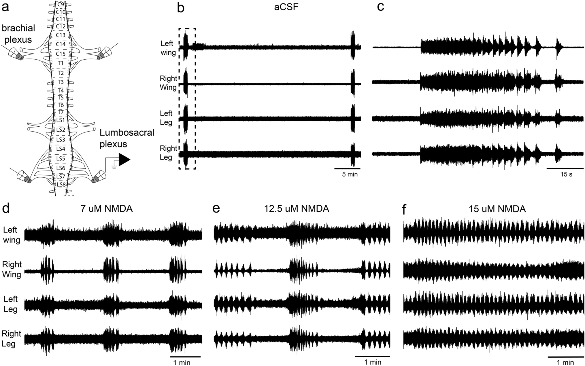


A typical example of a tetrode recording is shown in Fig. 2. Three different dynamic concentration changes in the 0–100% range were tested: a rectangular pulse with sharp on and off edges causing a jump and drop in concentration, a slow ramp up and ramp down forming the slopes of a trapezoidal concentration change, and slow oscillations with continuously rising and falling concentrations. Action potentials of varying amplitudes are evident in channels 2 and 4 (Fig. 2b–e). The recordings in channels 1 and 3 contain an amount of background noise impeding the detection and discrimination of weak spikes. In Fig. 2f, the superimposed, scaled and processed channel traces revealed the activity of four single neurons. A visual interpretation of their responses is provided by time histograms (Fig. 2g–j). There are three different ON neurons (Fig. 2g, h, j) with respect to the response rate as a function of time, and one OFF neuron (Fig. 2i). ON-neuron 1 (Fig. 2g) had a spontaneous discharge before stimulation that increased rapidly in response to both the concentration pulse and the trapezoidal concentration wave. During the up cycles of the sinusoidal concentration waves, the ON-neuron produced strong responses that consist of irregular, continuous discharges of action potentials without bunching or bursting of impulses. The faster rate of change during the concentration jump elicited lower impulse frequencies than the slower rates during oscillating concentration changes. ON-neuron 2 (Fig. 2h) was silent during the stimulus intervals and had a strong phasic response to the concentration jump, followed by a sustained low firing rate which outlasts the pulse, and then stops with a moderate discharge increase. The discharge was irregular but continuous during both the trapezoidal concentration wave and the up cycles of the concentration sinus. During the down cycles the ON-neuron became silent. OFF-neuron 3 (Fig. 2i) exhibited a low spontaneous discharge during the stimulus intervals that was reduced or absent during the odor stimulus. A strong excitatory response appeared as the concentration drop approached zero concentration, and a weaker one as the ramp down approached zero concentration, but during the down cycles of the sinusoidal concentration change a rapid peak discharge occurred. The faster rate of change during the concentration drop led to a stronger response than the slower rates during oscillating concentration changes. No activity was observed during the up cycles. ON-neuron 4 (Fig. 2j) had a spontaneous impulse discharge, a strong phasic response to the concentration jump, and moderate phasic responses to both the ramp up and the up cycles of the oscillations. The ON-neuron was not active during the down cycles.
Fig. 2
Example of an extracellular tetrode recording from four AL neurons responding to different dynamic changes in the concentration of the odor of lemon oil: a rectangular pulse, a trapezoidal wave and five sinusoidal waves. a Left column: time course of odor concentration illustrating a concentration jump from zero to 100%, followed 3 s later by a concentration drop back to the initial zero concentration. Middle column: time course of a 1-s ramp up from zero to 100% at a rate of + 100%/s, followed 5 s later by a 1-s ramp down to zero concentration at a rate of − 100%/s. Right column: time course of five continuous up and down cycles of sinusoidal concentration changes with a period of 3 s and at a constant amplitude of 100%, generating upward maximum rates of + 150%/s and downward maximum rates of − 150%/s. The negative values represent the downward direction of the concentration change. b–e Raw traces of the four tetrode channels 1 to 4 recorded at 20 kHz with bandpass filters at 0.1–3 kHz. Each microelectrode captures the activity of different neurons, so that overlapping spikes cannot be trivially resolved by detecting individual neurons in a distinct electrode, as in channels 1 and 3. f Sorting algorithm assigns the spikes to four single neurons which generate them; superimposed, color-coded activities of the four neurons in response to the different forms of concentration changes. g–j Response of the four neurons presented as time histogram (bin width, 200 ms) indicating that each neuron accentuates a different aspect of the changing concentrations. g ON-neuron 1 produces moderate activities for the duration of both the concentration pulse and the trapezoidal concentration wave, but strong responses during the up cycles of sinusoidal concentration waves. h ON-neuron 2 generates a phasic response to the concentration jump, a sustained discharge at low rates during the duration of both the concentration pulse and the trapezoidal concentration wave, and also produces sustained firing rates during the up cycles of the sinusoidal concentration waves. i OFF-neuron 3 generates a strong excitatory response to the concentration drop that appears at the pulse off, a weaker response at the end of the ramp down and stronger responses during the down cycles of the sinusoidal concentration wave. j ON-neuron 4 produces a moderate phasic response to the concentration jump, a weaker response during the ramp up and also weak responses during the up-cycles of the concentration waves. Pulse repetition intervals, 10 s; trapezoidal wave intervals, 10 s; V voltage
The four neurons convey more information than merely the presence and absence of the odor of lemon oil, the concentration amplitude, the direction of the concentration change, the onset and offset of the concentration change, and the duration of the stimulus period and the stimulus interval. As the three forms of concentration changes starts from the same initial concentration to the same end concentration, the amplitude of the concentration change alone may not determine the responses. The rate of concentration change became the obvious choice of parameters. The ability to respond to the rate of change is a prerequisite for “pulse slope detection”. The difficulty is to sufficiently accurately establish the rates of change during transient concentration changes provided by concentration pulses. To describe what information about the dynamics of a concentration pulse was transformed and processed in ON and OFF AL neurons, sensitivity to concentration pulses was characterized in terms of imp/s per % concentration change (Δ%) rather than rate of concentration change (%/s).
Pulses in odor concentrationA sequence of 7 concentration pulses of lemon-oil odor with descending amplitudes from 97 to 18% was tested three times, but for descriptive purposes a single test sequence is shown in Fig. 3. ON-neuron 1 produced a large phasic response to strong concentration jumps, followed by an irregular discharge throughout the pulse. An after discharge outlasted the concentration drop and resumed to the initial ongoing activity. At low concentration jumps, the phasic component was absent and the discharge rate rose monotonically during the pulse duration. The response magnitude decreased with decreasing jump concentration. ON-neuron 2 had a phasic discharge at moderate concentration changes, followed by a sustained, irregular activity with brief peaks after pulse off. At strong and low concentration jumps, the phasic component was often reduced and preceded a plateau of stable activity. The response magnitude remained almost unchanged when jump concentration decreased. The odorless pulse intervals suppressed spontaneous activity. OFF-neuron 3 responded to concentration drops with a rapid phasic discharge, followed by a gradual return to the spontaneous activity. At low concentration drops the phasic discharge was reduced or even absent. The response magnitude decreased with decreased drop concentration. ON-neuron 4 produced a strong phasic discharge to large concentration jumps and a weak phasic discharge to small concentration jumps, with a sharp transition between large and small jumps. The activity ceased during the pulse periods and was absent throughout the pulse intervals. The four AL neurons generate both dynamic and static responses to the same concentration pulse.
Fig. 3
Tetrode recording of the activity of the same four AL neurons shown in Fig. 2 during a sequence of rectangular pulses of decreasing odor concentration. Top row: concentration time course of seven 3-s concentration pulses with 10-s pulse repetition intervals. The record is continuous; due to the restricted space the pulse intervals are not shown in full-length. Next rows: instantaneous discharge rates of neurons 1–4 presented as time histograms (bin width, 200 ms). ON-neuron 1 produces a rapid discharge increase to the concentration jumps, followed by a slow decline and a turn off after pulse termination. Response magnitude decreases with decreasing pulse amplitude. ON-neuron 2 generates a moderate discharge increase to the concentration jumps but also peak discharge rates, followed by a moderate, maintained activity and a brief after-discharge outlasting pulse duration. Response magnitude remains unchanged when pulse concentration decreases. The OFF-neuron 3 produces a phasic response at the concentration drops, followed by a decline in the discharge rate. Response magnitude decreases with decreasing pulse amplitude. The ON-neuron 4 is a highly phasic neuron producing a rapid discharge increase to the concentration jumps followed by a rapid decline to zero before the pulse end is reached. Responses to high pulse concentrations are strong, but they are weak to low pulse concentration, with an abrupt transition between the two activity levels
The neurons’ sensitivity to transient upward and downward concentration changes was estimated by the linear regressions. Each regression was calculated for three consecutive series of odor pulses, and the response magnitude was determined by the cumulative impulse count during 3-s periods beginning with the onset of the jump or drop in concentration. The course of the regressions in the left diagram of Fig. 4a indicates that the impulse frequency of ON-neuron 1 increased with the size of the concentration jump (R2 = 0.96). The gain was 0.16 imp/s per % jump concentration, which means that an increase in 1 imp/s can be elicited by increasing the concentration jump by 6.2%. In ON-neuron 2, a linear function described the relationship between impulse frequency and jump concentration quite well (R2 = 0.84); the negative correlation indicates that the elevated activity decreased with increasing concentration jump. The gain was low, only − 0.08 imp/s per % jump concentration, meaning that the concentration jump must decrease by 12% to increase impulse frequency by 1 imp/s. OFF-neuron 3 was not affected by concentration jumps (R2 = 0.32). In ON-neuron 4, the linear regression fitted the relationship well (R2 = 0.82). The gain was 0.45 imp/s per % jump concentration, suggesting that jump concentration must be increased by 2.2% to raise the impulse frequency by 1 imp/s. The regressions in the right diagram of Fig. 4a show no dependence of the ON-neurons 1, 2 and 4 on drop concentration (R2 < 0.18), but OFF-neuron 3 increased impulse frequency with increasing drop concentration (R2 = 0.96). The slope of the regression indicates a gain value of 0.39 imp/s per concentration drop, requiring an increase in drop concentration of 2.5% for an increase of 1 imp/s. The relatively high activity of ON-neuron 2 reflects the sustained discharge, which outlasts the duration of the 3-s pulse periods.
Fig. 4
Response functions of the same four AL neurons shown in Figs. 2 and 3 obtained from a sequence of rectangular pulses of decreasing odor concentration delivered to antennal ORNs. a Left side: mean impulse frequencies plotted as linear functions of jump concentration (Δ%). The functions are positive and steep for ON-neurons 1 and 4, but they are negative and flat for ON-neuron 2 and OFF-neuron 3. The high R2 values for ON-neurons 1 and 4 indicate a strong dependence of impulse frequency on jump concentration, and the low R2 values for ON-neuron 2 and OFF-neuron 3 reveal a weak if any dependence. Impulse frequency of the ON-neurons 1 and 4 increases with increasing jump concentration, but in ON-neuron 2, impulse frequency slightly decreases with increasing jump concentration. The results from the regression equations are indicated under each diagram. Impulse frequency (F) determined by the number of impulses falling in the 3-s duration of the concentration pulse. Right side: mean impulse frequencies plotted as linear functions of drop concentration (− Δ%). The function for OFF-neuron 3 is positive and steep, but negative and flat for ON-neurons 1, 2 and 4. The high R2 for OFF-neuron 3 implies that the regression line is a perfect fit, and the low R2 for ON-neurons 1, 2 and 4 indicates no relationship. Impulse frequency of OFF-neuron 3 increases with increasing drop concentration. The results from the regression equations are indicated under each diagram. Impulse frequency (F) determined by the number of impulses falling in the 3-s period beginning with the concentration drop. b Left side: maximum impulse frequencies of the four neurons obtained during the 3-s pulse plotted as a linear function of jump concentration (Δ%). As expected from the mean-frequency plots, the maximum frequency of the two ON-neurons 1 and 4 increases with increasing jump concentration, but in contrast to the mean-frequency function, the maximum frequency of ON-neuron 2 is independent of jump concentration. Similar to the mean frequency, the maximum frequency of OFF-neuron 3 displays no dependence on jump concentration. Right side: maximum frequency values of the four neurons plotted as a linear function of drop concentration (− Δ%). As described for the mean frequencies, only the maximum frequency of OFF-neuron 3 increases with increasing drop concentration. The results from the regression equations are indicated above each diagram
The ON and OFF neurons exhibited peak discharge rates during the first second of the response to jumps and drops in concentration, respectively, in particular when the concentration change was large (Fig. 3). In Fig. 4b, peak frequency (bin width 0.2 s) of the four neurons was plotted as a function of the concentration changes, and the stimulus response functions were approximated by linear regressions. As peak frequency values were much higher than the cumulative impulse counts, the slopes of the two ON-neurons 1 and 4 became steeper by a factor 1.5 for concentration jumps (left graph), and the slope of the OFF-neuron 3 by a factor 2.5 for concentration drops (right graph). The R2 values indicate a similar good fit, suggesting that the scatter of the frequency values about the regressions remained the same. This observation raised the question of whether peak frequency values would reflect transient concentration changes with a higher accuracy than mean frequencies. The gain of the peak response of ON-neuron 1 was 0.24 imp/s per % concentration jump, and for ON-neuron 4, 0.79 imp/s per % concentration jump. As indicated by the reciprocal of the slope values, the concentration jump must be increased by 4.2% to raise the impulse frequency of ON neuron 1 by 1 imp/s; ON-neuron 4 was more sensitive, requiring an increase in concentration jump of only 1.3% (left graph). For the peak response of OFF-neuron 3, gain was 0.71 imp/s per % concentration drop, needing an increase in drop concentration of 1.4% for an increase of 1 imp/s (right graph).
As illustrated in Figs. 2 and 3, individual AL neurons responded quite differently to the same lemon-odor concentration pulse. They can be sub-divided into three groups according to the effect of the concentration amplitude. The first group consists of ON neurons whose responses to concentration jumps increased with jump concentration (Fig. 5a, left graph), the second group contained OFF neurons whose responses to concentration drops increased with drop concentration (Fig. 5a, middle graph), and the third group encompassed ON neuron that exhibited concentration-invariant responses to concentration jumps (Fig. 5a, right graph). The line graphs in Fig. 5a differ in their position or height on the frequency axis (y-intercept) indicating broad ranges of response minima and maxima elicited with low and high concentration pulses, respectively. However, the overall rate at which the response frequency of a neuron increased with further change in concentration was similar within a group. Linear regressions were used to quantify for each neuron the individual relationship between impulse frequency and pulse concentration. For the ON neurons showing a dependence on jump concentration (Fig. 5a, left graph), the mean gain value was 0.20 imp/s per % increase in jump concentration, and for the OFF neurons with a dependence on drop concentration (Fig. 5a, middle graph), the mean gain value was 0.21 imp/s per % increase in drop concentration. The reciprocal of the average slope of the ON neurons reveals that the concentration jump must be increased by 5.0% to raise the impulse frequency by 1 imp/s; the average slope of the OFF neurons suggests a required increase in the concentration drop of 4.7% to raise impulse frequency by 1 imp/s. The basic statistics are shown in Table 1.
Fig. 5
Response functions of 32 single ON and OFF neurons for a sequence of seven, triple-tested rectangular pulses of decreasing odor concentration delivered to antennal ORNs. a Left side: 16 ON-neurons exhibit an increase in impulse frequency with increasing jump concentration. Middle: 9 OFF-neurons generate an increase in impulse frequency with increasing drop concentration. Right side: 7 ON-neurons produce an excitatory response to concentration jumps that is invariant to jump concentration. The stimulus–response relationship shown for each neuron by a line graph was validated by fitting a linear regression and quoting R2 statistics, as outlined in the text. Pooling the responses across each neuron group and approximating the course of the cumulated responses by single linear regressions (orange lines) yields low R2 values, which indicate no correlation between the average ON-neuron’s response on jump concentration and the average OFF-neuron’s response on drop concentration. b Responses of the each neuron are normalized to its maximum frequency value and pooled across each neuron group for regression analysis to estimate the relationship between the cumulative normalized responses and pulse concentration. For the 16 ON-neurons showing a dependence on jump concentration (left side), the average gain value of normalized responses is 0.01 imp/s per % increase in jump concentration; for the 9 OFF-neurons depending on drop concentration (middle), the average gain value of normalized responses is 0.01 imp/s per % increase in concentration drop. R2 is 0.77 for the ON neurons, 0.82 for the OFF neurons. For the 7 ON-neurons showing no dependence on jump concentration (right side), R2 is 0.22. Impulse frequency (F) determined by the number of impulses falling in the 3 s of the pulse duration or the 3-s period beginning with the concentration drop. The results from the regression equations are indicated above each diagram
Table 1 Summary of data used to determine gain of the ON and OFF neurons for concentration pulsesWhile the responses of individual neurons within each group are variable in absolute terms, they nonetheless maintain a relatively constant relationship with one another during series of concentration pulses. This raises the question how the information of concentration pulses transmitted and passed by individual AL neurons is combined by target neurons to provide the maximal possible transfer of transient concentration changes to the brain. A simple combinatorial process would be summing or averaging the responses of individual AL neurons, in which target neurons in no way differentiate the inputs of the different AL neurons. If the neurons in the group have similar response characteristics, this simple summing of responses may greatly improve the definition of the stimulus above what possible by single neurons. To verify this, the regression analysis was performed by pooling the responses across the ON neurons and OFF neurons. However, approximating the course of the cumulative points by a single linear regression (Fig. 5a, orange lines; Table 1) eliminates any dependence of the ON-neurons’ responses on concentration jumps and of the OFF-neurons’ responses on concentration drops.
Another combinatorial process involves adjusting the responses of individual AL neurons to a common scale, without distorting differences in the ranges of the response magnitudes. Normalizing individual responses to the maximum frequency values of each neuron and then pooling the normalized responses across the neurons of each group yielded fitted regression lines with diagonal slopes of approximately 45° (Fig. 5b, orange lines; Table 1). The functions, however, did not go through the origin. Nevertheless, the relative impulse frequency and pulse concentration had a proportional relationship. The cumulative, relative impulse frequency provided a group estimate for the neurons’ response gain. For the ON neuron, the mean indicated that the normalized gain value was 0.01 imp/s per % concentration jump, for the OFF neuron the normalized gain value was − 0.01 imp/s per % concentration drop. As indicated by the reciprocal of gain values, an increase in the concentration jump by 1% results in an increase in the ON neurons’ relative activity of 1% of the full frequency scale, and an increase in the concentration drop by 1% produces an increase in the OFF neurons’ activity of 1% of the full frequency scale.
The analysis demonstrates that, for a given group of ON or OFF neurons, the differentiation of the pulse concentration is realistic if the measure of the group response was the actual discharge rate of individual neurons and the brain differentiated the inputs of the different fibers. Simple summing or averaging the responses of individual neurons, however, lost the resolution of pulse concentration. Even though the neuron samples were relative uniform in the gain for concentration pulses, they differ in their response threshold. This yields a high variability of the cumulative response, making it impossible to detect any relationship on concentration pulses. Optimally scaling the contribution of each neuron to the measure of the group response improved the pulse-concentration resolution.
The ON and OFF neuron responses not only represent pulse concentration but reflect, implicitly at least, information about the rate at which concentration changes. The common view, however, is that impulse frequency is the response to pulse concentration alone: almost all studies dealing with this issue showed that impulse frequency takes on different values with different values of pulse concentration. As the slope of the concentration pulse defines a change in concentration over time, the rate of change may also determine the neuron’s impulse frequency (beyond the concentration amplitude). The result will be a double dependence of impulse frequency on pulse concentration and the rate of changes required to reach the level of pulse concentration. Describing what information on the dynamics of concentration changes is contained in the activity of AL neurons requires changing the two parameters of the odor stimulus, namely, concentration and its rate of change, independently of each other. We did this by oscillating the changes in odor concentration.
Oscillations in odor concentrationFigure 6 illustrates the responses of the same four neurons shown in Figs. 3 and 4 during constant-amplitude oscillations with periods of 3, 6, 60 and 120 s. The upper two panels show the time course of the oscillating concentration and the corresponding oscillating rate at which concentration changes (Fig. 6a, b). The maxima of the oscillating rate of change are one quarter of the full period in advance of the maxima of the oscillating concentration. The lower four panels show the rate of discharge of the 4 neurons using a bin width of 0.2 s, which results in 30 bins for the relatively brief 6-s oscillation period (Fig. 6c–f). ON-neuron 1 was continuously active through the full cycles of the oscillations, ON-neurons 2 and 4 were active through the up cycles and fell silent through the down cycles, and OFF-neuron 3 was active through the down cycles but not through the up cycles. The maximum frequency of each neuron decreased with the duration of the oscillation period.
Fig. 6
Simultaneously recorded responses of the same 4 AL neurons shown in Figs. 2, 3 and 4 to oscillating concentration changes. a Time course of odor concentration oscillating at constant amplitude (0–100%) over four different period durations. Different time scales are used to demonstrate complete oscillation periods. b Time course of the rate of concentration change. The maxima of the oscillating rate-of-change are one quarter of the period duration in advance of the maxima of the oscillating concentrations. c–f Time course of the neurons’ impulse frequencies. c Impulse frequency of ON-neuron 1 is high during brief oscillation periods (3 and 6 s) and low during long oscillation periods (60 and 120 s); frequency maxima are in phase with concentration maxima during brief periods (3 and 6 s), but ahead of the concentration maxima and behind the maxima of the rate of change during long oscillation periods (60 and 120 s). d Frequency maxima of ON-neuron 2 decrease with increase period duration, showing at the same time an increasing phase advance to the concentration maxima. During the longer periods (60 and 120 s), the discharge peaks at the beginning of the up cycle of the concentration change. e OFF-neuron 3 produces a peak discharge at the beginning of the down cycle of the concentration change, and the frequency maxima decrease with increasing duration of the oscillation period. f Frequency maxima of ON-neuron 4 are ahead of the concentration maxima and slightly behind the maxima of the rate of change. The phase advance and the frequency maxima increase with the period duration. Dotted vertical lines indicate the phase shift between time courses of odor concentration, impulse frequency and rate of concentration change. The same instantaneous concentration within a given oscillation period (horizontal line in a) can be accompanied in each neuron by two different values of impulse frequency (vertical lines)
As shown in Fig. 6c, the impulse-frequency profile of ON-neuron 1 was smooth during brief oscillation periods (3 and 6 s), and frequency rose up to 60 imp/s when the concentration increased and declined to almost zero when the concentration decreased. During long periods (60 and 120 s), the profile was irregular and the frequency maxima were about one-third compared to brief periods. The mean frequency during the up cycles of the 60-s and 120-s periods was similar: 6.49 ± 6.19 imp/s (n = 300) and 6.89 ± 6.23 imp/s (n = 600), respectively. The irregularity in the spike train was quantified using the coefficient of variation (CV, defined as the ratio between SD and mean frequency). The variation values were 0.95 for the 60-s period and 1.16 for the 120-s period. During brief oscillation periods (3 and 6 s), the frequency maxima were in phase with the concentration maxima, during long oscillation periods (60 and 120 s), the frequency maxima were in advance of them. The impulse-frequency profile of ON-neuron 2 (Fig. 6d) was also smooth during brief oscillation periods (3 and 6 s). The frequency maxima were in phase with the concentration maximum during the 3-s period but ahead of it during the 6-s period. During the longer 60- and 120-s periods, frequency rose faster than concentration and the frequency maxima for the two periods were quite similar, namely, 21 imp/s for the 60-s period and 18 imp/s for the 120-s period. The mean frequency values during the two up cycles differed (p < 0.01), they were 6.31 ± 5.03 imp/s (n = 160) for the 60-s period and 3.97 ± 4.14 imp/s (n = 270) for the 120-s period. The spike train irregularity (CV) for the up cycle of the 60-s period was 0.79 and 1.04 for the up cycle of the 120-s period. During the longer 60- and 120-s periods, the frequency maxima were ahead of the concentration maxima. OFF-neuron 3 (Fig. 6e) produced a continuous and smooth discharge during periods of 3 and 6 s just as the oscillating concentration passed the minimum values and then increased. During the longer periods of 60 and 120 s, a phasic peak discharge occurred right at the minimum concentration. In ON-neuron 4 (Fig. 6f), the discharge increased smoothly to a maximum, which was in advance of the maximum values of the concentration during both the 3- and 6-s periods. At longer periods of 60 and 120 s, the phasic peak discharges was also ahead of the maximum values of the instantaneous concentration but occurred just at the maximum values of the rate of change. Note that the amplitude of maximum frequency values increased with the period duration.
The phase relationship between the oscillations in impulse frequency and the oscillations in the instantaneous concentration has the tendency to vary with the duration of the oscillation period. With increasing oscillation period the frequency maxima tended to lead the concentration maxima and to lag behind the rate-of-change maxima. In Fig. 7, impulse frequencies of the ON and OFF neurons were plotted as a function of the instantaneous concentration and its rate of change. The frequency curves approached closed circles similar to Lissajous figures, in which two oscillating parameters are plotted, one as a function of the other. The shape of these figures is determined by the ratio of the two oscillating parameters, or more specifically, the ratio of their amplitudes and their phase differences. Multiple regressions were calculated to determine the simultaneous effect of the instantaneous concentration (b slope) and the rate of change (a slope) on the response frequency during different oscillation periods.
Fig. 7
Impulse frequency of the same 4 AL neurons in Fig. 5 during 4 periods of oscillating odor concentration plotted as a function of the instantaneous concentration and the rate of change. Multiple regressions that utilize three-dimensional planes were calculated to determine the gain of responses for the instantaneous odor concentration and the rate of concentration change. a Impulse frequency values of ON-neuron 1 closely fit the slopes of the regression planes (R2 ≥ 0.78), indicating a double dependence on the instantaneous concentration and its rate of change. Increasing the duration of the oscillation period leads to a decrease in the gain for the instantaneous concentration and an increase in the gain for the rate of change. b ON-neuron 2 shows a similar double dependence like ON-neuron 1. Impulse frequency and gain values are lower, the values of R2 (≥ 0.82) for periods of 3 and 6 s represent the relatively small distance between the data values and the fitted values. For 60 and 120 s periods, impulse frequency values are not close to the regression planes, hence the relationship is good (R2 = 0.58) and low (R2 = 0.29), respectively. The p values (p < 0.001) for each independent variable, the instantaneous concentration and its rate of change indicate statistically significant correlation with impulse frequency. c OFF-neuron 3 produces short-duration responses at the falling concentration cycles, suggesting limited usefulness of regression planes for describing the relationship between the impulse frequency and the two parameters of the oscillations in concentration over the entire range of rates. R2 values are good for periods of 3, 6 and 60 s (R2 ≥ 0.57), and low (R2 = 0.12) for the long period of 120 s. The p values (p < 0.01) indicate statistically significant correlation of the impulse frequency and the instantaneous concentration for brief periods of 3 and 6 s, and strong evidence for a double dependence on the instantaneous concentration and its rate of change for long periods of 60 and 120 s. Note that the slopes for the instantaneous concentration are orientated in the opposite direction to the ON neurons. d Impulse frequency of ON-neuron 4 displays a pronounced excitatory response of brief duration to the rising concentration cycles, pointing to limited usefulness of regression planes for describing the relationship between the impulse frequency and the two parameters of the oscillations in concentration for the entire range of rates tested. R2 values are medium for brief periods (R2 ≥ 0.52) and low for long periods (R2 ≥ 0.23). The p values (p < 0.001) indicate a statistically significant correlation between the impulse frequency and the rate of concentration for periods of 3 and 6 s, and between the impulse frequency and the instantaneous concentration for periods of 60 and 120 s
In ON-neuron 1, the coefficients of determination (R2 ≥ 0.78) indicate a strong linear relationship between the impulse frequency, the instantaneous concentration and the rate of concentration change for each of the 4 oscillation periods (Fig. 7a). According to the regression slopes, the gain for the instantaneous concentration decreased with the duration of the oscillation period, from 0.59 imp/s per % for the 3-s period to 0.11 imp/s per % for the 120-s period. Conversely, the gain for the rate of change increased with the period duration, from 0.03 imp/s per %/s for the 3-s period to 2.01 imp/s per %/s for the 120-s period. Although the two parameters, instantaneous concentration and its rate of change, cannot be set in direct relationship to each other, their effects on impulse frequency can be by determining that increment of each parameter which results in the same increment in impulse frequency. The measurements showed that an increase of 1 imp/s can be elicited during an oscillation period of 3 s either by a 1.7% increase in the instantaneous concentration (provided the rate of change is constant) or by a rate of change of 33%/s. During a 120-s oscillation period, it takes an increase of 9% in the instantaneous concentration to increase impulse frequency by 1 imp/s, or a rate of change of 0.49%/s. Thus, during brief oscillation periods, impulse frequency could be influenced more by changing the instantaneous concentration by 1% than by changing the rate of concentration change by 1%/s. Conversely, however, during long oscillation periods, impulse frequency could be influenced more by changing the rate of concentration change 1%/s than by changing the instantaneous concentration by one additional degree.
In ON-neuron 2, the coefficients of determination (R2 ≥ 0.82) for the brief oscillation periods of 3 and 6 s indicate a strong linear relationship between the impulse frequency and the two components of the odor stimulus. For the 60-s period, the multiple regression provides a good fit to the frequency values (R2 = 0.58), but for the 120-s period (R2 = 0.29), a quadratic relationship may fit the data better than a linear one (Fig. 7b). Checking the regression analysis, however, revealed a significant dependence of the impulse frequency on both the instantaneous concentration and the rate of change (p < 0.01). This was accompanied by a large variance in the y-intercept (p < 0.02), meaning that the height of the plane is not interpretable. The large variance is explained by the discontinuous activity: the rapid increase in the discharge rate, which occurred directly at the increase in concentration, was followed by remarkably irregular spike trains, declining slowly to zero (Fig. 6c). As indicated by the regression slopes, the gain values for the two brief periods (3 and 6 s) were the same, as was also the case for the two long periods (60 and 120 s). During the brief 3-s period, the gain for the instantaneous concentration was 0.22 imp/s per % and the gain for the rate of change was 0.05 imp/s per %/s. An increase of 5% in the instantaneous concentration has the same effect on impulse frequency as a 20%/s increase in the rate of change. Both result in a 1 imp/s increase in impulse frequency. During the long 60-s period, the gain for the instantaneous concentration was 0.04 imp/s per % and the gain for the rate of change was 0.95 imp/s per %/s. An increase of 1 imp/s can be elicited either by a 25% increase in the instantaneous concentration or by a 1.05%/s rate of change. During brief periods, impulse frequency can be altered more by changing the instantaneous concentration by 1% than by changing the rate of concentration change by 1%/s. Conversely, during long periods, impulse frequency can be altered more by changing the rate of concentration change by 1%/s than by changing the instantaneous concentration by one additional degree.
In OFF-neuron 3, the coefficients of determination (R2 ≥ 0.57) for oscillation periods of 3, 6 and 60 s indicate that the regression planes represent a good approximation of the relationship between the impulse frequency, the instantaneous concentration and the rate of concentration change (Fig. 7c). The statistical evaluation of the regression analysis, however, revealed that during brief periods (3 and 6 s) the impulse frequency was not influenced by the rate of change (p < 0.48), only by the instantaneous concentration (p < 0.01). During the 3-s period, the gain for the instantaneous concentration was − 0.14 imp/s per %, meaning that a 7% decrease in the instantaneous concentration is required to increase the impulse frequency by 1 imp/s. During the long periods of 60 and 120 s, the horizontal axes of the regression planes and the low coefficient of determination (R2 ≥ 0.12) suggest a poor if any dependence of impulse frequency on the two parameters of the odor stimulus. They were, however, well-correlated with impulse frequency (p < 0.01). During the long 60-s period, the gain for the instantaneous concentration was − 0.03 imp/s per % and the gain for the rate of change was − 0.21 imp/s per %/s. An increase of 1 imp/s can be elicited either by a 33% decrease in the instantaneous concentration or by a rate of change of − 4.7%/s. Impulse frequency can be influenced more by changing the rate of concentration change by 1%/s than by changing the instantaneous concentration by one additional degree.
In ON-neuron 4, the coefficients of determination ranged between a good fit (R2 ≥ 0.52) for brief periods (3 and 6 s) and a poor fit (R2 ≥ 0.23) for the long periods (60 and 120 s), but the p values (< 0.01) for brief periods indicates a dependence on the rate of change, and for long periods on the instantaneous concentration. During brief periods, the gain for the rate of change was 0.02 imp/s, indicating that an increase of 1 imp/s can be elicited by a 50% increase in the rate of change. During long periods, the gain for the instantaneous concentration was 0.03 imp/s, suggesting that a 1 imp/s increase can be elicited by a 33% increase in the instantaneous concentration. During brief oscillation periods, the gain of response can be altered by changing the rate of concentration change, but during long oscillation periods by changing the instantaneous concentration.
Figure 7a, b shows that the gain for the rate of change of ON-neurons 1 and 2 tended to be low during brief oscillation periods and increased during long oscillation periods. Conversely, the gain for the instantaneous concentration tended to be high during brief periods and decreased during long periods. Thus, during long oscillation periods, ON neurons 1 and 2 increased the gain for the rate of change at the expense of the gain for the instantaneous concentration. This trade-off indicates that the neurons do not simply transform fluctuations in odor concentration, but balance—from instant to instant—their gain according to stimulus conditions. OFF-neuron 3 exhibits a similar gain control during long periods, but during brief periods the gain for the instantaneous concentration is not traded for the rate of change. Thus, the gain for the instantaneous concentration is not influenced by variations in the duration of the oscillation period. In ON-neuron 4, no gain control and resulting trade-off was found. During brief oscillation periods, the gain for the rate of change is not affected by the instantaneous concentration; and during long periods, the gain for the instantaneous concentration is not affected by the rate of change.
The increase in the period duration from 1 to 120 s results in a decrease in the rate of concentration change by a factor of roughly one hundred. Gain control can, therefore, be interpreted as being an adaptation to variations in the range of concentration rates due to variations in the period duration. This can be illustrated by plotting the gain values obtained from the regression planes for each ON and OFF neuron against the oscillation period (Fig. 8). The resulting gain functions of individual neurons differ with respect to several parameters. These include the rate at which both gain values change with increases in period duration (gain slope) and the size of the maximum gain, with due consideration of sign. In general, the gain for the rate of change has steeper functions and higher gain maxima than the gain for the instantaneous concentration, sign ignored. Furthermore, the gain values show a high variability. In spite of this variability, simple inspection of Fig. 8 points to general trends in some variable in relation to another. To help compare these trends, the relationships between the gain functions of the ON and OFF neurons and the period duration were categorized as strong (group 3), moderate (group 2), or non-existent (group 1), shown by the colored gain functions (Fig. 8). Grouping was done manually with a sense of proportion, and the subdivision between strong and moderate gain values for the rate of change corresponds with a value set at 2 imp/s per %/s for the 120-s period. If a neuron’s gain maximum for the rate of change was closely above or below this value, the gain slope was used to verify grouping. These criteria seem admittedly somewhat arbitrary, but nevertheless cover all neurons of the total recorded population, reveal general trends in the data, differences in the groups and overlap.
Fig. 8
Effect of the duration of the oscillation period on the gain of a 18 ON neurons and b 18 OFF neurons. Gain values are obtained for each period from regression planes by calculating impulse frequency as a function of instantaneous concentration and its rate of change. Both types of neurons can be subdivided into 3 groups, each with 6 neurons, according to the dependence of the gain for the rate of concentration change on the period duration: strong dependence (blue), moderate dependence (red), and no dependence (green). Mean values and standard error of the means obtained for each group are indicated. Left side: dependence of the mean gain for the rate of change on the duration of the oscillation periods. Middle: dependence of the mean gain for the instantaneous concentration on the duration of the oscillation periods. Right side: for each period, the mean gain for the rate of change is plotted as a function of the mean gain for the instantaneous concentration. The negative values for gain reflect the downward direction of the concentration change, yielding an increase in impulse frequency of the OFF neurons. Differences in mean gain values between adjacent neuron groups at the same period or adjacent oscillation periods of the same neuron group were assessed by ANOVA, *p < 0.05; **p < 0.01; ***p < 0.001
Across the entire range of oscillation periods, the average gain values of group-1 ON and OFF neurons are lower than the average gain values of groups 2 and 3 ON and OFF neurons (p < 0.01). During the 120-s period, group 2 ON and OFF neurons have on average a lower gain than group-3 ON and OFF neurons (p < 0.01), and the gain of group-2 ON neurons is on average lower during the 60-s versus 120-s period (p < 0.01). In addition, during the 60-s period, group 2 ON neurons are on average lower than group-3 ON neurons (p < 0.05). The diagrams indicate that the gain for rate of change and the gain for instantaneous concentration are inversely related to the period duration. Neurons with high gain values for the rate of change during long periods consistently show high gain values for the instantaneous concentration during brief periods. Thus, the ON and OFF neurons trade the gain for the rate of change at the expense of the gain for the instantaneous concentration (Fig. 8a, b, right graphs).
The mean gain values of group-3 ON neurons for the instantaneous concentration decreased from 0.40 ± 0.20 imp/s per % for the 3-s period to 0.07 ± 0.05 imp/s per % for the 120-s period (p < 0.001), and the mean gain values of group-3 OFF neurons decreased from − 0.16 ± 0.19 imp/s per % to − 0.05 ± < 0.01 imp/s per %, respectively (p < 0.1), sign ignored. The mean gain values of group-3 ON neurons for the rate of change increased from 0.05 ± 0.11 imp/s per %/s for the 3-s period to 3.61 ± 1.05 imp/s per %/s for the 120-s period (p < 0.001), and their mean gain values increased from 0.01 ± < 0.01 imp/s per %/s to − 2.67 ± 1.25 imp/s per %/s, respectively (p < 0.001), sign ignored. The gain for the rate of change is increased more than one hundred times in the presence of low rates of change during long oscillation periods, and the gain for the instantaneous concentration is increased about ten times in the presence of fast rates of change during brief oscillation periods. These effects occur at a speed equal to the fluctuating changes in odor concentration, indicating a substantial contribution to moment-to-moment odor processing.
留言 (0)