
記住我


Neuro-ophthalmology can benefit from synchronous telehealth by addressing workforce shortages, expanding access to care for patients limited by geographical distance, accommodating patients with physical impairment, offering neuro-ophthalmic education and training to a broader audience, and improving efficiency of care delivery for applicable neuro-ophthalmic conditions [13••].
Tele-Neuro-Ophthalmology Addresses Neuro-Ophthalmology Workforce Shortages and Expands Access to Neuro-Ophthalmic CareWithin the USA, there is an alarming shortage of neuro-ophthalmologists, with a projected need for an additional 180 neuro-ophthalmologists (working 0.5 clinical full-time equivalent) to meet the population demands of the country [14••]. This workforce shortage has resulted in prolonged wait times and geographic limitations in access to neuro-ophthalmic care, with pre-COVID median wait times of 6 weeks for new appointments [14••]. Telehealth services offer partial amelioration by affording patients in geographically distant regions access to neuro-ophthalmic care without requiring exhaustive commutes [13••]. Triaging suitable conditions and shifting established patients not requiring an internal eye examination to telemedicine vacates limited in-person clinic slots in favor of patients needing direct examination [13••]. Telemedicine also offers stable established patients the opportunity to address their concerns without waiting for an in-person visit [13••].
Asynchronous Tele-Neuro-Ophthalmology Improves Patient AccessTime-sensitive patient access to neuro-ophthalmology from specialists who frequently request our diagnostic testing and interpretation of tests is a common conundrum. Oftentimes, our colleagues from neurosurgery, otolaryngology, and endocrinology urgently seek visual fields to triage if a skull-base or sellar mass is sight-threatening and therefore requires emergent surgery. Pre-pandemic, some neuro-ophthalmic practices employed specific procedure-only slots within a weekly clinic template to accommodate these urgent requests whereby the ophthalmic technician performs the diagnostic testing and electronically sends the imaging for the neuro-ophthalmologist to interpret formally without having to physically examine the patient.
Kung et al. recently performed a retrospective chart review of their remote interpretation of visual fields performed in 2012 (n = 180), well before the COVID-19 pandemic. They found that the most frequent referring departments were endocrinology (79; 44%), neurology (51; 28%), and neurosurgery (43; 24%) [11•]. Indications for the visual field included parasellar lesion (107; 59%), seizure disorder (26; 14%), meningioma (19; 11%), vascular lesion (11; 6%), and other (17; 9%). Seventy-eight patients (43%) had an intervention prior to the VF, while 49 (27%) were preoperative VFs. Eighty-seven (48%) of the VFs were interpreted as abnormal [11•].
Of all the 180 remote interpretations of visual fields, 87% had VF interpretations consistent with the clinical question posed by the referring provider based on prior clinical history and imaging. The median wait time for patients receiving remote VF interpretations was 1 day at this single institution [11•]. With such rapid turnaround, remote interpretation of visual fields may offer specific referring departments an efficient method of obtaining visual field interpretations than in-office neuro-ophthalmology examinations. This can improve timely multi-disciplinary decision-making, particularly in brain tumor operative management cases. Other practices have utilized remote interpretation of data workflows whereby the visual fields are performed off-site at the referring physician’s office (e.g., neurosurgery practices). Then, the visual fields are remotely sent via the electronic medical record to the neuro-ophthalmologist for interpretation (H Moss, Personal Communication, February 2021).
eConsults (also called interprofessional consults) are another form of asynchronous communication utilized within neuro-ophthalmology. Like remote interpretation of testing, the neuro-ophthalmologist does not see or examine the patient. Rather, the requesting physician asks the neuro-ophthalmologist a specific clinical question via review of the clinical records. While this can be accomplished in formal written form, within the digital age, the requesting physician often utilizes an EMR platform to send the neuro-ophthalmologist their query for back-and-forth response.
Xu et al. published a retrospective review of a single institution’s experience with eConsults (n = 80) during a 3-year study period [15•]. The median time from eConsult request to completion was 4 days (range 0–34 days). The most frequent eConsult questions were vision/visual field disturbances (35.0%) cases, optic neuropathies (32.5%), and optic disc edema (21.3%). At the time of eConsult, 44% of cases provided sufficient prior information for the neuro-ophthalmologist to provide a definitive management decision. In over 56% of the eConsults, further diagnostic testing was recommended. In-person neuro-ophthalmology consultation was recommended in 30.0% of cases. Sixty-one (76.3%) eConsults provided diagnostic and/or treatment direction, and 12 (15.0%) provided reassurance. eConsults increase access to timely neuro-ophthalmic care and provide diagnostic and treatment direction to requesting physicians when sufficient information is provided at the time of eConsult. In addition, with 15% of cases providing reassurance, this, in essence, triages otherwise referred in-person visits and opens additional scheduling template slots for the most appropriate patient cases.
Elements of the Neuro-Ophthalmic Exam Amenable to Synchronous TelehealthA prospective survey was conducted on patient and physician experiences with consecutive tele-neuro-ophthalmology visits at three tertiary care academic institutions in the USA during the early COVID period [16•]. Neuro-ophthalmologists generally indicated that the synchronous telehealth history and examination were sufficient for decision-making (87% of visits) [15•]. Exam elements indicated by neuro-ophthalmologists that were easy to conduct include.
These impressions were corroborated by the 2021 NANOS survey, which found similar results [2••].
Tele-Neuro-Ophthalmology Expands Neuro-Ophthalmic Education OpportunitiesThe Accreditation Council for Graduate Medical Education (ACGME) requires neurology and ophthalmology training programs to provide sufficient training in subspecialties applicable to their respective fields, including neuro-ophthalmology [17, 18]. Because of the workforce shortages in neuro-ophthalmology, some US training programs have had to obtain neuro-ophthalmology faculty from outside institutions to satisfy ACGME requirements [13••]
Synchronous telemedicine offers trainees from remote institutions exposure to geographically distant neuro-ophthalmologists [13••]. Asynchronous telehealth curricula such as the Neuro-Ophthalmology Virtual Education Library (NOVEL) and the Eccles Health Sciences Library at the University of Utah and the Neuro-Ophthalmology Illustrated Curriculum developed by NOVEL and NANOS fill curriculum gaps and provide education to learners across a broad range of interests [19].
Tele-Neuro-Ophthalmology and Physician WellnessThe American Medical Association (AMA) conducted a survey to evaluate relationships between pandemic-related stress and work intentions in a sample of US health care workers [20]. Among 2914 physician respondents at 124 institutions, approximately 1 in 3 physicians surveyed intend to reduce work hours. One in 5 physicians intend to leave their practice altogether within the next 2 years. Burnout, fear of exposure, COVID-19-related anxiety/depression, and workload were predictors of intent to leave [20]. Medscape examined burnout rates in over 13,000 physicians and noted that burnout among neurologists and ophthalmologists was 46% and 40%, respectively [21]. The top cited reasons for burnout among physicians included bureaucratic tasks, lack of respect from administrators/employees, too many hours at work, and lack of control/autonomy over one’s life. When asked what would help improve burnout, respondents stated that a more manageable work schedule, lighter work schedule, more autonomy/control over life, and increased compensation would help [21].
While it has been suggested in opinion pieces that telemedicine may provide a reduction in physician burnout and improve wellness, few studies have been performed. Aklureishi et al. performed an observational, cross-sectional study of healthcare providers (nearly 75% of physicians). They noted that video visits took longer to prepare (33%) and conduct (62/200, 32%) than in-person visits [22]. Clinicians who conducted a higher percentage of their telemedicine by video were less likely to feel overwhelmed, with no difference in reported burnout. Interestingly, clinicians 50 or older were “less overwhelmed” than those younger than 50 years, which may be related to clinical experience. Clinicians who received more video visit training modalities were less likely to feel overwhelmed by the conversion to video visits (P = 0.007) or burnt out (P = 0.009). In addition, those reporting a higher ability to navigate a video visit were also less likely to feel overwhelmed by video visits (P = 0.02) or burned out (P = 0.001). More than half “enjoyed conducting video visits” (60%) and wanted to continue using video visits in the future (75%) [23]. Similar to the neuro-ophthalmology surveys cited above, the top telemedicine barriers were patient-related: lack of technology access, lack of skill, and reluctance, with clinicians noting an increased need for increased support staff to ease the workflow. More research is required to understand if telehealth facilitates physician wellness. Nonetheless, Aklureishi’s study suggests that experienced clinicians and those with sufficient telehealth training and operational support are less likely to experience burnout [22].
Reimbursement and regulations on telehealth services vary from country to country. In the USA, the Centers for Medicare and Medicaid Services (CMS) determine the parameters of telehealth eligibility for Medicare patients. Private insurers and state Medicaid programs may have different requirements and fee schedules.
Prior to the COVID-19 pandemic, CMS allowed telehealth in restricted contexts, only allowing patients to receive synchronous telehealth care if they lived in designated “Healthcare Provider Shortage Areas” [23], and both the physician and patient were located in approved facilities. Other triage-associated telehealth services, such as eConsults, remote interpretation of diagnostic tests, and short synchronous and asynchronous physician–patient communications (phone, video, or electronic) were also available [24].
The COVID-19 PHE temporary waivers removed restrictions on some interstate licensure requirements, geographic restrictions on eligible patients, added reimbursement for audio-only synchronous telehealth, and waived location requirements for medical professionals and patients. These waivers allowed tele-neuro-ophthalmic care for patients living hours away from a neuro-ophthalmologist [10]. There are continued advocacy efforts to codify these changes to improve access to care.
The Future of Tele-Neuro-OphthalmologyThe practice of neuro-ophthalmology will never return to pre-pandemic patterns. Based on the authors’ 2021 survey of neuro-ophthalmologists 1 year into the pandemic, utilization of tele-neuro-ophthalmology continued despite lower COVID case numbers, lower COVID-related hospitalizations, and the advent of the COVID vaccination [2••]. While 30% of respondents never utilized telehealth, over 60% adopted tele-neuro-ophthalmology. Eighty-two percent of adoptees indicated that they plan to continue, but the survey did not directly query respondents as to their rationale for continued telehealth use [2••].
Digital Transformation: Tele-Neuro-Ophthalmology 2.0 and BeyondAs tele-neuro-ophthalmology enters its next iteration, incorporating other digital technologies is expected to enhance healthcare and its intersection with telehealth delivery (Fig. 1). Digital transformation is defined as the process of improving an entity through the combination of digital technologies, including information, computing, communication, and connectivity [25]. Within ophthalmology and, by extension, neuro-ophthalmology, digital transformation can potentially improve patient access and healthcare delivery through artificial intelligence (AI), machine learning, deep learning (DL), and virtual reality and augmented reality (VR and AR) [26]. Through these digital transformation technologies, some existing barriers within current tele-neuro-ophthalmology delivery may be surmountable, including improving physician workflow and utilizing forms of deep learning for screening and triaging patient based on DL analysis of ophthalmic images and using augmented reality for home monitoring of vision field progression.
Fig. 1
The interplay of synchronous and asynchronous telehealth with incorporation of artificial intelligence. Synchronous telehealth is live/interactive telehealth that can be either clinic or home based. It includes video or phone visits. Asynchronous telehealth is a “store-and-forward” approach where testing/information is obtained and then shared through a portal for a physician to review later. Hybrid telehealth incorporates both synchronous and asynchronous modalities including capturing a visual field or fundus photograph asynchronously for the neuro-ophthalmologist to review at a later time point. A video visit is then scheduled for the neuro-ophthalmologist to synchronously review with the patient the testing results and care management plan. An example of a potential artificial intelligence (AI)-driven future state of neuro-ophthalmology is where asynchronous data is obtained via augmented reality (AR) home monitoring of a visual field defect. Deep learning (DL) algorithms then interpret the change in the visual field defect from previously obtained visual fields of the patient. If there are significant changes, DL can then pivot to either alert scheduling of an urgent synchronous video visit or a routine video visit with the patient
Artificial Intelligence and Deep LearningAI is the broad umbrella concept under which machine and deep learning reside (Fig. 2). AI is defined as any technique that is used to mimic sentient behavior [27]. AI is utilized in everyday life, from voice commands on smart devices to robotic vacuum cleaners.
Fig. 2
Artificial intelligence, machine learning, and deep learning defined. AI is the broad umbrella concept under which machine and deep learning reside. AI is defined as any technique that is used to mimic sentient behavior. Machine learning (ML) is a form of AI focused on data analysis by the construction and study of algorithms that process data to develop prediction or decision models. Deep learning (DL) is a form of machine learning that mimics human learning by layering multiple automated learning algorithms over the data in an iterative process to achieve the desired level of accuracy
Machine learning (ML) is a form of AI focused on data analysis. ML is the construction and study of algorithms that process data to develop prediction or decision models. Unlike traditional computer programming, ML does not need programmers to write step-by-step scripting for a computer to follow. Instead, they train computers to recognize patterns based on data [28]. One example of ML is the spam filter on e-mail systems, which can learn by direct input from humans and from automated pattern-recognition algorithms.
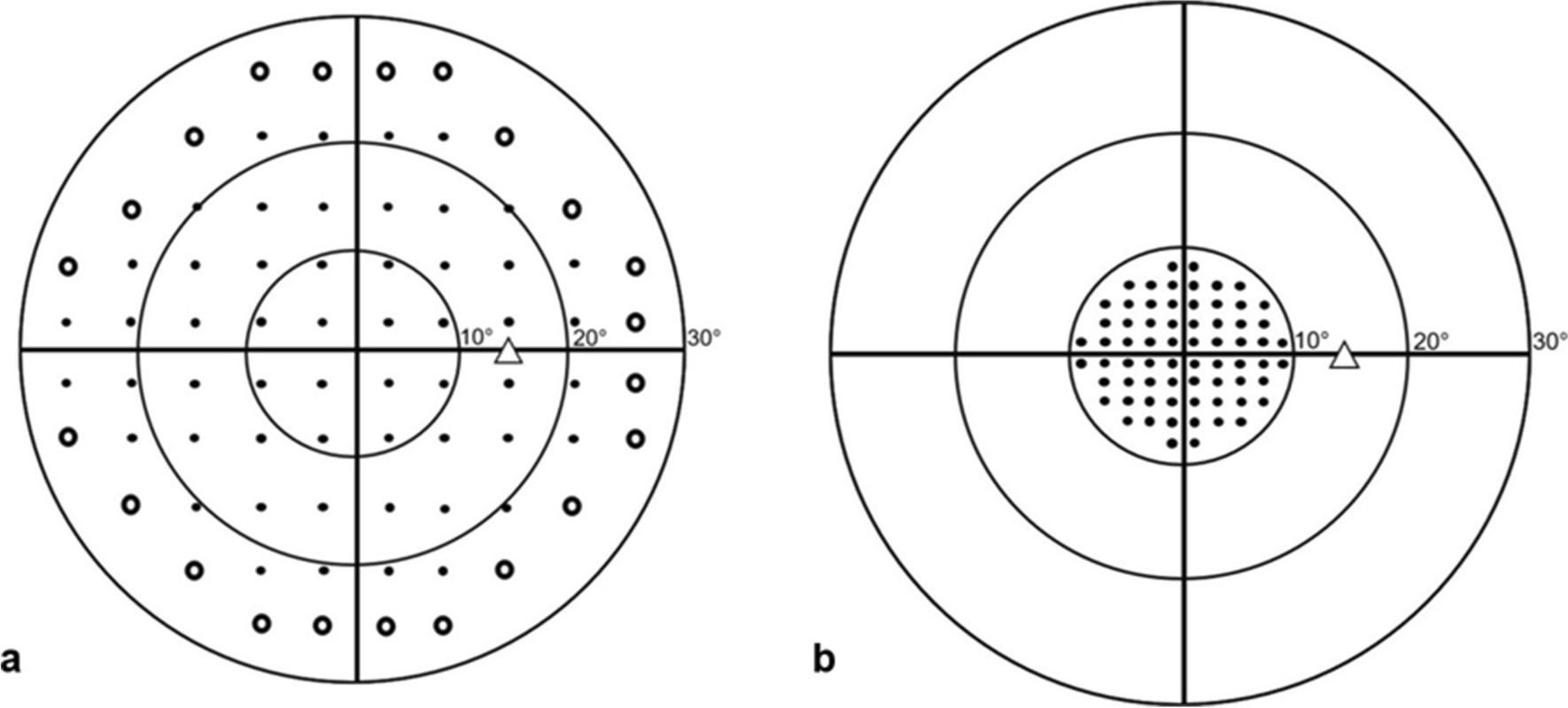
Deep learning (DL) is a form of machine learning that mimics human learning by layering multiple automated learning algorithms over the data in an iterative process to achieve the desired level of accuracy (Fig. 1) [29]. As a result, DL is especially powerful with image recognition and highly applicable to image-heavy specialties, including ophthalmology. Within neuro-ophthalmology, DL applications can analyze ophthalmic images, including digital fundus photography, visual fields, and optical coherence tomography. DL can assist in automated screening and diagnosis. DL has been utilized to distinguish abnormal optic discs from normal controls, including papilledema and the degree of optic nerve swelling, and to differentiate various forms of non-glaucomatous optic neuropathies [30, 31••, 32,33,34]. Recently, DL applications were proven to be at least as accurate as expert neuro-ophthalmologists in identifying papilledema [35]. Given the current workforce challenges within neuro-ophthalmology, one can envision a future practice model in which mature DL applications triage screening fundus photographs of referred patients to prioritize the most severe cases of papilledema over mild cases. Additionally, patients with anomalous but normal optic nerves may not even require a referral to neuro-ophthalmology. With direct ophthalmoscopy instruction declining in medical school curricula, many non-ophthalmology-based physicians lack confidence in viewing the fundus [36]. Proven DL applications may support non-ophthalmology-based doctors by funneling patients with potential sight-threatening papilledema to the neuro-ophthalmologist and deferring those with normal optic nerves to comprehensive ophthalmology. Automated DL telescreening is already utilized by non-eye health professionals in diabetic retinopathy screening [37••]. This workflow integrates virtual technologies throughout a patient’s continuum of care: patient visit registration, digital capture of the funduscopic images, DL-guided screening algorithms, and then synchronous video telemedicine to communicate the results to the patient.
Virtual and Augmented RealityVirtual reality (VR) and its subset, augmented reality (AR), comprise another aspect of digital transformation that will impact Tele-Neuro-Ophthalmology 2.0 [37••]. VR is where the user, via a wrap-around headset, is fully immersed in a virtual environment. VR is used in ophthalmology for patients with visual loss/impairment where an attached camera records the patient’s environment and displays or magnifies that image into the patient’s visible visual field. Safety concerns are a limitation; patients can not see their real environment while ambulating because the headsets are fully occlusive and immersive [37••].
Augmented reality (AR), on the other hand, integrates technology with real life, for example, smartphone language applications projecting translations over the native text. A developing neuro-ophthalmology application of AR is glasses with embedded cameras that can map out a patient’s scotoma or visual field defect, create a custom remapping of the missing images, and overlay that image onto functioning areas of a patient’s vision dynamically, allowing the patient to continue functioning within the real environment [37••, 38]. Other uses of AR include home monitoring units developed for patients with age-related macular degeneration to detect retinal fluid changes with the data then transmitted via the cloud to healthcare providers for interpretation [37••]. These technologies present potential translation opportunities for neuro-ophthalmic patients from a diagnostic monitoring and therapeutic perspective by allowing patients to engage visually with their environment. Additional applications of AR that impact the neuro-ophthalmic surgeon include the development of 3D glasses (heads up surgery) to improve ergonomics and enhance visualization during ophthalmic surgery. This AR technology allows surgeons to view projected operative images directly in front of them, aiding greater maneuverability [37••]. Lastly, VR and AR applications have potential to extend into neuro-ophthalmic medical education by training medical students and residents in pertinent anatomy and surgical techniques through surgical simulators, which have already demonstrated efficacy and validity in improving surgical performance and reducing complication rates. Ophthalmoscopy simulators have also demonstrated efficacy and validity evidence in improving ophthalmoscopy skills in the clinical setting [39].
留言 (0)