
記住我
Breastfeeding is the gold standard for infant feeding. Exclusive breastfeeding is recommended for the first 6 months of life, continuing with complementary foods for at least the first year of life and beyond. These recommendations are strongly supported by multiple medical and professional childcare organizations, including the American Academy of Pediatrics (AAP) [1], the American Academy of Family Physicians (AAFP) [2], the American College of Obstetricians and Gynecologists (ACOG) [3], the World Health Organization (WHO) [4] and the Canadian Pediatric Society (CPS) [5], the American Academy of Breastfeeding (ABM) [6] and the Spanish Association of Pediatrics [7] based on short- and long-term benefits for mother and child. Suboptimal breastfeeding is associated with an increased risk of infant and child morbidity and mortality, and an increased risk of certain chronic conditions [8,9,10,11,12].
With the initiation of vaccination campaigns against SARS-CoV-2, many questions were raised about its compatibility with breastfeeding. Vaccination is now recommended for all postpartum individuals, including those who are breastfeeding. Mothers should not discontinue breastfeeding if they choose to be vaccinated. Although breastfeeding women were not included in the initial large vaccine trials, the available vaccines are unlikely to pose a risk to the breastfeeding infant. These vaccines do not contain infectious viruses and the minimal amount of vaccine that passes into breast milk [13, 14] and is then ingested by the infant is likely to be inactivated by the infant’s digestive system. In addition, maternal antibodies to SARS-CoV-2 induced by maternal vaccination can pass into breast milk and may offer passive protection to the infant [15,16,17,18,19,20].
Our main objective was to determine the evolution of IgG and IgA antibodies directed against SARS-CoV-2 protein S in the blood and breast milk of lactating women vaccinated with different vaccines.
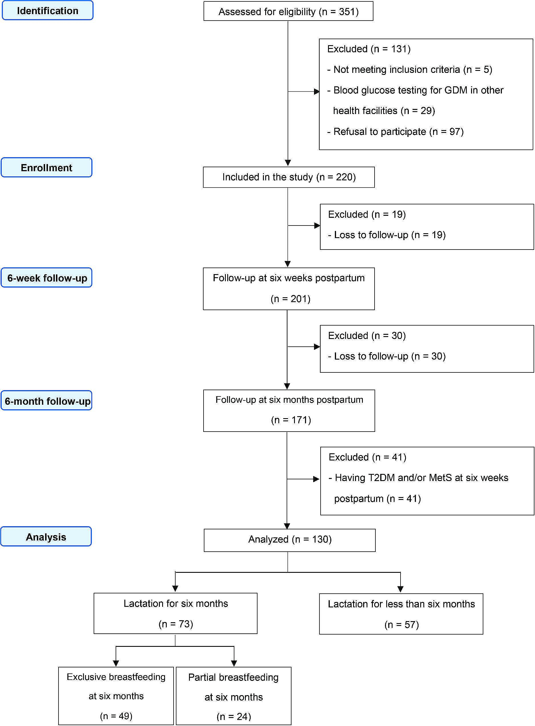
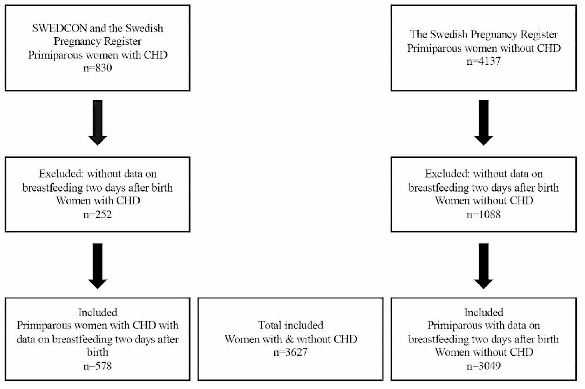
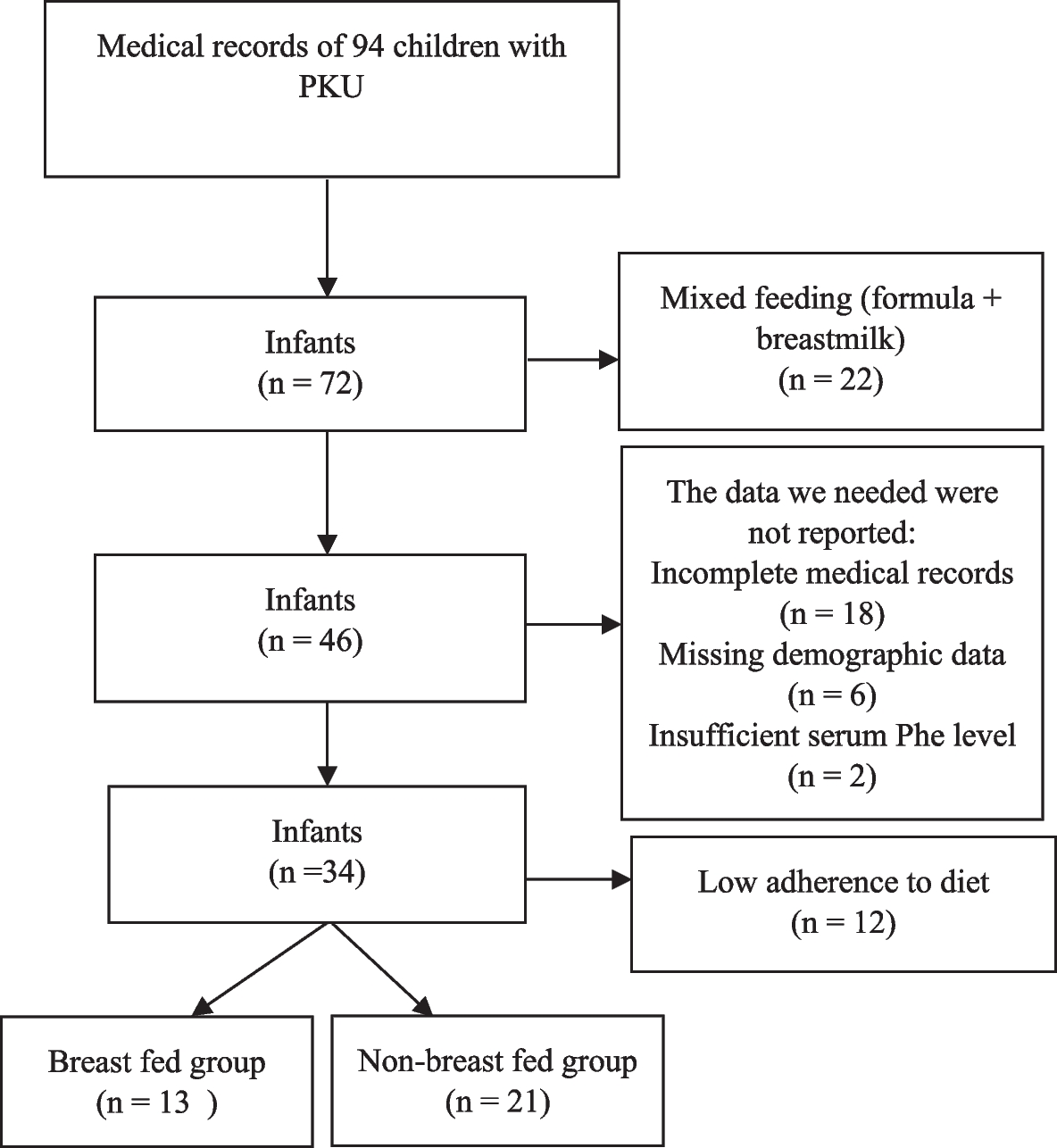
Design and settingA cohort study was conducted, including 110 uninfected, vaccinated breastfeeding mothers and an initial control group of 23 additional infants who had no previously documented infection and had not been vaccinated against SARS-CoV-2. Once baseline values were established in the unvaccinated uninfected unvaccinated women (where the arithmetic mean was 0.02 AU/mL [range: 0.00–0.04] in the determination of antibodies in breast milk [20]) it was not necessary to further analyze this control group and they were excluded from the 6-month follow-up. All lactating women who received both doses of the vaccine were included in the study, together with women vaccinated with ChAdOx1-S, who received a single dose. It is of note that vaccination with ChAdOx1-S was halted during the study because of the appearance of severe episodes of vaccine-induced immune thrombotic thrombocytopenia; for this reason, the second dose for breastfeeding mothers who received the first dose of ChAdOx1-S was delayed. Thus, the women recruited in our study had received two doses of either mRNA-based vaccines (BNT162b2 or mRNA-1273) or just one dose of ChAdOx1-S vaccine. Recruitment of breastfeeding mothers into the study was informed by a mass institutional mailing. All women interested in participating underwent a structured interview for data collection and collection of blood and breast milk samples after informed consent. The study was conducted at the Hospital Universitario Marqués de Valdecilla, Santander, Spain. The initial sample recruitment period was from 1 April 2021 to 30 April 2021 with a follow-up after 6 months. Our previously published study [20] refers to the initial period from April 2021 in which the safety of the vaccines and the determination of the antibody production response 1 month after vaccination are evaluated. In this new study, we report on the six-month follow-up, carried out in October 2021. During the follow-up period, 48 women stopped breastfeeding, which is why the sample at 6 months was reduced to 62 women. Blood and milk samples were taken 1 month and 6 months after the last vaccination dose. See Fig. 1 of participants & samples.
Fig. 1 Sources of information and data gathering
The main study variables were: mothers’ age, educational level, employment, medical history of gynecological interest, current pharmacological treatment, type of breastfeeding (exclusive or mixed with formula), infant’s age, type of vaccine received (BNT162b2, mRNA − 1273, or ChAdOx1-S) and batch, dates of vaccination, adverse effects in the mother (none, local pain, fever, malaise, lymphadenopathy, headache, nausea and others) and adverse effects in the infant after each dose and history of SARS-CoV − 2 infection.
Collection and processing of breast milk and serum samplesThe preservation and processing of the serum and breast milk samples followed the same conditions as the initial samples collected in the previous study period [20]. In the initial period, serum and breast milk samples were collected from all 110 lactating mothers in the study. Samples were collected 30 days after the second dose of vaccine (mean 30.3 days, SD 0.56), irrespective of the type of vaccine received. In the second recruitment period during the month of October 2021, serum and breast milk samples were once again collected from 62 of the 110 lactating mothers from the initial period. Following the same processing conditions, at least 5 mL of venous blood without anticoagulants and 1 mL of milk were collected. The blood sample was centrifuged at 3000 rpm (rpm) for 10 min at room temperature and the sera were divided into aliquots in cryogenic vials and stored at − 20 °C until use. Breast milk was centrifuged at 2000 rpm at 4 °C for 25 min and the supernatant was divided into aliquots in cryogenic vials and stored at − 20 °C until use. Prior to processing, breast milk samples were thawed, centrifuged at 2000 rpm for 15 min, fat was removed, and the supernatant was transferred to a new tube. Centrifugation was repeated twice to ensure removal of all cells and fat.
All serum and breast milk samples were tested in parallel on two different SARS-CoV-2 antibody test platforms, which are described in detail below.
Detection of IgG and IgA antibodies by ELISAThe detection of IgG and IgA isotype antibodies present in serum derived from venous blood samples and breast milk samples against SARS-CoV-2 was carried out by ELISA following the protocol published by IrsiCaixa [21] and used in several publications as described below. A list of reagents and consumables and their details can be consulted in this reference [21] for its reproducibility by other interested groups.
Briefly, serum samples were pre-diluted 1:100 using phosphate buffered saline (PBS) and breast milk samples were used without any dilution.
Nunc MaxiSorp 96-well plates (Thermo Fisher Scientific, Waltham, MA, USA) were coated using optimized concentrations of the capture antibody (2 μg/mL) (MA1–21315, Thermo Fisher Scientific) diluted in PBS for 17–24 hours at 4 °C. The capture antibody used was a His Tag monoclonal antibody (HIS.H8). The washing and blocking cycle was performed with 1x PBS + 1% bovine serum albumin (BSA) for 2 h at room temperature. After a further wash cycle, S2 antigen + RBD (Sino Biological, Beijing, China) diluted in blocking buffer was added to one half of the plate and blocking buffer was added to the other half, followed by incubation for 17–24 hours at 4 °C. Specifically, the antigens used were His recombinant proteins (SARS-CoV-2 (2019-nCoV) Spike S2 ECD-His Recombinant Protein, and SARS-CoV-2 (2019-nCoV) Spike RBD-His Recombinant Protein). Serum and breast milk samples were added and incubated for 1 h at room temperature. Subsequently, incubation with peroxidase-conjugated anti-IgG and anti-IgA detection antibodies (Jackson Immunoresearch, West Grove, PA, USA) was carried out for 30 min at room temperature. Specifically, the detection antibodies used were peroxidase AffiniPure F(ab´)2 fragment goat anti-human IgG, Fcγ fragment specific; peroxidase AffiniPure F(ab´)2 fragment goat anti-human IgM, Fc5μ fragment specific; and peroxidase AffiniPure goat anti-human IgA, α chain specific. Finally, the substrate solution and the corresponding stop solution were added. The resulting absorbance was determined at 492 nm spectrophotometrically using the plate reader Infinite M200 (Tecan Magellan™). The specific signal associated with each sample was calculated by background subtraction as follows: AU specific signal = OD (+Ag) - OD (−Ag), where OD (+Ag) is the optical density (OD) obtained in the wells containing the antigens and the OD (−Ag) is the OD obtained in the control wells where no antigen was added. Arbitrary units were used because the units were defined by a measurement procedure that is not traceable to an international unit or an international certified reference material [22].
Detection of anti-S1 IgG antibodies by chemiluminescent microparticle immunoassay (CLIA)Serum and breast milk samples were tested in parallel with the Alinity SARS-CoV-2 IgG II Quant Assay by the Alinity i immunoassay system (Abbott, Abbott Park, IL, USA) for the determination of IgG antibodies directed against SARS-CoV-2 S1 protein (RBD). This assay is validated for application in human serum and plasma, although several studies have already applied this assay in breast milk samples and our working group has previously referenced it [20]. For the interpretation of the value as a positive result in the determination of antibodies, a value above 50 AU/mL in serum samples was considered positive, following the manufacturer’s indications. For the determination of antibodies in breast milk, the arithmetic mean of the values obtained in the milks of the control group (unvaccinated lactating mothers) was subtracted from the analytical result of each sample. This control group consisted of a total of 23 unvaccinated and uninfected lactating mothers, also referred to in our previously published study [20]. In this manner, possible analytical interferences in the determination of antibodies in breast milk are eliminated, as the sample is heterogeneous in nature.
Statistical analysisComparisons of antibody concentrations between all three vaccines was performed using ANOVA. The correlation between different types of antibodies at recruitment and at the 6 months follow-up was studied with the linear correlation coefficient, stratifying for the administered vaccine. Differences in antibody concentration between time 0 and time 6 months was carried out with the paired t tests. We considered the possibility of adjusting for a number of confounders. However, age, body mass index and weight gain in pregnancy were far from associated with the type of vaccine, whereas nationality, educational level, occupational situation and method of fertilization had little or no variability; therefore, we discarded their use for adjustment. All statistical analyses were performed using the Stata 16/SE package (Stata Corp, College Station, Tx, US).
留言 (0)