
記住我


Newborn screening (NBS) by quantifying T-cell receptor excision circles (TREC) in dried blood spots (DBS) has made early detection of severe combined immunodeficiency (SCID) a reality: the median age at diagnosis in large cohorts has come down from 4.5 months [1] in the pre-NBS era to 3.5 weeks [2]. Survival has increased as well, but this has not only been the case for patients diagnosed by NBS, but also for patients diagnosed clinically [3, 4]. Infections acquired before undergoing hematopoietic stem cell transplantation (HSCT) remain a major cause of morbidity and mortality in both groups [3]. According to recent data, more than a half of SCID patients identified by NBS have developed at least one infection prior to HSCT [2]. Bacterial and fungal infections can be controlled pre-HSCT in most cases, but viral infections tend to persist and remain active [3]. While for most viruses, only careful hygiene can be offered as a preventive measure, monoclonal antibodies are available for the prophylaxis of respiratory syncytial virus (RSV) infection [2]. Their use is officially restricted to certain patient groups [5]. No studies evaluating the effectiveness of anti-RSV monoclonal antibodies in patients with SCID have been conducted thus far. However, the inability to prevent RSV in SCID can cost a life, as illustrated by the following case.
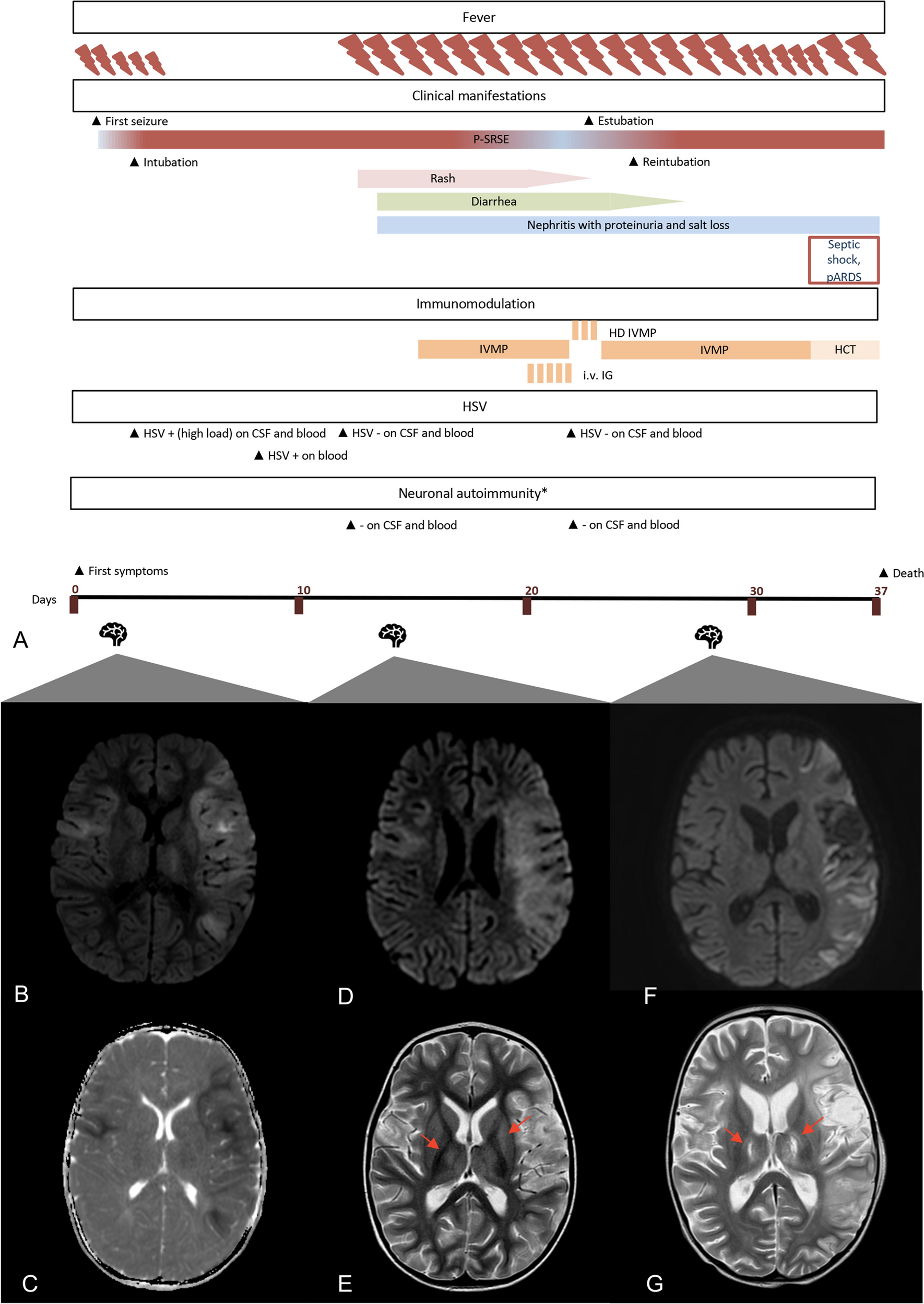
We report on a male newborn born during the local flu season with abnormal findings in his DBS reported on the tenth day of life. His TREC and kappa deleting excision circle (KREC) levels were 1 and 0 copies per punch, respectively (reference: TREC > 5 copies/punch; KREC > 9 copies/punch). He was reviewed at a regional pediatric immunology unit on the following day confirming SCID diagnosis with decreased levels of CD3 + T cells (1617/μl) and CD4 + T cells (1551/μl) as well as very low levels of naive CD4 + T cells (15/μl) and almost absent B cells (1/μl). He had a skin rash, which in combination with an expanded CD4 + memory T cell pool (1536/μl) indicated Omenn syndrome or maternal engraftment. Two days before the first review, his sister and parents had developed symptoms of a respiratory infection. The boy did not have any respiratory symptoms despite testing positive for human coronavirus OC43 in a multiplex panel for respiratory pathogens in a nasopharyngeal aspirate (NPA) sample. He was admitted to the local hospital, received antimicrobial prophylaxis for Pneumocystis jirovecii and other fungal infections, and broad-spectrum antibiotics for a possible bacterial skin infection. After a few days, he was transferred to our hospital, the national reference center for children with inborn errors of immunity.
Unfortunately, not only did the rash worsen within a few days after the transfer, but the child also developed respiratory symptoms. A rapid RSV test was negative, while RSV was detected in a multiplex polymerase chain reaction (PCR) test from the NPA, the results of which were not available until later. Due to rapid clinical deterioration, he was transferred to the pediatric intensive care unit (PICU) and was started on intravenous immunoglobulin substitution and off-label palivizumab. He soon required invasive ventilation and was given steroids to suppress pulmonary inflammation or a possible proliferation of auto-reactive lymphocytes. At this time, results from an urgent trio whole-exome sequencing revealed two compound heterozygous mutations in recombination activating gene 1 (RAG1): c.[1331C > T] and c.[2018_2025del] (NM_000448.2), the prior a known pathogenic and the latter a novel mutation leading to a premature stop codon. The recovery from this episode was slow and only partial with a continued requirement of supplemental oxygen. Over the following weeks, he tested negative for RSV in NPA a few times, but subsequently tested positive again (Fig. 1). Shortly thereafter, his respiratory situation deteriorated and antiviral therapy with off-label ribavirin and nitazoxanide were started. The additional administration of these antiviral drugs did not lead to an improvement, and he required invasive ventilation again. His recovery was once again only partial. A similar episode occurred at the age of 3 months, followed by a partial recovery.
Fig. 1
Timeline depicting major symptoms and their severity, timing of prophylactic measures, main infection surveillance findings, main treatment measures and their relative doses as well as key events during transplantation. Darker colors represent a more pronounced symptoms and higher doses. Black symbols stand for pathological and grey symbols for normal results. CMV, cytomegalovirus; CSA, ciclosporin A; HCoV-OC43, human coronavirus OC43; HSCT, hematopoietic stem cell transplantation; IVIG, intravenous immunoglobulin, RSV, respiratory syncytial virus
At 4.5 months of age, after reduced intensity conditioning with fludarabine (7.2 mg/kg), alemtuzumab (0.6 mg/kg) and treosulfan (30 g/m2), the patient received a peripheral blood stem cell transplant with a T cell add back from a 10/10 matched unrelated donor. In addition to ciclosporin A, which he was already receiving, mycophenolate mofetil was given for prophylaxis of graft-versus-host disease. On day 16 after transplantation, neutrophil engraftment was documented.
Soon after engraftment, there was a further severe respiratory deterioration requiring invasive ventilation. Despite persisting detection of RSV from nasopharyngeal fluid, other possible differentials were considered. The steroid dose was increased to treat possible engraftment syndrome, but then reduced again to allow for a better T cell response from the graft, when there was no improvement. Etanercept was attempted for a potential idiopathic pneumonia syndrome, defibrotide for a possible pulmonary veno-occlusive disease and finally steroid pulse therapy. All these measures had no effect on his clinical condition, and after almost a month of invasive ventilation, it was decided to end active therapy. The patient passed away at the age of 6 months. His lung autopsy demonstrated syncytial giant cells with cytoplasmic inclusions typical for RSV. There were no signs of an organizing pneumonia, and no other organisms could be detected in the tissue despite extensive investigations. The cause of death was found to be uncontrolled RSV pneumonia.
Regular infection surveillance was performed throughout the course of the disease. Apart from two positive NPA PCR results for human coronavirus OC43, a single positive blood PCR for CMV and an isolated tracheal aspirate culture sample with growth of Pseudomonas non-aeruginosa, no pathogens other than RSV could be detected. Despite empirical treatment against CMV and Pseudomonas, it was unlikely that these pathogens contributed significantly to the clinical deterioration. Inflammation was monitored regularly, but C-reactive protein levels remained normal almost through the entire course of the disease.
Although impossible to prove, this boy’s life might have been saved by the initiation of palivizumab prophylaxis immediately after confirmation of the SCID diagnosis. Direct evidence of the efficacy of anti-RSV monoclonal antibodies in SCID is unlikely to ever be available due to the obvious limitations in conducting such studies. Not only does the rarity of this disease limit the numbers of potential study participants, but also not giving prophylaxis to the control group would raise significant ethical issues. Most European countries, including the majority of countries with established SCID NBS programs, recommend the use or at least considering the use of anti-RSV monoclonal antibodies in SCID for RSV prophylaxis in their local guidelines despite the lack of direct evidence of its effectiveness [5]. These are also recommended as routine RSV prophylaxis in SCID in the USA [2]. To the best of our knowledge, no other patient detected by SCID NBS has experienced a fatal RSV infection. This is an indirect indication of the efficacy of anti-RSV monoclonal antibodies for RSV prevention in SCID patients. It is therefore of utmost importance to make them available for SCID patients in all parts of the world. In particular, countries that can afford newborn screening programs should provide access to monoclonal antibodies for RSV prevention for all SCID patients.
留言 (0)