Animal
Sixty-six adult Sprague-Dawley (SD) rats (female; 8 weeks old; 200–250 g) purchased from Animal Laboratory Supplies (Xiangya School of Medicine, Central South University, Changsha, Hunan, China) were used in this study (n = 22 rats per group). All animals were housed in comfortable conditions under a 12-h light/12-h dark cycle at 23 ± 2 °C, humidity level of 60%–70%, with free access to food and water. All surgical procedures were performed under general anesthesia using a solution of 2% sodium pentobarbital (80 mg/kg) administered by intraperitoneal injection. Oxybuprocaine hydrochloride (0.4% Oxybuprocaine; Santen, Japan) was applied as a topical anesthetic and 0.5% levofloxacin hydrochloride (Santen, Japan) was applied to prevent postsurgical infection. Animals under general anesthesia were placed on a heating blanket until they woke up.
Experimental design
In all rats, both eyes were used as experimental specimens and untreated controls. The intraocular pressure (IOP) was measured in both awake animals’ eyes using a rebound tonometer (Tonolab, Icare, Vantaa, Finland) without topical anesthesia, on three consecutive days to establish a baseline by the same inspectors at the same time of the day (between 8 a.m. and 10 a.m.) and under similar lighting conditions. The IOP readings were taken 2 min after the surgery under general anesthesia. For the 1st week, IOP was measured daily, and for the rest of the experimental period, IOP was measured three times per week. AL and refraction recordings were taken in both eyes 3 days before the operation and at 1, 2, 4, and 24 weeks after the operation. The flash visual evoked potential (fVEP), retrograde tracing, Western blot, MASSON staining, and transmission electron microscope (TEM) were also recorded simultaneously from both eyes at 4 weeks post-surgery. Non-invasive in vivo assessment of eye structures was performed using the optical coherence tomography (OCT) system, and magnetic resonance imaging (MRI) at 4 weeks. After the AL, IOP, and refraction recordings, the first batch of rats was euthanized at 1 and 2 weeks (n = 6). Furthermore, after fVEP, MRI, and OCT measurements were taken, the second batch of rats was euthanized at 4 weeks (n = 13) and the third group at 24 weeks (n = 3) for Western blot analysis.
Surgical technique
Procedures for the surgical technique have been described in detail previously [11, 12]. Briefly, a glass microneedle (33G, Hamilton) was positioned parallel to the vessel axis, inserted into the vessel lumen and a volume of 50 µL of lauromacrogol (Polyoxyethylene lauryl ether 1%; Tianyu, Shanxi, China) was dissolved in distilled water and slowly injected into the superior scleral vein of one eye during the SI induction. During the CS induction, a CS (7-0 nylon) was placed around the equator of the eye at approximately 1.5 mm behind the limbus (Additional file 2: Fig. S1). The suture was anchored by placing it below the conjunctiva, avoiding the major episcleral drainage veins evenly spaced around the globe at five to six anchor points. The congestion of the vortex veins could be prevented by the anterior position of the suture. The eyes of the experimental animals were treated with antibiotic eye drops after the operation. The control group was accepted without any disposal. Both eyes were performed with the same treatment in experimental groups.
Axial length and refraction measurement
AL was measured by an ophthalmic A-scan ultrasonic diagnostic instrument (ODM-2100S, Maida, Tianjin, China). The ultrasonic probe was used to touch the corneal surface gently (frequency = 10 Hz and resolution = 0.01 mm). Then, the pupils were dilated with the instillation of eye drops containing a mixture of 0.5% tropicamide and 0.5% phenylephrine hydrochloride (Santen, Japan). Refractions were measured by streak retinoscope (YZ24B; 66 Vison Tech, Suzhou, China). The average AL and refraction were calculated and recorded.
Magnetic resonance imaging
Details of the MRI protocol have been published previously [13]. The MRI scans (7.0T BioSpec 70/20 USR; Bruker, Billerica, MA, USA) were applied to measure AL and access the shape of the eyeball with a three-dimensional (3D) MRI. According to the positioning image, T1 and T2 imaging scanning parameters included the acquisition sequence [T1 = multislice-multiecho (MSME) and T2 = rapid acquisition with relaxation enhancement (RARE)], the thickness of acquisition layer (T1 = T2 = 0.6 mm), relaxation time (T1 = 10,188.3320 ms and T2 = 5344.0875 ms), and time to echo (T1 = 14 ms and T2 = 36 ms). The 3D imaging was performed by using commercially available software (OsiriX MD; Food and Drug Administration cleared, Pixmeo, Geneva, Switzerland).
Flash visual evoked potential
The fVEP was performed using the device (GT-2008V-VI; GOTEC, Chongqing, China) for functional evaluation of retinas (the stimuli intensity = 10.0 cd·s/m2, the flash frequency = 1 Hz, and the number of flashes = 64 times). After dark adaptation for 12 h, pupils were dilated with the instillation of eye drops, the fully anesthetized animals were then placed on the examination table, and the fVEP of the unilateral eye was examined. The average value after 63 flashes was taken to calculate the P2 wave.
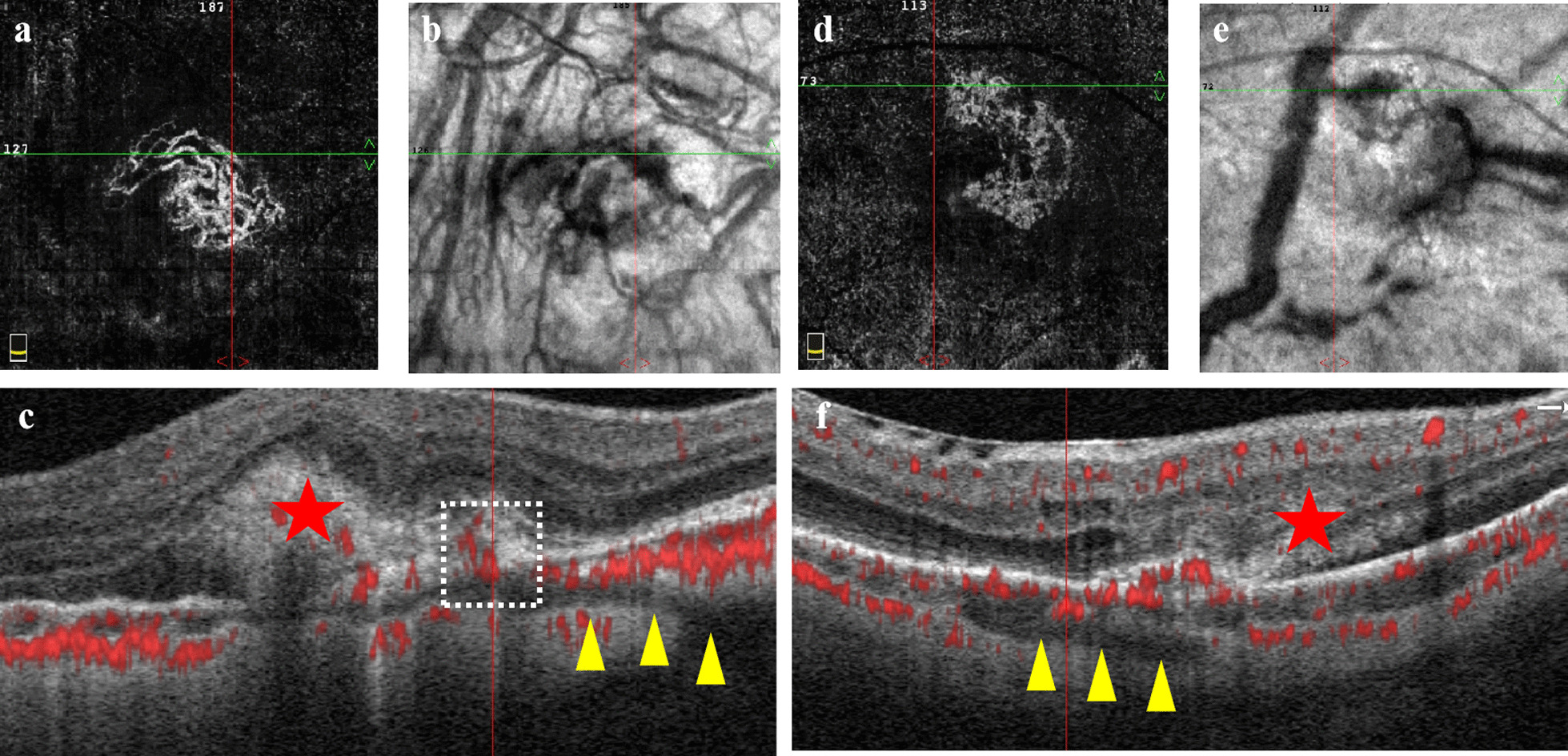
Optical coherence tomography
Following the fVEP recording, the retinal structure was imaged using OCT (Micron IV, Pleasanton, CA, USA) as previously reported [14]. The ganglion cell complex (GCC) included the retinal nerve fiber layer (RNFL), ganglion cell layer (GCL), and inner plexiform layer (IPL) [15]. In brief, the corneal surface was protected using a 0.9% saline solution before the inspection. OCT images were obtained from a position located horizontally at one disc diameter superior to the optic disc. Meanwhile, ocular fundus monitoring was performed when the rats were fully anesthetized. The acquired retinal spectral domain OCT images were quantitatively analyzed using Image J v.1.8.0 after segmentation of each sublayer. During all experimental procedures, the physical condition of the rats was frequently monitored by inspection and gentle palpation.
Retrograde tracing
To observe the survival of retinal ganglion cells (RGCs) in OHT eyes, retrograde tracing was performed as described previously with slight modifications [16]. Briefly, a surgical blade was used to make an incision to expose the bregma and posterior fontanelle on anesthetized rats. The localizations of the bilateral SC are 6 mm posterior to the bregma and 1.8 mm lateral to the midline. Two small holes (2.5 mm in diameter) were made using a mini drill on both sides of the midline. A syringe (5 μL, Hamilton, USA) was vertically inserted to a depth of 4 mm and used to slowly inject 0.5 μL of tracer (4% fluorogold dissolved in 0.9% saline, USA) into the ambilateral SC. Placement of the syringe in the target zone and tracer injection took 10 and 3 min, respectively. Following the completion of the two-site injection, 4-0 surgical threads (Ethicon, Inc., Somerville, USA) were used to suture the incision. During the whole procedure, a heating lamp was used to keep the rats warm. The mean density of retinal ganglion cells (MD-RGCs) was performed using Image J v.1.8.0 with a semi-automated method [17].
Tissue preparation and staining
The rats were euthanized, and the eyeballs were enucleated instantly and immersed in 4% paraformaldehyde for 2 h at room temperature, and then embedded in paraffin. The paraffin sections of 4 μm was cut through the papillary optic nerve plane and stained with hematoxylin-eosin (H & E) and Masson. In Masson-stained images, collagen was dyed blue, the blue area of the posterior segment (except cornea) of the eyeball wall across ONH was taken as the numerator and the total staining area of that eyeball wall was taken as the denominator. Thus, the collagen volume fraction of the posterior segment was calculated by the percentage of the ratio between the numerator and denominator [18].
Western blot analysis
According to the previous regions of the globe, the sclera within 4 mm outside the corneal limbus and the sclera within 4 mm from the optic nerve was defined as the peripheral sclera and the posterior sclera, respectively, by a vernier caliper (1–150 mm; SPIFFFLYER, Germany) with a resolution of 0.01 mm. The position of the peripheral sclera corresponded to the equatorial part and the central one corresponded to the PPS according to the division of the eyeball wall [1, 19]. The minced tissue samples were ground using a grinder (KZ-II, Servicebio, Wuhan, China) set at the power setting model (frequency = 60 Hz and time = 60 s). Sample protein concentration was determined by bicinchoninic acid (BCA) protein quantitative detection kit. For Western blotting, 5× sodium dodecyl sulfate (SDS) loading buffer was added to each sample, and samples were placed in a boiling water bath for 5 min before being loaded into wells of 10% sodium dodecyl sulfate polyacrylamide gel electrophoresis (SDS-PAGE) gel. After electrophoresis, proteins were transferred onto a polyvinylidene fluoride membrane. After blocking with 5% bovine serum albumin, the proteins on the membranes were immunoblotted overnight at 4 °C with the anti-alpha smooth muscle actin (αSMA, 1:1000; #19245; Cell Signaling Technology, USA) antibody. After three washes with tris buffered saline with Tween 20 (TBST), the membranes were incubated with the appropriate horseradish peroxidase-conjugated secondary antibody (1:5000; Jackson ImmunoResearch) at room temperature for 1 h. Western blot bands were detected using an enhanced chemiluminescence solution (Millipore, Bedford, MA). Densitometric analysis was performed using Image J v.1.8.0 software.
Transmission electron microscopy
The peripheral sclera and retina regions were located as previously mentioned [1]. Histopathological tissue preparation followed the measurement of the external diameter of eyeballs in vitro by the vernier caliper. For analysis, the globe was divided into peripheral sclera corresponding to the equatorial part and the central one corresponding to the PPS. The nasal and temporal regions were combined to focus on the anterior patterns. Tissue (diameter = 1 mm) harvested from animals was fixed by electron microscope fixative (2.5% Glutaric dialdehyde solution, Servicebio, China). The prepared niosomal formulations were characterized for their shape using TEM (HT770, HITACHI, Japan) at 80 kV. A TEM micrograph was taken at suitable magnification after staining.
Statistical analysis
The data are presented as mean ± standard deviation of results from at least three independent experiments [the data are presented as mean ± standard error of the mean (SEM) in Table 1 and Additional file 1: Table S1]. The statistical significance of the experimental differences between groups was analyzed using the Student’s t-test, and comparisons among groups were analyzed using one-way analysis of variance (ANOVA). Statistical analyses were performed on GraphPad Prism 8.0 (GraphPad Software, La Jolla, CA, USA).
Table 1 Intraocular pressure measurements at different time points in each group (mmHg) 


留言 (0)