


記住我


Parkinson’s disease (PD) is the second most common neurodegenerative disease, characterised by clinical manifestations of resting tremor, bradykinesia, and/or muscle stiffness (Liddle, 2018; Fayyad et al., 2019). Currently, the diagnosis of PD depends largely on clinical manifestation scales, imaging examination and the experience of clinicians (Su et al., 2020). However, the scales are often subjective and easily affected by symptoms. Functional neuroimaging is very expensive and can only be done at a few centers (Bhidayasiri and Martinez-Martin, 2017). It is urgent to find a reliable and economical biomarker for the early diagnosis of PD.
Gastrointestinal dysfunction, particularly constipation, is the most common non-motor symptom of PD and often precedes the motor disability onset by decades (Lin et al., 2019). Similarly, accumulation of alpha-synuclein (α-syn) in the enteric nervous system (ENS) may begin 20 years before the onset of degenerative changes in the central nervous system (CNS) and motor symptoms associated with PD (Hasegawa et al., 2015). Previous studies on pathological changes in the intestines of patients and animal models of PD suggest the possibility that gut alteration is associated with PD pathogenesis (Choi et al., 2018). Thus, we speculate that α-syn appears in the ENS first, and then can be transferred to the CNS.
Recent studies have revealed that gut microbiota and their metabolites affect brain function through neural, endocrine, and immune pathways (Cryan and Dinan, 2012). Meanwhile, the body also regulates the composition of the gut microbiota through this pathway to maintain the balance of intestinal microecology–that is, the microbiota–gut-brain axis (Elfil et al., 2020). The gut microbiota widely participates in the synthesis/release of various hormones and gut-brain axis–related neurotransmitters, which then regulates brain function and host behaviour. In summary, changes in the gut microbiota composition and function play an important role in the occurrence and progression of PD. Researchers have found differences in the gut microbiota composition between patients with PD and healthy controls (HC), but the results are not consistent. For example, Scheperjans et al. (2015) pointed to a relatively lower abundance of Prevotellaceae in advanced PD. Bedarf et al. (2017) found Prevotellaceae was markedly lower in patients with early-stage PD. However, Keshavarzian et al. (2015) found that there was no statistically significant difference between PD and HC in Prevotellaceae.
In addition to our previously published studies on 16S rRNA (Zhang et al., 2020), we included more studies and used meta-analysis to preliminarily determine the altered gut microbiota between PD and HC. Furthermore, metagenomics was used to validate the differential gut microbiota in the meta-analysis results. The possible pathogenic mechanisms of these differential gut microbiota in PD are also discussed.
Materials and methods Meta-analysis Literature retrieval, inclusion, and exclusionTo conduct a meta-analysis of studies related to the gut microbiota in PD, we conducted a comprehensive and systematic literature search using the following English and Chinese databases (for paper published up to October 2021): PubMed, Web of Science, the Chinese National Knowledge Infrastructure (CNKI) databases and the Wanfang database. The search strategy to identify all potential studies involved using combinations of the following terms: (Parkinson’s disease OR Parkinson disease OR Parkinsonism) AND (microbes OR microbiome OR microbiota OR bacteria) in the Title/Abstract. We also searched the articles referenced in the included articles to identify any studies we may have missed with the above-mentioned search strategy. For the articles retrieved through the above-described search strategy, we included studies that met the following criteria: (1) the study was a randomised controlled trial (RCT) comparing differences in the intestinal flora of patients with PD and HCs; (2) the study used faecal samples; (3) the abundance of the microbiome was expressed as the average proportion of each microbiome; (4) differences between patients with PD and HCs were presented with the mean difference (MD) and the 95% confidence interval (CI); and (5) studies with scores > 5 stars as assessed by the nine-star Newcastle–Ottawa Quality Assessment Scale (NOS). We excluded studies with the following criteria: (1) studies without assessment of the measurement indexes; (2) only patient studies, family-based studies, intervention studies and review articles; and (3) studies with scores below five as assessed by the NOS; (4) studies were not considered or explicitly excluded participants who received antibiotics and probiotics 1–3 months before the trial.
Data extractionWe extracted the following data from the included studies: (1) general basic information (author, publication date, and location); (2) characteristics of patients with PD and HCs (gender and age); (3) diagnostic criteria, sample sizes, and microbiological experimental methods (detection); and (4) microbiome abundance effect size (MD and 95% CI); (5) clinical features of PD patients (UPDRS total score, score of UPDRS Part III and H&Y stage).
Quality assessmentDisagreements were solved through discussion or involvement of a third investigator, if necessary. We included studies with five or more stars after a general evaluation with the NOS in the meta-analysis. Detailed criteria are provided in Supplementary Table 1 (Stang, 2010).
Statistical analysisAt least five studies of the same gut microbiota have reported differences between PD and HC. Meta-analysis was conducted using Review Manager Version 5.3 (Cochrane collaboration) to generate forest plots and funnel plots. We calculated mean difference (MD) and 95% confidence interval (CI) of microbiota abundance as summary statistics and generated forest plots. At the same time, the degree of heterogeneity was evaluated by the inconsistency index (I2) test. The fixed-effects model was used when I2 < 50%; otherwise, the random-effects model was applied. Heterogeneity is represented by funnel plots. Differences were considered statistically significant when the FDR corrected p-value was <0.05.
Validation of differential gut microbiotaFecal samples from 39 PD patients and corresponding 39 healthy spouses were collected for shotgun metagenomic sequencing. PD patients were diagnosed according to the 2015 PD diagnostic criteria of the MDS (Li J. et al., 2017). The inclusion criteria of healthy spouses were healthy individuals without PD symptoms. All participants who received or injected antibiotics or probiotics within the last 3 months were further excluded. SPSS (version 25) was used for statistical analysis. First, the relative abundance of gut microbiota was extracted from the metagenomic data. Comparison of two independent groups was performed by Student t-test for normally distributed data, and Mann-Whitney U-criteria (M-U test) for not normally distributed data. The results of the meta-analysis were then verified. Finally, SPSS software was used to draw receiver operating characteristic (ROC) curve.
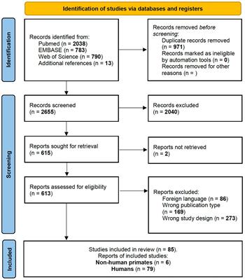
ResultsThe literature screening process is summarised as a flow chart (Figure 1). We retrieved 1,647 potential records, of which we excluded 337 duplicates. In addition, we excluded 1,270 studies whose content was inconsistent with our study, 10 studies that lacked control groups, had incomplete data, or used non-fecal samples, 15 studies that could not provide quantitative data on gut microbiota abundance, 2 studies that did not specify exclusion criteria for the use of antibiotics and probiotics (Specific inclusion and exclusion criteria for inclusion studies are in Supplementary Table 2), and 1 study conducted quantitative PCR detection technique. Hence, we included 14 studies in the meta-analysis. The main features of these studies are summarised in Table 1. Based on the NOS scale, all 14 case-control studies are high quality (Supplementary Table 1).
Figure 1. Flow diagram of the assessment of studies used in the meta-analysis.
Table 1. Characteristics of the 14 studies included in the systematic review.
A total of 1,886 subjects were included in the meta-analysis, 997 were PD patients (age range: 60.0–76.5 years; percent of female subjects ranged: 20.7–76.9; H&Y: 1–3; UPDRS-III score: 12.6–33.7; UPDRS total score: 30.5–49.0) and 889 were healthy controls (age: 62.4–79.5 years; percent of female subjects ranged: 28.6–62.5). All 14 studies had detected gut microbiota by next-generation sequencing (NGS) (see Table 1 for the demographic data).
Meta-analysis of standardised MDWe extracted continuous data from the 14 included studies for the meta-analysis. We analysed alterations in the abundance of f_Prevotellaceae, f_Bifidobacteriaceae, f_Lachnospiraceae, f_Ruminococcaceae, f_Verrucomicrobiaceae, and f_Christensenellaceae in patients with PD. The meta-analysis revealed lower abundance of f_Prevotellaceae (MD = –0.41, 95% CI = –0.68 to –0.15; I2= 78%; p < 0.00001; 9 studies, Figures 2A, 3A) and f_Lachnospiraceae (MD = –0.26, 95% CI = –0.51 to –0.01; I2= 71%; p < 0.0001; 7 studies, Figures 2B, 3B) in patients with PD compared with HCs. On the other hand, there was higher abundance of f_Ruminococcaceae (MD = 0.70, 95% CI = 0.15 to 1.25; I2= 94%; p < 0.00001; 10 studies, Figures 2C, 3C), f_Christensenellaceae (MD = 0.32, 95% CI = 0.02 to 0.63; I2= 63%; p = 0.03; 6 studies, Figures 2D, 3D), f_Bifidobacteriaceae (MD = 0.33, 95% CI = –0.21 to 0.87; I2= 94%; p < 0.00001; 8 studies, Figures 2E, 3E) and f_Verrucomicrobiaceae (MD = 0.40, 95% CI = 0.24 to 0.57; I2= 53%; p = 0.02; 8 studies, Figures 2F, 3F) in patients with PD compared with HCs.
Figure 2. Forest plots of alterations in gut microbiota in patients with Parkinson’s disease (PD) compared with healthy controls (HC). (A) f_Prevotellaceae, (B) f_Lachnospiraceae, (C) f_Ruminoccoccaceae, (D) f_Christensenellaceae, (E) f_Bifidobacteriaceae, and (F) f_Verrucomicrobiaceae.
Figure 3. Funnel plots of alterations in gut microbiota in patients with Parkinson’s disease (PD) compared with healthy controls (HCs). (A) Prevotellaceae, (B) Lachnospiraceae, (C) Ruminoccoccaceae, (D) Christensenellaceae, (E) Bifidobacteriaceae, and (F) Verrucomicrobiaceae.
Validation of the six differential gut microbiota in the above meta-analysis resultsWe extracted the differential family level gut microbiota and found that among the six different microbiota obtained by meta-analysis, only Bifidobacteriaceae was also at high levels in the validation cohort of the metagenome. And three species from f_Bifidobacteriaceae included s_Scardovia_inopinata, s_Bifidobacterium_dentium, and s_Scardovia_wiggsiae were also high (Table 2).
Table 2. Altered families, genera, and species in the metagenomic validation cohort.
We used the abundance of f_Bifidobacteriaceae and the abundance of s_Scardovia_inopinata, s_Bifidobacterium_dentium, and s_scardovia_wiggsiae generated ROC curves separately (Figure 4). The AUC were 63.1 and 71.2%, respectively. The ROC curve showed that the three species (71.2%) from f_Bifidobacteriaceae had good predictive efficiency for PD.
Figure 4. The receiver operating characteristic (ROC) curve for predicting the occurrence of Parkinson’s disease (PD) in 78 samples: The dotted line represents the performance of the probabilistic model, the blue line represents the classification performance of f_Bifidobacteriaceae, the red line represents the classification performance of the s_Scardovia_inopinata, s_Bifidobacterium_dentium, and s_Scardovia_wiggsiae.
DiscussionWe included 14 case-control studies in this meta-analysis and found the abundance f_Prevotellaceae, f_Lachnospiraceae were reduced, the abundance f_Bifidobacteriaceae, f_Ruminococcaceae, f_Verrucomicrobiaceae, and f_Christensenellaceae were elevated in PD. Furthermore, additional metagenomic verification showed that only f_Bifidobacteriaceae is higher in PD. At the same time, three species from f_Bifidobacteriaceae also showed a high abundance, including s_Scardovia_inopinata, s_Bifidobacterium_dentium, and s_Scardovia_wiggsiae (Figure 5).
Figure 5. An overview of the pathogenesis of three species from Bifidobacteriaceae in Parkinson’s disease (PD).
F_Bifidobacteriaceae is considered to be one of the important probiotics that participates in several protective physiological functions. Whereas, 8 studies in the meta-analysis consistently reported that patients with PD have increased abundance of f_Bifidobacteriaceae. It suggests that the elevated of f_Bifidobacteriaceae in PD may not be an accidental phenomenon. We speculate that some species from f_Bifidobacteriaceae may have played a potential promoting role in the pathogenesis of PD.
Protein functional analysis was performed using the UniProt Knowledgebase (UniProtKB) at UniProt. The search found s_Scardovia_inopinata may have the function of aspartate-semialdehyde dehydrogenase (Homologous gene: asd, 373 amino acids), which enzyme is one of the nine enzymes present in microbiota responsible for lysine biosynthesis. All is known that the accumulation of α-syn in the brain forms Lewy body, which leads to the death of dopaminergic neurons (DN) and is thought to be one of the important causes of PD. In fact, the protein sequence of α-syn has a remarkable amount of lysine (Shi et al., 2019). Likewise, not only in the brain, but also in the presence of α-syn in ENS. Therefore, based on the possible function of s_Scardovia_inopinata, we speculate that the aggregation of α-syn in ENS may come from the lysine produced by s_Scardovia_inopinata, which is further converted aldehydes.
In PD, in addition to the progressive degeneration of the substantia nigra pars compacta and other pigmented nuclei of the brain (Khan et al., 2019). Studies (Sanjari et al., 2017) have shown that gamma – aminobutyric acid ergic (GABAergic) and other neurotransmitter systems also have dysfunction in PD. A study on s_Bifidobacterium_dentium found that s_Bifidobacterium_dentium can converted glutamate to GABA by the enzyme glutamate decarboxylase [GAD; Enzyme Commission number (EC) 4.1.1.15] (Luck et al., 2021). s_Bifidobacterium_dentium not only secretes GABA in vitro microbial medium, but also increases the level of GABA in the stool of mice treated with s_Bifidobacterium_dentium compared with the bacteria-free control group. Clinical studies have found that increased levels of GABA correlate with the degree of gait disorder in PD (Siucinska, 2019). Therefore, we speculated that s_ Bifidobacterium_dentium could promote PD by increasing the level of GABA. The study also found that in addition to s_Bifidobacterium_dentium, thirty out of 83 Bifidobacteria species contained succinate-semialdehyde dehydrogenase (EC 1.2.1.16) for converting succinate to GABA. This may explain why f_Bifidobacteriaceae, known as probiotics, promote PD.
Finally, in a study investigating the relationship between serum inflammatory factors and gut microbiota in PD with end-stage renal disease, s_Scardovia_wiggsiae was found to have a significant positive correlation with the inflammatory factor indole sulfate (IS) (Asgharian et al., 2022). A study on the concentration of toxins in the blood and cerebrospinal fluid of PD patients found that IS also increased and affected the development of PD through inflammation and oxidative stress (Sankowski et al., 2020). Recent studies have suggested that oxidative stress plays an important role in the process of neurodegeneration in PD. We speculated that the increase of s_Scardovia_wiggsiae could lead to the increase of IS level in blood and cerebrospinal fluid and aggravate the degree of oxidative stress, which may lead to the occurrence of PD. Furthermore, the ROC curve showed that the three species (71.2%) from Bifidobacteriaceae had good predictive efficiency for PD.
In addition to diseases, studies have shown that physiological aging is related to intestinal ecological disorders. A study to investigate the impact of aging on the gut microbiota and to probe the bacterial taxonomic composition of fecal samples from younger (25–49 years) and older (61–100 years) persons showed that the f_Bifidobacteriaceae, known as probiotics, showed a decrease in abundance with age (Mueller et al., 2006). These results suggest that the protective mechanisms of human body gradually decline with aging, which leads to the decrease of beneficial microbiota and the increase of harmful microbiota. Therefore, its elevated in PD further indicates the potential pathogenic effect of f_Bifidobacteriaceae, but the specific mechanism needs further study. Due to several factors, including the delivery mode, feeding during infancy, eating habits, culture, geographical area, age and gender, among others, the structure of intestinal microflora varies among individuals (Zhuang et al., 2017). However, even taking these factors into account, we found common changes in the abundance of f_Bifidobacteriaceae in patients with PD across different studies conducted in different geographical regions. Thus, it is highly likely that these alterations in the gut microbiota result from PD itself.
LimitationsOur meta-analysis had some limitations. First, there was statistical bias among the included studies, as the majority of the subjects included were patients with PD who came to the hospital for medical treatment and physical examination. Second, we discussed only the structure and composition of the gut microbiota, but transcriptomic and proteomic studies would provide greater insight into the function of the gut microbiota. Third, all the included studies used 16S rRNA amplicon sequencing techniques to analyse the diversity of the gut microbiota. However, 16S rRNA primers for different regions may result in inconsistent results because their corresponding flanking conservative regions not only have a significant binding affinity, but also differ in the resolution of each variable region in the taxon, resulting in deviations due to the detection technique. Finally, we extracted only MD and 95% CI from the studies and did not analyse raw data to eliminate the method biases. These factors need to be improved in future studies.
ConclusionOur meta-analysis identified six differential gut microbiota in PD patients. Through validation, only Bifidobacteriaceae was also found to have high levels in the metagenomics validation cohort. Meanwhile, three species from the Bifidobacteriaceae, including Scardovia_inopinata, Bifidobacterium_dentium, and Scardovia_wiggsiae were also high. Our findings suggest that high levels of Bifidobacteriaceae may be involved in the pathogenesis of PD. The high levels of three species from Bifidobacteriaceae in patients may affect PD by participating in the aggregation of α-syn, the increase of GABA and IS levels. The three species (71.2%) from Bifidobacteriaceae had good predictive efficiency for PD. In the future, interventions of the above gut microbiota may help to prevent PD and improve disease progression.
Data availability statementPublicly available datasets were analyzed in this study. This data can be found here: 16S rRNA sequence data from the Genome Sequence Archive (CRA001938); metagenomic study (PRJNA588035).
Ethics statementThe studies involving human participants were reviewed and approved by Ethics Committee of Xiangyang No.1 People’s Hospital. The patients/participants provided their written informed consent to participate in this study.
Author contributionsPW and JTi: conceptualisation. JTi: methodology. QZ: software, data curation, and funding acquisition. JTa and HW: validation. YZ: formal analysis. SZ and HW: investigation. PW: resources, writing – original draft preparation, writing – review and editing, supervision, and project administration. MS: visualisation. All authors contributed to the article and approved the submitted version.
FundingThis study was supported by National Natural Science Foundation of China (81703496) and the Key Project of Xiangyang Science and Technology Bureau (2020YL31 and 2021YL21).
Conflict of interestThe authors declare that the research was conducted in the absence of any commercial or financial relationships that could be construed as a potential conflict of interest.
Publisher’s noteAll claims expressed in this article are solely those of the authors and do not necessarily represent those of their affiliated organizations, or those of the publisher, the editors and the reviewers. Any product that may be evaluated in this article, or claim that may be made by its manufacturer, is not guaranteed or endorsed by the publisher.
Supplementary materialThe Supplementary Material for this article can be found online at: https://www.frontiersin.org/articles/10.3389/fnint.2022.1054627/full#supplementary-material
Supplementary Table 1 | Scores of the 14 studies included in this meta-analysis based on the Newcastle–Ottawa Quality Assessment Scale (NOS).
Supplementary Table 2 | Inclusion and exclusion criteria for the 14 studies included in this meta-analysis.
Footnotes ReferencesAho, V., Pereira, P., Voutilainen, S., Paulin, L., Pekkonen, E., Auvinen, P., et al. (2019). Gut microbiota in Parkinson’s disease: Temporal stability and relations to disease progression. EBioMedicine 44, 691–707. doi: 10.1016/j.ebiom.2019.05.064
PubMed Abstract | CrossRef Full Text | Google Scholar
Asgharian, M., Gholizadeh, P., Samadi, K. H., Ghojazadeh, M., Samadi, A., Soleymani, J., et al. (2022). Correlation of inflammatory biomarkers with the diversity of Bacteroidaceae, Bifidobacteriaceae, Prevotellaceae and Lactobacillaceae families in the intestinal microbiota of patients with end stage renal disease. Adv. Med. Sci. 67, 304–310. doi: 10.1016/j.advms.2022.07.004
PubMed Abstract | CrossRef Full Text | Google Scholar
Barichella, M., Severgnini, M., Cilia, R., Cassani, E., Bolliri, C., Caronni, S., et al. (2019). Unraveling gut microbiota in Parkinson’s disease and atypical parkinsonism. Mov. Disord. 34, 396–405. doi: 10.1002/mds.27581
PubMed Abstract | CrossRef Full Text | Google Scholar
Bedarf, J. R., Hildebrand, F., Coelho, L. P., Sunagawa, S., Bahram, M., Goeser, F., et al. (2017). Erratum to: Functional implications of microbial and viral gut metagenome changes in early stage L-DOPA-naïve Parkinson’s disease patients. Genome Med. 9:39. doi: 10.1186/s13073-017-0451-z
PubMed Abstract | CrossRef Full Text | Google Scholar
Bhidayasiri, R., and Martinez-Martin, P. (2017). Clinical assessments in Parkinson’s disease: Scales and monitoring. Int. Rev. Neurobiol. 132, 129–182. doi: 10.1016/bs.irn.2017.01.001
PubMed Abstract | CrossRef Full Text | Google Scholar
Choi, J. G., Kim, N., Ju, I. G., Eo, H., Lim, S. M., Jang, S. E., et al. (2018). Oral administration of Proteus mirabilis damages dopaminergic neurons and motor functions in mice. Sci. Rep. 8:1275. doi: 10.1038/s41598-018-19646-x
PubMed Abstract | CrossRef Full Text | Google Scholar
Cryan, J. F., and Dinan, T. G. (2012). Mind-altering microorganisms: The impact of the gut microbiota on brain and behaviour. Nat. Rev. Neurosci. 13, 701–712. doi: 10.1038/nrn3346
PubMed Abstract | CrossRef Full Text | Google Scholar
Elfil, M., Kamel, S., Kandil, M., Koo, B. B., and Schaefer, S. M. (2020). Implications of the gut microbiome in Parkinson’s disease. Mov. Disord. 35, 921–933. doi: 10.1002/mds.28004
PubMed Abstract | CrossRef Full Text | Google Scholar
Fayyad, M., Salim, S., Majbour, N., Erskine, D., Stoops, E., and Mollenhauer, B. (2019). Parkinson’s disease biomarkers based on α−synuclein. J. Neurochem. 150, 626–636. doi: 10.1111/jnc.14809
PubMed Abstract | CrossRef Full Text | Google Scholar
Hasegawa, S., Goto, S., Tsuji, H., Okuno, T., Asahara, T., Nomoto, K., et al. (2015). Intestinal Dysbiosis and lowered serum lipopolysaccharide-binding protein in Parkinson’s disease. PLoS One 10:40. doi: 10.1371/journal.pone.0142164
PubMed Abstract | CrossRef Full Text | Google Scholar
Hill-Burns, E. M., Debelius, J. W., Morton, J. T., Wissemann, W. T., Lewis, M. R., Wallen, Z. D., et al. (2017). Parkinson’s disease and Parkinson’s disease medications have distinct signatures of the gut microbiome. Mov. Disord. 32, 739–749. doi: 10.1002/mds.26942
PubMed Abstract | CrossRef Full Text | Google Scholar
Hopfner, F., Kunstner, A., Muller, S. H., Kunzel, S., Zeuner, K. E., Margraf, N. G., et al. (2017). Gut microbiota in Parkinson Disease in a Northern German cohort. Brain Res. 1667, 41–45. doi: 10.1016/j.brainres.2017.04.019
PubMed Abstract | CrossRef Full Text | Google Scholar
Keshavarzian, A., Green, S. J., Engen, P. A., Voigt, R. M., Naqib, A., Forsyth, C. B., et al. (2015). Colonic bacterial composition in Parkinson’s disease. Mov. Disord. 30, 1351–1360. doi: 10.1002/mds.26307
PubMed Abstract | CrossRef Full Text | Google Scholar
Khan, A. U., Akram, M., Daniyal, M., and Zainab, R. (2019). Awareness and current knowledge of Parkinson’s disease: A neurodegenerative disorder. Int. J. Neurosci. 129, 55–93. doi: 10.1080/00207454.2018.1486837
PubMed Abstract | CrossRef Full Text | Google Scholar
Li, C., Cui, L., Yang, Y., Miao, J., Zhao, X., Zhang, J., et al. (2019). Gut microbiota differs between Parkinson’s disease patients and healthy controls in northeast China. Front. Mol. Neurosci. 12:171. doi: 10.3389/fnmol.2019.00171
PubMed Abstract | CrossRef Full Text | Google Scholar
Li, F., Wang, P., Chen, Z., Sui, X., Xie, X., and Zhang, J. (2019). Alteration of the fecal microbiota in North-Eastern Han Chinese population with sporadic Parkinson’s disease. Neurosci. Lett. 707:134297. doi: 10.1016/j.neulet.2019.134297
PubMed Abstract | CrossRef Full Text | Google Scholar
Li, J., Jin, M., Wang, L., Qin, B., and Wang, K. (2017). MDS clinical diagnostic criteria for Parkinson’s disease in China. J. Neurol. 264, 476–481. doi: 10.1007/s00415-016-8370-2
PubMed Abstract | CrossRef Full Text | Google Scholar
Li, W., Wu, X., Hu, X., Wang, T., Liang, S., Duan, Y., et al. (2017). Structural changes of gut microbiota in Parkinson’s disease and its correlation with clinical features. Sci. China Life Sci. 60, 1223–1233. doi: 10.1007/s11427-016-9001-4
PubMed Abstract | CrossRef Full Text | Google Scholar
Lin, A., Zheng, W., He, Y., Tang, W., Wei, X., He, R., et al. (2018). Gut microbiota in patients with Parkinson’s disease in southern China. Parkinsonism Relat. Disord. 53, 82–88. doi: 10.1016/j.parkreldis.2018.05.007
PubMed Abstract | CrossRef Full Text | Google Scholar
Lin, C. H., Chen, C. C., Chiang, H. L., Liou, J. M., Chang, C. M., Lu, T. P., et al. (2019). Altered gut microbiota and inflammatory cytokine responses in patients with Parkinson’s disease. J. Neuroinflammation 16:129. doi: 10.1186/s12974-019-1528-y
PubMed Abstract | CrossRef Full Text | Google Scholar
Luck, B., Horvath, T. D., Engevik, K. A., Ruan, W., Haidacher, S. J., Hoch, K. M., et al. (2021). Neurotransmitter profiles are altered in the gut and brain of mice mono-associated with Bifidobacterium dentium. Biomolecules 11:1091. doi: 10.3390/biom11081091
PubMed Abstract | CrossRef Full Text | Google Scholar
Mueller, S., Saunier, K., Hanisch, C., Norin, E., Alm, L., Midtvedt, T., et al. (2006). Differences in fecal microbiota in different European study populations in relation to age, gender, and country: A cross-sectional study. Appl. Environ. Microbiol. 72, 1027–1033. doi: 10.1128/AEM.72.2.1027-1033.2006
PubMed Abstract | CrossRef Full Text | Google Scholar
Peng, Y., He, Y., Wang, L., and Wang, B. (2021). Changes of intestinal flora and serum amino acid levels in elderly patients with Parkinson’s disease at different stages. Chin. J. Geriatr. 40, 153–157.
Ren, T., Gao, Y., Qiu, Y., Jiang, S., Zhang, Q., Zhang, J., et al. (2020). Gut microbiota altered in mild cognitive impairment compared with normal cognition in sporadic Parkinson’s disease. Front. Neurol. 11:137. doi: 10.3389/fneur.2020.00137
PubMed Abstract | CrossRef Full Text | Google Scholar
Sanjari, M. H., Zare-Shahabadi, A., Rahmani, F., and Rezaei, N. (2017). Neurotransmission systems in Parkinson’s disease. Rev. Neurosci. 28, 509–536. doi: 10.1515/revneuro-2016-0068
PubMed Abstract | CrossRef Full Text | Google Scholar
Sankowski, B., Ksiezarczyk, K., Rackowska, E., Szlufik, S., Koziorowski, D., and Giebultowicz, J. (2020). Higher cerebrospinal fluid to plasma ratio of p-cresol sulfate and indoxyl sulfate in patients with Parkinson’s disease. Clin. Chim. Acta 501, 165–173. doi: 10.1016/j.cca.2019.10.038
PubMed Abstract | CrossRef Full Text | Google Scholar
Scheperjans, F., Aho, V., Pereira, P. A., Koskinen, K., Paulin, L., Pekkonen, E., et al. (2015). Gut microbiota are related to Parkinson’s disease and clinical phenotype. Mov. Disord. 30, 350–358. doi: 10.1002/mds.26069
PubMed Abstract | CrossRef Full Text | Google Scholar
Shi, L., Huang, C., Luo, Q., Rogers, E., Xia, Y., Liu, W., et al. (2019). The association of iron and the pathologies of Parkinson’s diseases in MPTP/MPP+-induced neuronal degeneration in non-human primates and in cell culture. Front. Aging Neurosci. 11:215. doi: 10.3389/fnagi.2019.00215
PubMed Abstract | CrossRef Full Text | Google Scholar
Su, D., Yang, S., Hu, W., Wang, D., Kou, W., Liu, Z., et al. (2020). The characteristics of tremor motion help identify Parkinson’s disease and multiple system atrophy. Front. Neurol. 11:540. doi: 10.3389/fneur.2020.00540
PubMed Abstract | CrossRef Full Text | Google Scholar
Tan, A. H., Chong, C. W., Lim, S. Y., Yap, I., Teh, C., Loke, M. F., et al. (2021). Gut microbial ecosystem in Parkinson disease: New clinicobiological insights from multi-omics. Ann. Neurol. 89, 546–559. doi: 10.1002/ana.25982
PubMed Abstract | CrossRef Full Text | Google Scholar
Zhang, F., Yue, L., Fang, X., Wang, G., Li, C., Sun, X., et al. (2020). Altered gut microbiota in Parkinson’s disease patients/healthy spouses and its association with clinical features. Parkinsonism Relat. Disord. 81, 84–88. doi: 10.1016/j.parkreldis.2020.10.034
PubMed Abstract | CrossRef Full Text | Google Scholar
Zhuang, X., Xiong, L., Li, L., Li, M., and Chen, M. (2017). Rome foundation Asian working team report: Real world treatment experience of Asian patients with functional bowel disorders. J. Gastroenterol. Hepatol. 32, 28–38. doi: 10.1111/jgh.13471
留言 (0)