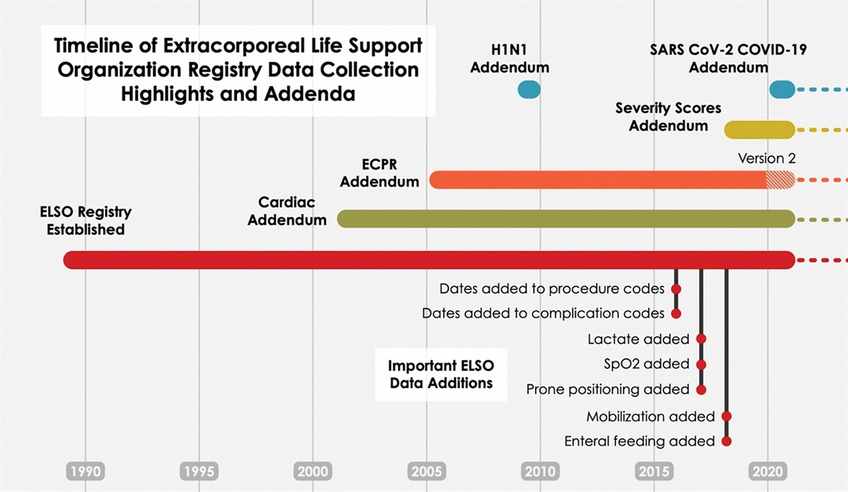
Outcome improvements with mechanically assisted circulation have paralleled advances in device design. In a relatively short order, the field transitioned from pulsatile to continuous flow devices allowing simplification of design, reduction in components prone to wear, and improved hemocompatibility and durability. An important stop in this journey was the preclinical and clinical observation that preservation and maintenance of normal human cardiovascular physiology and end-organ function were not dependent upon a “normal” pulse pressure, a vital step in miniaturization and simplification of left ventricular assist devices (LVADs).1 As a result, a clinically meaningful mechanical therapy for patients with advanced heart failure now exists. More than 10,000 LVADs are placed annually with 2 and 5 year survival rates of 79% and 58%, respectively.2,3
Although newer generation devices have been proven to reduce postimplant morbidity, patients and practitioners recognize the limitations of current LVADs. Serious adverse events including infection, stroke, bleeding, and residual heart failure remain common and result in frequent interactions with the healthcare system.4,5 In fact, a recent analysis from the MOMENTUM-3 trial suggested that the incidence of postimplant heart failure may be increasing.6,7 Large clinical datasets preclude a deeper understanding of postimplant heart failure because they lack sufficient patient-level detail to allow dissection of the etiology of this outcome. It has often been assumed that these patients have failure of the unsupported right ventricle either resulting from a primary heart muscle disease that impairs systolic or diastolic performance or alternatively that the LVAD, through its complex influence on ventricular interaction, is changing the geometry and function of the right ventricle and tricuspid valve.8 However, another explanation for residual heart failure is insufficient support of the systemic circulation by the LVAD. Although continuous flow LVADs are preload dependent, the static setting of impeller rotational speed (that typically occurs infrequently during clinical visits, at rest, and in a recumbent position) renders the device incapable of responding to many routine physiologic perturbations including reductions in left ventricular preload, the need for increased flow in response to exercise, and detection of interventricular septal shift that may contribute to right ventricular dysfunction. In support of the potential for real-time adjustment of device speed, Mancini and colleagues have recently shown that maximal exercise performance is so markedly limited in continuous flow LVAD patients that peak oxygen consumptions would qualify patients for transplantation.9
A next critical step in recapitulating normal cardiovascular physiology, reducing postimplant heart failure, and enhancing exercise performance following continuous flow LVAD support is development of a “Smart Pump” that integrates innovative tools and algorithms to detect changes in cardiac structure and physiologic demand to inform automated changes in device performance.
In this issue of the Journal, Palagani et al. present an important step toward the clinical reality of Smart Pump technology.10 These investigators developed and tested a series of sensors that could be easily integrated into current LVAD technology that would allow real-time assessment of left ventricular size and inform adjustments in pump speed based upon changes in left ventricular volumes. The authors integrated a transmitter in the apical sewing ring and placed a series of detectors in the outflow graft of a ventricular assist device to enable determination of left ventricular volumes. Through a series of in vitro experiments and complex modeling, the positioning of the sensors was optimized. The authors demonstrated that the safety profile of this technology would be high based upon the electromagnetic radiation absorption of tissue. Further, this approach was found to be highly accurate with minimal drift in measured values over a 3 month follow-up period. Finally, the proposed design would allow the apical transmitter to be powered by the LVAD controller with wireless energy transfer to the outflow sensors.
Although the current study provides a glimpse into the potential of predicting left ventricular size and septal position using resonantly coupled sensors, there are several potential limitations that need to be addressed before clinical application. First, the current series of experiments were performed in a static (nonbeating) system. The impact of ventricular volume changes during the cardiac cycle, heterogeneous baseline cardiac structure, and LVAD implantation technique variability will require revalidation of the approach in in vivo systems. The structure of the failing heart is inconsistent and dynamic—any system intended to detect and respond to changes in ventricular volumes must be capable of adapting not only to abrupt changes in loading conditions but also progressive remodeling and reverse remodeling seen in VAD patients—for example, how would automated algorithms respond to the development of postimplant de novo aortic or mitral valve insufficiency? The impact of right ventricular dilation also needs to be considered in the construct, as it would also be anticipated to increase the distance between the apical transmitter and outflow graft detectors. Automated algorithmic changes in device performance based upon misinterpretation of the relative contributions of right and left ventricular volumes could have detrimental impacts on the sufficiency of circulatory support. Finally, integration of the system into current LVAD technology may be more complicated than anticipated, particularly the algorithms that convert changes in ventricular volumes to changes in device performance.
It would seem that the dynamic nature of ventricular remodeling and physiologic demand would be well-suited for machine learning approaches that would integrate large volumes of data over time allowing the system to learn and make refined adjustments in device performance.
The current study gazes into the future of autonomous LVAD function, a technology-enabled approach to LVAD management that is appealing and would obviate the need for clinicians to pretend that intermittent LVAD adjustments based upon static measurements of surrogate data will improve patient outcomes or functional capacity. It is uncertain today that application of resonantly coupled sensors to measure left ventricular volumes will translate to sufficiently sophisticated management algorithms and result in improved outcomes. The latter question will only be answered through larger-scaled, well-controlled clinical trials—in other words, the pudding will be in the proof that we can convert these fundamental observations into clinically meaningful outcomes.
1. Purohot SN, Cornwell WK, Pal J, et al.: Living without a pulse: The vascular implications of continuous-flow left ventricular assist devices. Circ Heart Fail. 11: e004670, 2018.
2. Mehra MR, Uriel N, Naka Y, et al.: A fully magnetically levitated left ventricular assist device- final report. New Engl J Med. 380: 1618–1627, 2019.
3. Mehra MR, Goldstein DJ, Cleveland JC, et al.: Five-year outcomes in patients with fully magnetically levitated vs. axial flow left ventricular assist devices in the MOMENTUM 3 randomized trial. JAMA. 328: 1233–1242, 2022.
4. Nolina EJ, Shah P, Kiernan MS, et al.: The Society of Thoracic Surgeons Intermacs 2020 annual report. Ann Thorac Surg. 111: 778–792, 2021.
5. Chuzi S, Ahmad FS, Wu T, et al.: Time spent engaging in health care among patients with left ventricular assist devices. JACC Heart Fail. 10: 321–332, 2022.
6. Vidula H, Taleda K, Estep JD, et al.: Hospitalization patterns and impact of a magnetically levitated left ventricular assist device in the MOMENTUM 3 trial. JACC Heart Fail. 10: 470–481, 2022.
7. Cogswell R, Rogers JG: Residual heart failure on mechanically assisted circulation: A call to action. JACC Heart Fail. 10: 482–484, 2022.
8. Ali HR, Kiernan MS, Choudhary G, et al.: Right ventricular failure post-implantation of left ventricular assist device: Prevalence, pathophysiology, and predictors. ASAIO J. 66: 610–619, 2020.
9. Moss N, Rakita V, Lala A, et al.: Hemodynamic response to exercise in patients supported by continuous flow left ventricular assist devices. JACC Heart Fail. 8: 291–301, 2020.
10. Palagani Y, Sorkin E, Bonde R, et al.: Resonantly coupled high-efficiency sensors for assessment of ventricular chamber size for autonomous control of left ventricular assist device. ASAIO J. 69: 50–58, 2022.


留言 (0)