1.1. General Aspects
Artificial intelligence (AI) is acknowledged as a dramatic technological development, mainly due to the variety of applications in which its techniques can be used.
The tools of artificial intelligence have proved their effectiveness in modelling and optimization, with the most used being artificial neural networks (ANNs), evolutionary algorithms, and fuzzy systems. Their medical application in ophthalmology is a promising approach based on estimations and could enhance clinical examinations [
1,
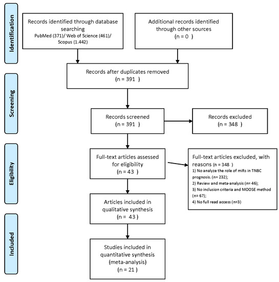
2].An artificial neural network is a simplified model of the biological brain. In its most general outline, a neural network is a machine designed to shape how the brain solves a particular problem or performs a function for a specific purpose (
Figure 1).
In neural networks, information is no longer stored in well-defined areas, as in the case of standard computers, but is stored diffusely throughout the network. Memory storing is done by assigning appropriate values of the weights of synaptic connections between network neurons.
An artificial neural network takes over the biological characteristics of the biological network:
-
Information is stored and processed throughout the network—it is global, not local.
-
A key feature is the plasticity—the ability to adapt, to learn
-
Knowledge is stored in inter-neural connections (synaptic weights).
-
The ability to generalize—artificial neural networks can find the correct answers for slightly different inputs than those for which they were initially trained.
-
The ability to synthesize—can give correct answers for input affected by noise/inaccurate/partial.
Neural networks require processing units called neurons and channels through which information flows—interconnections. They are distinguished by a wide range of parameters (weights) based on the top neural connection. Each processing neuron first accounts for the weighted sum of all interconnection signals in a previous layer, to which it adds a basic term, resulting in an output via an activation function. The most employed networks consist of the following categories of layers: input, hidden, and output layer. The input layer is defined by the network input data, the output layer by the output data, and the topology of a network is outlined by the hidden layers (set of intermediate layers and of neurons within these layers) [
1,
3].By adjusting the parameters in the connections between neurons, the network becomes able to learn from a set of numerical data suitable to the desired input and output variables. Therefore, based on wide range of examples, a key feature of RNA is that it can synthesize a specific pattern of the analyzed issue It is obvious that an essential advantage of neural networks is that they do not call for identifying a problem-solving algorithm, so it is not necessary to know the relevant laws, because the network alone learns from examples. Modelling based on neural networks consists of rendering the subordination between the output and input variables [
1,
2,
4,
5,
6]. An essential feature of neural networks is their ability to generalize; after “learning” the behavior of the process, they can make projections regarding the “unseen” data (that were excluded from the training set), which defines their ability to generalize. It is the verification stage of the model represented by the network [
7].
The neural network training is performed on a specific structure. The training process consists of establishing the network configuration, including architecture and topology.
Several methods are known for determining the topology of a neural network, of which the one based on successive tests is the most widely used. In this case, different topologies are tested until an acceptable error level is reached. This method, however, does not guarantee that the best network topology has been found. That is why evolutionary methods that have the chance to generate a favorable neural network are considered more efficient [
8].
Multilayer perceptron (MLP) is the most acknowledged and extensively employed type of neural network. Most often, its processing units are interconnected in a feedforward way, meaning that the interconnections are not disposed in loops.
The advantage of using other artificial intelligence tools, such as machine learning, deep learning, and convulsional neural networks, is shown in the following sub-chapters, as they are useful through image analysis in the early diagnosis of some diseases. The field of machine learning has expanded to include deep learning and advanced neural networks such as the convolutional neural network (CNN). This is an advanced network, a multi-layered variant of DL, which simulates the interconnection of neurons of the human brain to analyze an input image (recognition, processing, and classification). Le-NET, AlexNet, VGG, GoogLeNet, and ResNet are some of the CNN algorithms.
1.2. ANN Ophthalmology Reviews
Having several medical applications, artificial intelligence (AI) tools started being employed in ophthalmology, such as detecting visual function deficits, thus playing a key role in diagnosing eye diseases and in predicting the evolution of these common and disabling diseases. Although relatively young in the field of ophthalmology, AI technologies are constantly expanding and have a significant impact on the scientific research and the improvement of clinical practice.
Artificial intelligence tools, and especially artificial neural networks, are progressively involved in detecting and customized control of ophthalmic diseases. The precise data of the explorations and, particularly, the emergence of new imaging methods (OCT) have led to the increase in interest in the use of these tools by multidisciplinary teams. The combination of AI technologies and optical coherence tomography (OCT) proved to be trustworthy in detecting retinopathies or in enhancing the diagnostic conduct of retinal diseases.
As in medicine, there are two types of approaches based on neural networks in ophthalmology: setting the database by image processing or using data from medical records. The first case is more common due to the possibility of using a lot of information from the image. Consistent databases can be both quantitatively and qualitatively generated. The second option is less used, precisely due to the difficulties in achieving a database suitable for processing.
There are a number of reviews in the literature that attempt to present the use of artificial intelligence tools in ophthalmology. We consider it necessary to refer briefly to these in order to highlight and justify our own contribution in this field and the originality of the approach.
In a short review [
9], Grewal S., P., et al.’s Deep learning in ophthalmology: a review, deep learning tools were implemented for various diseases (cataracts, glaucoma, age-related macular degeneration, diabetic retinopathy) based on several diagnostic investigations (digital photographs, optical coherence tomography, and visual fields). Deep learning (DL) refers to machine-learning methods that use winding neural networks (CNNs), which employ storing image-processing filters to remove different types of image characteristics. The relevance and convenience of DL research in ophthalmology as daily clinical practice as part of digital ophthalmologic diagnostic tools is pointed out here. The approach reviews the benefits of DL techniques as a secure tool to render ocular data acquired from digital photographs and visual fields, and its contribution to early diagnosis of diseases such as AMD, DR, and glaucoma. Apart from its advantages for both patient and clinician, DL has also limitations related to modern technology and its best applications.The mini-review of Kenji Karako et al. [
10] describes applications of neural networks in medicine. However, as it addresses the classification of data types necessary for the neural networks training, this is also of interest for the field of ophthalmology. The first general consideration is that medical neural network applications can be divided into two types: automated diagnosis and physician aids.
Neural networks are being trained by employing different medical images to change diagnosis by a physician because diagnosis is often based on imaging, and a winding neural network activates image analysis. The network has the capacity to diagnose disease more accurately and even faster than a physician. Physicians can only identify a restricted amount of patients, while a neural network automated diagnosis can detect a substantial number of patients without time limitations.
The second type of applications refers to the use of data from medical records for neural network training, aiming to support the physician by generating a diagnostic rule.
A consistent and well-structured review is one by Lu W. et al. [
11]—Applications of Artificial Intelligence in Ophthalmology: General Overview. The ophthalmological approach starts with the idea that the quantity of the image data is quickly increasing; therefore, investigating and changing these efficiently becomes a priority. AI methods can do this task very well, attempting to examine medical data and leading to remarkable development in establishing a diagnosis. Useful algorithms from the category of CML (Conventional Machine Learning), such as Decision trees, Random Forest, Support vector machines, Bayesian classifiers, k-nearest neighbors, k-means, Linear discriminant analysis, as well as CNN (Convolutional Neural Networks), are reviewed.
Various ophthalmic imaging methods in AI diagnosis were mentioned as potential sources for generating databases: fundus images, optical coherence tomography, ocular ultrasound B-scan, slit-lamp image, and visual field.
The main diseases discussed in detail from this point of view—the processing of characteristic images with AI tools—are diabetic retinopathy, glaucoma, age-related macular degeneration, and cataract.
AI applications can greatly contribute in order to provide support to patients in remote areas by sharing expertise, knowledge and methods. Clinicians also receive valuable help in diagnosis.
A comprehensive review (Artificial intelligence and deep learning in ophthalmology) by Daniel Shu Wei Ting et al. [
12] deals with deep learning (DL) applied in ophthalmology for image recognition, as applied to fundus photographs, optical coherence tomography, and visual fields, reaching a powerful classification performance in detecting diabetic retinopathy and retinopathy of prematurity, the glaucomatous disc, macular oedema, and age-related macular degeneration. DL in ocular imaging may be employed linked to telemedicine as a potential solution to screening, diagnosing, and monitoring major eye diseases for patients in primary care and community environments. Considering the approaches of different researchers, the contribution of DL is explained in detail for diseases such as diabetic retinopathy, age-related macular degeneration, DM, choroidal neovascularization, glaucoma, and retinopathy of prematurity. The review of the 72 bibliographic references ends with highlighting and discussing potential challenges.A short article (AI papers in ophthalmology made simple) by Sohee Jeon [
13] points out, without many examples, the essential considerations for AI (particularly deep learning, DL) applications in ophthalmology. Three directions are briefly described: 1. Depending on the specific research question AI input may comprise clinical data, medical images, or genomics. 2. In AI processing, the most familiar type of DL method applied for medical images is winding neural network (CNN). 3. For AI output, the AI efficiency is measured by comparing it with a reference standard, which is often an extensively endorsed gold standard or ground truth. Having a key role in validating an algorithm, the reference standard is often grounded in compliance with several professionals, consultant ophthalmologists, fellowship-trained subspecialists, certified nonmedical professional graders, or optometrists who have engaged in extensive training and accreditation with restored and substantial outcomes. The results section quantifies the development of the AI system by relating to separating procedures, such as the area under the curve (AUC), sensitivity (or the genuine positive rate or withdrawal), peculiarity (corresponding to 1—false positive rate), positive predictive value (PPV), and negative predictive value (NPV).Another review (Artificial Intelligence: The Big Questions, Review of ophthalmology, 2021) by Christine Leonard [
14] presents the general problems of AI and the latest innovations and challenges, with some examples in ophthalmology, including detecting diabetic retinopathy, devices for diabetic eye-disease screening, cataract surgical videos (an AI algorithm to be able to automatically detect what steps are being performed in a surgical video at any given moment), predicting macular thickness from fundus photos, etc.A short review (Artificial intelligence and deep learning in ophthalmology—present and future) by Moraru Angrea Dana et al. [
15] explains Artificial Intelligence terms and focuses especially on deep learning and using OCT images for generating databases with the purpose of diagnosing DMO (diabetic macular oedema) and AMD (age-related macular degeneration) while using various algorithms such as (MLC, SVM, MLP, RBFNN). Other diseases, such as Diabetic Retinopathy and Retinopathy of prematurity, where deep learning was used are also mentioned. The review also focuses on limitations and future prospects, highlighting the importance of these technologies and how useful they can be, provided we can manage to standardize databases.
Different surveys in the literature review show the remarkable benefit of these AI tools in ophthalmology in evaluating the visual field, optic nerve, and retinal nerve fiber layer, thus ensuring a higher precision in detecting advances in glaucoma and retinal shifts in diabetes. All the studies that refer to these medicinal and opthalmological AI applications were analyzed in this review.

留言 (0)