1. IntroductionSensorineural hearing loss is one of the most common sensory deficits in the world and will affect up to 10% of the world population by 2050, according to the estimates of the World Health Organization [
1]. Hearing disability has a large impact on the daily quality of life; it can lead to social isolation and even give rise to dementia, yet possibilities for effective treatment remain limited [
2,
3]. In the majority of cases, sensorineural hearing loss is due to structural damage of the cochlea, which is often treated with a cochlear implant in patients with severe hearing loss. Unfortunately, the surgical insertion of the stimulating electrode array often traumatizes the delicate intracochlear microstructures, which cannot be visualized intraoperatively. Targeted regenerative inner ear therapies are being researched, but their translation into clinical practice relies on the ability to diagnose the underlying pathology and administer the treatments to specific regions of the cochlea without collateral damage [
4,
5]. As such, the efficacy of current and future treatments largely depends on the ability to visualize the intracochlear anatomy.The human cochlea is a very small (4 mm × 7 mm × 10 mm) and complex organ, deeply embedded inside the human skull bone, prohibiting direct visualization of its internal structure [
6]. In current clinical practice, magnetic resonance imaging (MRI) or computed tomography (CT) is used for intracochlear diagnostics and preoperative planning. Unfortunately, these techniques fall short in imaging most intracochlear microstructures due to their limited resolution (0.5 mm) [
6]. MicroCT and histology can provide high-resolution visualization of the internal cochlear anatomy, but these destructive methods are not viable for future (in vivo) clinical use [
7,
8].Recently, the use of optical coherence tomography (OCT) in hearing research has been rising in both morphological and functional studies [
9,
10,
11,
12,
13,
14,
15,
16,
17]. OCT is a nondestructive high-resolution imaging technique with similar working principles to ultrasonography. It uses low-coherence infrared light instead of sound waves, resulting in a higher micrometer-scale resolution. It was first applied in the field of ophthalmology, and due to its success, the use of OCT rapidly increased in other medical fields, such as oncology, cardiology, and dermatology [
18,
19,
20]. A main advantage of OCT is the possibility to perform transmembrane OCT imaging, which happens through the round window membrane (RWM), providing the ability to image the first 1–3 mm of the most basal portion of the inner ear, namely the proximal hook region [
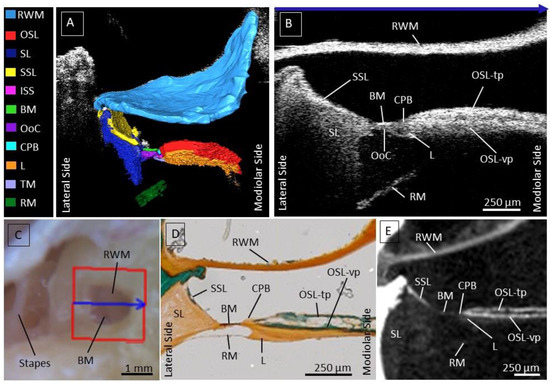
14]. This eliminates the need to disrupt the cochlear homeostasis by opening it, which makes it a promising tool for atraumatic cochlear implantation, inner ear therapy, and diagnostics. In our previous work, we demonstrated that relevant structures such as the basilar membrane (BM), Osseous Spiral Lamina (OSL), Secondary Spiral Lamina (SSL), and spiral ligament (SL) can be identified in the proximal hook region [
14]. However, the interpretation of human intracochlear OCT images is non-trivial for researchers and clinicians who are not yet familiar with this novel technology.
This study aims to extensively augment the knowledge of intracochlear structures by nondestructive transmembrane OCT imaging and to provide otological clinicians and researchers with an atlas of the human cochlear microanatomy. Likewise, we investigated whether abnormalities of the intracochlear structures can be visualized using OCT in fresh and fresh-frozen human temporal bones.
4. Discussion
The objective of this study was to provide an OCT-based atlas of the human proximal hook region to aid future clinicians and researchers with the interpretation of transmembrane OCT images of the human cochlea. We performed transmembrane OCT imaging in a series of seventeen human cadaveric cochleae and analyzed the characteristics of the visualized intracochlear structures, which are relevant for inner ear diagnostics and therapies. Our results demonstrate that transmembrane OCT imaging enables nondestructive, high-resolution, 3D visualization of the intracochlear microstructures, which are crucial for hearing function, as well as the detection of abnormalities in these structures. To the best of our knowledge, this was also the first OCT imaging study performed on a large sample size of human cochleae.
We were able to consistently visualize important intracochlear structures in the proximal hook region through the RWM. The RWM is a crucial entry point to the cochlea for inner ear therapies, functional studies, and, most likely intracochlear diagnostics. The proximal hook region is characterized by complex anatomy and is critical for the implementation of inner ear therapies. In particular, the OSL, BM, SSL, and SL, which are structures of interest during cochlear implantation [
26,
27], could be visualized using transmembrane OCT imaging. Damage occurring at these structures during an electrode insertion can harm residual hearing, causing mechanical-induced hearing loss, inflammation, fibrosis, or ossification of the cochlea [
27,
28].Pathologies of the inner ear inducing hearing loss mostly affect the OoC, making it a target structure for the development of gene and inner ear therapies and accurate diagnostics. Based on OCT-based visualization of important landmarks such as the TM, ISS, and TC, we could determine the position of the inner and outer hair cells together with the supporting epithelium surrounding them. At the current resolution, it was not possible to visualize the cells of the sensory epithelium individually; however, in the future, this can be tackled by using functional OCT [
11,
29,
30,
31]. On the other hand, improving the resolution could be achieved by increasing the spectral bandwidth of the light source and enhancing the resolution of the optical spectrometer that records the reflected light’s interference spectrum. However, spreading the signal over a larger detection array requires a higher optical output to maintain a similar signal-to-noise ratio (SNR). Some of these challenges have been addressed in a recent study, but these solutions have not yet been implemented in commercially available OCT systems [
32].Recently, the CPB, a soft tissue structure between the OSL and BM, has been identified in humans, differentiating it from the cochlear partition composition of rodents [
25]. In this study, we were able to visualize the CPB in the proximal hook region using OCT imaging. Here, the CPB was a remarkably short structure, supporting the most lateral attachment of the spiral limbus. These observations are in line with the description of Raufer et al. [
25], stating that the CPB becomes wider from base to apex in the human cochlea. The ability of transmembrane OCT to visualize CPB nondestructively in the proximal hook region of the human cochlea is highly promising for the functional study of inner ear mechanics [
25].In addition, we were able to detect intracochlear abnormalities with unprecedented detail using OCT in an intact human cochlea, which is highly relevant for intracochlear diagnostics and therapy. Out of seventeen specimens, the sensory epithelium was apparent in four fresh samples. In contrast to the other samples, these were not frozen before being imaged with OCT. In one sample, we observed normal sensory epithelium within a few hours post-mortem, but it disappeared after being frozen for six months, suggesting that freezing may have a destructive effect on the sensory epithelium. as it is remarkable that the sensory epithelium was mostly left in fresh samples, compared to fresh frozen samples. This is a significant finding, and on top of that, the sensory epithelium was most likely to be preserved in fresh samples compared to fresh-frozen samples. These findings highlight the importance of using fresh samples for reliable morphological and functional studies of the human cochlea and suggest that further research is needed on the effects of freezing and prolonged frozen storage on the quality of human temporal bones. Additionally, we also observed degenerated sensory epithelium in two fresh cochlear samples. We do not have any otologic background information for these samples, but these results might demonstrate the ability of transmembrane OCT to detect pathological changes in the human organ of Corti, which in clinical practice could be related to ototoxic drugs, noise exposure, aging, genetic factors, or other factors. Furthermore, OCT imaging could be a promising tool to monitor in vivo the efficacy of future regenerating therapies [
33].Other abnormalities were detected at the TM, which is believed to play a key role in hair cell activation in response to acoustical stimulation and undergo significant structural changes and degeneration with aging [
34,
35,
36]. We were able to visualize detachment of the TM from the limbus, a structural indication that is potentially linked to (the onset of) age-related hearing loss and hence relevant for future studies regarding future inner ear (gene) therapies and nondestructive (preventive) diagnostics [
35,
36]. While no otologic background information about the donors is known, the median age of the donors was 77 (range between 55 and 90 years) and thus might show signs of age-related hearing loss. However, why hypothesize that aging will not significantly affect the location of the visualized intracochlear structures using OCT but further clinical application in hearing-impaired subjects would be necessary to investigate the effect of aging on the integrity of intracochlear structures.Additionally, we were also able to evaluate the RM through the RWM. The RM separates the SM from the SV and normally is tense between its attachment at the OSL and the lateral wall. Included in our intracochlear OCT atlas are both a straight tensed RM and a flaccid RM. The cause of the latter appearance is not known, yet in certain pathologies such as Meniere’s disease, endolymphatic hydrops in the SM can cause distension of the RM [
37]. In OCT images of the isolated cochlea, it is possible that the tension caused by the fluid in the scalae is altered, causing bulging of the RM.Since OCT imaging was performed through the RWM, any overlying pseudomembrane was also imaged when present [
38,
39,
40,
41]. A pseudomembrane was noted in 18% of all samples, comparable to the findings of Sahin et al. [
41]. Since the RWM is the main access point for transmembrane OCT imaging, the presence of a pseudomembrane could negatively affect the visualization of intracochlear structures. It is not yet clear what the cause of pseudomembranes might be and hence unpredictable whether it is present in a patient who qualifies for inner ear surgery and diagnostics [
39]. Regarding inner ear therapy, a pseudomembrane could also negatively affect the diffusion of drugs administered by transtympanic injection into the cochlea [
42].Our results illustrate that transmembrane OCT imaging is a promising tool for clinical practice to nondestructively investigate the intracochlear anatomy in high resolution and in real time. The anatomy of the hook region is individually highly variable, and OCT can be crucial as a tool to anticipate the patient-specific anatomy, decreasing the risk for traumatic CI insertions [
43,
44,
45]. With the rising interest in residual hearing preservation and atraumatic electrode insertion during CI surgery, understanding the round window area and the anatomy of the proximal hook region is of utmost importance [
46]. Additionally, the hook region is also highly relevant regarding safe intratympanic injections, precise diagnostics, gene therapy, and in vivo studies of human intracochlear mechanics [
47,
48,
49]. OCT meets the needed requirements for in vivo high-resolution imaging, with the advantage of in-depth transmembrane imaging, facilitating the anatomical investigation of the cochlear base without the need for opening the cochlea and disrupting its integrity [
14,
32,
50,
51].OCT imaging does have certain limitations. First, we used extracted cochleae, providing the advantage of determining the optimal imaging angle, which might be unachievable in clinical settings. One solution might be using an endoscopic-based OCT through a transcanal approach, which might account for the limited degree of freedom one experiences within an entire skull [
32,
51]. To achieve comparable imaging results to a rigid OCT system, an endoscopic OCT system would need to be specially designed and optimized for imaging the cochlea. This might involve using a smaller, more flexible endoscope and a light source with a broader spectrum to improve the resolution and contrast of the images. It would also require advanced imaging algorithms and specialized image processing techniques to improve the overall quality of the images produced [
32].Furthermore, due to the limited imaging depth of the OCT, we were only able to image the approximate hook region. Because of this, the apex of the cochlea remains unreachable with OCT. In the future, fiberoptic tools might provide a solution for this, such as imaging of the intracochlear space during the insertion of a cochlear implant or during the injection of intracochlear therapies [
50]. Finally, certain factors may negatively affect the quality of transmembrane OCT imaging: a thickened RWM, pseudomembrane, and remaining debris or fluid on top of the RWM. Lowering the scanning frequency, removing the debris, and aspiration of the fluid can overcome these limiting factors. In many cases, the pseudomembrane also can be carefully removed by the surgeon. The origin of a pseudomembrane is not yet known, but it might be related to middle ear infections. However, clinical evidence for this correlation is still lacking, and further research is needed to investigate this. A mucosal pseudomembrane found adjacent to the surface of the RWM may impact its permeability. They may either inhibit diffusion by acting as an additional barrier, protecting the RWM [
41,
52]. On the other hand, several pathologies, such as Menière’s disease or chronic otitis media, can cause the thickening of the RWM itself, which might negatively affect transmembrane OCT imaging and its future applications [
53,
54]. Additionally, the presence of blood in a clinical setting on top of the RWM could form an additional challenge, as blood is highly scattered. This could be helped with the aspiration of the blood drops and better control of the blood pressure, as it is currently conducted during cochlear implant surgery.In summary, various studies investigated intracochlear anatomy using transmembrane OCT imaging; most of these were based on animal research [
14,
29,
30,
55,
56,
57]. Here, we provide an extensive imaging atlas of the human intracochlear anatomy in the proximal hook region, which would help future otologic researchers and clinicians to familiarize themselves with the features of intracochlear structures on OCT images. We were able to consistently visualize relevant intracochlear microstructures at a very high resolution and illustrated both normal and abnormal anatomical appearances using transmembrane OCT imaging. Being able to disentangle normal and abnormal composition of intracochlear structures, together with the fact that OCT is nondestructive and can be used in real-time in vivo, makes it a highly promising tool for clinical practice. OCT is a step forward towards aiding hearing-impaired patients in getting a safe insertion of inner ear therapies and enabling microstructural inner ear diagnostics, which is currently not possible.

留言 (0)