1. IntroductionDiabetic ketoacidosis (DKA) is an acute life-threatening complication occurring mainly at the onset of type 1 diabetes mellitus [
1,
2], with an incidence rate that spans from 13 to 80% [
3,
4,
5]. Being a form of systemic inflammatory state [
6,
7], inflammatory markers such as blood leukocytes and C-reactive protein (CRP) play a key role in the pathogenesis [
8,
9]. Although complete blood counts (CBCs) are a part of the routine evaluation in diabetic patients, white blood cell (WBC) fractions did not receive significant attention from diabetes specialists in the past [
10]. In recent years, however, there has been growing interest regarding the neutrophil-to-lymphocyte ratio (NLR) as a marker of systemic inflammation in cardiac diseases, neoplasms, and obesity, as well as in diabetes-related complications such as diabetic foot ulcers and retinopathy [
11,
12,
13,
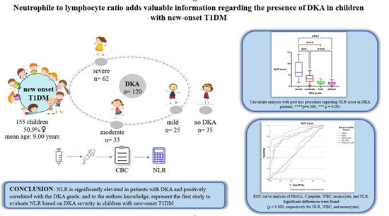
14]. Against this background, our aim was to study the association between the NLR and DKA severity among children with new-onset T1DM. 2. Materials and Methods 2.1. Patient Recruitment
This cross-sectional study included data from one of the largest Romanian reference centers for pediatric T1DM. We reviewed 181 consecutive T1DM patients charts from the Pediatric Emergency Hospital “Louis Turcanu” in Timisoara, Romania, between 1 January 2015 to 30 June 2022, in accordance with the principles of the Declaration of Helsinki (1975, revised in 2013). Ethical approval was obtained from the ethics committee.
Inclusion criteria for cases were noted as new-onset T1DM in children aged 0 to 18 years, with or without diabetic ketoacidosis. Diagnosis of type 1 DM was established according to the American Diabetes Association (ADA) criteria of 2021. Exclusion criteria were as follows: infectious states, any other medical conditions that could alter hematological parameters, and patients with other types of diabetes.
Patients with DKA had a plasma glucose level > 11 mmol/L, a urine ketone level defined as moderate to high (+ to +++), and an arterial pH value
15]. 2.2. Biochemical Assays
Laboratory tests, including routine biochemistry tests and arterial gas analysis, were performed in the hospital laboratory. Blood samples were drawn at admission before the initial therapy, to avoid posttreatment changes in CBC parameters, and collected for differential WBC counts in tubes with EDTA and processed using a Sysmex XN-550 (Sysmex Corporation, Kobe, Japan) automatic blood counting system. Glycated Hb (HbA1c) was measured using a high-performance liquid chromatography kit supplied by Cobas E 411–Roche, Japan. Peptide C was evaluated using automated chemiluminescent assay (Cobas E 411–Roche, Tokyo, Japan). Neutrophil-to-lymphocyte ratios (NLR) were calculated.
2.3. Statistical Analysis
All data analysis was performed using the standard computer program Statistical Package for the Social Sciences (SPSS) for Windows, version 28 (SPSS Inc., Chicago, IL, USA) and GraphPad Prism9. The Shapiro–Wilk test was used to test the normality of the data distribution. Normally distributed variables were expressed as mean ± standard deviation (SD) and non-normally distributed variables were expressed as medians with interquartile ranges. Intergroup comparisons were performed by using an independent-sample t test and one-way ANOVA for normally distributed continuous data and Chi-Square tests for categorical variables. Non-normally distributed data were compared among multiple groups using the Kruskal–Wallis test. GraphPad Prism version 9 was used for univariate analysis with a post hoc procedure regarding NLR scores in DKA patients. Multiple regression analysis was performed to evaluate the association between the NLR or WBC parameters and the occurrence of DKA in T1DM patients. Receiver operating characteristic (ROC) curve analysis was plotted to compare the discrimination performance of HbA1c, C peptide, and CBC parameters in predicting DKA severity. The optimal threshold values were obtained using Youden’s index (sensitivity + specificity − 1, ranging from 0 to 1) and the maximized area under the curve (AUC). A p value (two-tailed) < 0.05 was considered statistically significant.
4. DiscussionType 1 diabetes mellitus (T1DM) represents one of the most frequent chronic illnesses affecting children [
17]. Previous studies [
16,
17,
18,
19,
20,
21,
22,
23] have indicated an increase in both the frequency and severity of DKA cases in recent years. In our research, 81% of cases with T1DM presented with DKA, almost half of which were severe.WBC counts, fractions, and indices, among which the NLR has received attention in recent years, were correlated with inflammation-associated diseases such as systemic hypertension [
24], intracranial atherosclerosis [
25], neoplasia [
26], obesity [
14], and type 2 diabetes [
27,
28,
29].The shifts in the percentage formula of white blood cells (increase in total WBCs, neutrophils, and monocytes; decrease in lymphocytes and eosinophiles) were similar to those cited in the literature [
17,
30].Aside from systemic inflammation [
31,
32,
33], the NLR, a well-characterized systemic inflammatory response marker [
34], can also reflect both innate and adaptive immune (dys)function [
9,
35,
36]. This simple ratio, which combines the predictive power of both increased neutrophil and decreased lymphocyte counts, has the advantage of being ubiquitous, cost effective, and also more stable compared with the absolute count [
9,
30,
37]. Results from the present study are consistent with previous publications [
10,
30,
38], in that WBC count and the NLR were found to be higher in patients with DKA.Median NLR scores in our case were significantly different between groups, increasing from those without ketoacidosis (1.11; 0.80–1.80) to mild (1.58; 1.17–1.93), moderate (3.71; 1.98–4.85), and severe (5.77; 4.04–9.63) ketoacidosis groups. Our results regarding pediatric patients are consistent with a previous study addressing adults with DKA, which regards the NLR as a possible marker of the underlying severity of acute systemic inflammation in uninfected DKA patients [
6]. Aside from the obvious effect of hemoconcentration on the NLR, the potential relationship between hyperglycemia and an increased NLR has been addressed in previous studies [
39]. One possible explanation is that WBCs that are activated by advanced glycation end-products produce pro-inflammatory cytokines [
29]. However, our study did not reveal statistical differences among the four groups in terms of mean HbA1c levels. This is consistent with some studies regarding children with DKA [
40,
41,
42], and in opposition with other studies [
17]. Another explanation is the fact that, in DKA, acute hyperglycemia promotes the accumulation of reactive oxygen species (ROS) which can damage peripheral blood lymphocytes’ DNA. This in turn may cause the apoptosis of lymphocytes and affect their proliferation [
6,
43,
44].In the present study, with new-onset T1DM children grouped according to blood pH, multivariate logistic regression analysis was performed in order to assess whether confounding exists between age, sex, HbA1c, C peptide, and NLR regarding blood pH. The NLR displayed a good discriminatory power regarding association with DKA, through correlation with blood pH.; age at onset, and, to a lesser extent, C peptide added statistically significantly to the prediction. This is consistent with a previous published study regarding adult T1DM patients [
10] but, to our knowledge, was not yet reported in children. An upside to examining children is their lack of many confounding factors that can affect NLR levels, such as common medications and comorbidities present in adult patients with diabetes.
Assessing the ROC curve, the presence of DKA in our study lot was associated with an elevated NLR, monocytes, and WBCs. The area under the curve was largest for the NLR, with values above 1.84 being most frequently present in children with DKA (sensitivity of 80.2% and specificity of 80%). Regarding C peptide, plasma values were negatively correlated with the presence of DKA, mainly values below 0.690 ng/mL (sensitivity of 68.2% and specificity of 60%).
There were some limitations in the present study. Firstly, the sample size was relatively small, which could limit the power of the analyses. Secondly, our patients are only from one hospital, so that selection bias cannot be ruled out. Additionally, only one measurement of CBC and subsequent NLR calculation were used in the analysis: those upon admission. As such, there was no monitoring of the dynamic trend of the NLR. We look forward to additional multicenter studies with large samples.

留言 (0)