1. IntroductionCritical illness is defined as the presence of organ dysfunction following an acute insult, requiring medical interventions to restore homeostasis. Immune dysfunction is common in critical illness, not only in the setting of infection, but also after trauma, single organ infarction and neurocritical illness, among others [
1].The immune organ, in a simplified description, is a system capable of interacting with the self and the external environment, distinguishing between the two and hence guaranteeing a defense from external agents. It consists of an innate arm that allows a quick response to endogenous and exogenous pathogenic stimuli, and an adaptive arm that, upon antigenic recognition, produces a pathogen-specific immune response, developing memory of the recognized antigens. Communication between these two arms takes place through direct contact between cells, as well as through free molecules with an autocrine, paracrine, or systemic effect, which includes cytokines and chemokines, among others [
2]. The function of the immune system is, however, not limited to pathogen response but has a fundamental role in tissue regeneration processes, both frequently disturbed during critical illness.Notwithstanding, in-depth monitoring of the immune system in critically ill patients has never become common practice, despite both broad research in the subject for the last 40 years and common use of immune modulators like corticosteroids [
3]. Despite, rudimental monitoring of the immune response to track response to antibiotic therapy is already in used, immune monitoring could be further enhanced to guide modulation strategies, like corticosteroid therapy widely used in multiple critical ill scenarios or the administration of monoclonal antibody therapy, such as tocilizumab (anti-IL-6 receptor) recently used during the COVID-19 pandemic [
4]. In sum, multilevel immune monitoring is an important step towards the much-needed personalization of therapeutic interventions (
Figure 1) [
5].
In this review, we will briefly introduce the acute inflammatory response in critical illness, discuss why monitoring is needed and list some of the most promising markers based on flow cytometry techniques.
2. Why Monitor the Immune System during Critical Illness?
Tissue and organ lesion and injury are the hallmark of patients admitted to the ICU. Critical care physicians are familiar with brain, heart, lung, gastrointestinal or kidney monitoring, but not with immune monitoring, nor how it reflects adequate or inadequate immunological function.
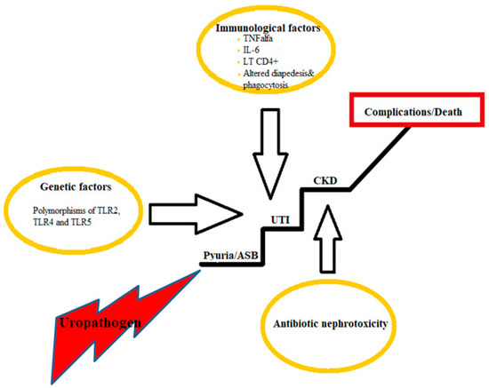
Alarmins released from injured and dying cells (DAMPs -damage-associated molecular patterns) together with molecules originated from microbes (PAMPs—pathogen-associated molecular patterns) determine immune cell activation [
6]. Responding immune, epithelial, or endothelial cells express alarmin receptors (PPR—pattern recognition receptors) which include TLR (toll-like receptors), C-type lectin receptors, nucleotide-binding oligomerization domain-like receptors, retinoic-acid-inducible gene-I-like receptors and RAGE [
6,
7]. One of the most studied is TLR4, that recognizes LPS molecule. Genetic polymorphisms associated with this molecule have been associated with an increased risk of critical illness such as sepsis or multiple organ failure after trauma [
8,
9]. Aside from TLR4, many other PPR have been implicated in the pathophysiology of critical illness (
Figure 2) and strategies to modulate them might, in the future, help reprogram the immune response in the context of critical illness.The downstream intracellular signaling of DAMPs or PAMPs involves different proteins, a major one being MyD88, resulting in the subsequent activation of NF-kB and the transcription of early cytokine genes, such as IL-1B, IL-8 or IL-6, and interferon genes [
6].Neutrophils are the most prevalent cells in circulation and constitute the first effectors of the innate inflammatory response. Upon activation, neutrophils express chemokine receptors such as CXCR2, which recognize molecules released by endothelial cells attracting neutrophils into the interstitial space [
10]. Furthermore, neutrophils can rapidly be recruited by release from capillary beds, such as in the spleen and lung, where they have a slower transit time [
11,
12].Neutrophils are armed with potent antimicrobial molecules that, when released freely like in degranulation or formation of extracellular webs, can also cause tissue damage. The latter response, whilst being fundamental for neutrophil function, is at the base of the pathophysiological process of pathologies such as ARDS [
13] or ischemia-reperfusion injury [
14].
Simultaneously, once activated, dendritic cells migrate to the lymph nodes, and present antigen to CD4 T lymphocytes, recruiting the adaptive immune response. These later produce cytokines, like IL-8 or IL-17, that further recruit neutrophils, thus amplifying the immune response. In addition to dendritic cells, monocytes change their phenotype from M2 (anti-inflammatory) to M1 (pro-inflammatory), produce pro-inflammatory cytokines and present antigens thus contributing to set off the adaptive immune response.
This response consists of antibody production, depending on B cell maturation and differentiation into plasma cell. T cell associated responses involve either cytotoxic (CD8) T cells, particularly relevant in viral infections, or T-helper (CD4) cells, fundamental to cytokine production and for shaping myeloid and B cell responses. Recruitment of each cell type depends on their upregulation of chemokine receptors, which also reflects the cell’s function, and the local tissue production of chemokines. This allows the infiltration of different cells in tissues vital for the response against foreign agents or necrotic tissue.
In parallel to the above, tissue regeneration is promoted. This supports a shift to regulatory mechanisms, with an increase in regulatory T cells (Tregs) in tissues, in Th2 mediated response [
15], and the production of cytokines such as TGFβ. Tissue regeneration and complete recovery depends on stopping the inflammatory process. Dysregulation at this stage, enabling chronic inflammation, may contribute to the persistence of organ dysfunction and accumulation of fibrotic tissue [
16].Immune dysregulation can occur at several moments of critical illness combining immune activation and suppression mechanisms. Cytokine storm with consequent multiple organ failure is considered a pathogenic hallmark in distributive shock associated with sepsis, trauma, or ischemia-reperfusion injury. In this context, high levels of cytokines such as IL-1, IL-6, IL-18, IL-8 or TNF contribute to organ lesion by recruiting neutrophils, NK cells and other lymphocytes to the tissues, by activating the endothelium and overall inducing a new metabolic immune state, as recently reviewed in the context of COVID-19 [
17].After this acute phase, there is either complete recovery, death, or progression towards an occult persistent immune dysregulation. This later phenomenon can occur in all settings of critical illness [
18], but it was in sepsis where it was primarily described. Immune dysregulation has been associated with an increased risk of nosocomial infections [
19], as well as cognitive dysfunction, ICU acquired muscle weakness and increased mortality [
20]. It is clinically represented by a group of patients with prolonged critical illness (more than 14 days of hospitalization in the ICU, persistence of organ dysfunction with evolution to chronicity in some cases) [
21]. Patients with this immune dysfunction are also represented in cohorts of patients with post-intensive care syndrome (PICS), as defined in 2012 by Moore, and immune dysfunction may partially explain its development [
22].The persistent immune dysregulation is characterized by a persistent activation of the innate system, by DAMPS or PAMPS. If the initial stimulus of the septic episode starts through PAMPS such as LPS, immune activation mediated by PAMPS might persist due to reactivation of viruses such as CMV, EBV, HHV-6, or TTV, occurring in more than 40% of septic patients [
23], or due to nosocomial bacterial infection. At the same time, there is the release of DAMPs such as S100, nuclear or mitochondrial DNA, HMGB1, RAGE, IL-33, adenosine, amongst others, that perpetuate the inflammatory cascade in response to injured tissue [
24].This persistence of alarmins causes an exhaustion of the immune system, which is expressed by the absence in lymphopenia recovery, a decrease in the expression of human leukocyte antigen DR (HLA-DR) in monocytes, and an increase in sPD-L1, which might support a state of cellular anergy and a decreased response to new pathogens. This immune-suppressive state can be seen as an adequate adaptive response to persistent cellular activation and an attempt to enter the repair process. Whatever the interpretation, this immunological picture has been called “immune paralysis” and is characterized by (a) a lower ability to present antigen, (b) a lower production of inflammatory cytokines and (c) a reduced clearance of bacteria. This phenomenon determines a greater risk of nosocomial infections and a persistent chronic inflammatory state. It also contributes to ongoing muscle catabolism marked by high levels of GLP-1 (15), whose pathophysiology is related to mitochondrial damage and release of mitochondrial DNA and molecules derived from reactive oxygen species. This process of prolonged immune dysregulation is also associated with accumulation of myeloid suppressor cells, which can be monocytic (M-MDSC) or polymorphonuclear (PMN-MDSC). These cells can also suppress lymphocyte function and decrease cytokine production, contributing to decreased pathogen clearance. The increase in myeloid suppressor cells is associated with a greater infection risk, ICU length of stay, and greater mortality [
25,
26].In addition to changes described in the innate system, multiple alterations in lymphocytes are also well-known. These range from persistent lymphopenia to modification in the profile of CD4 and CD8 T lymphocytes as well as of B cells [
27], promoting dysregulated immunoglobulin [
28], interferon and cytokine production and clearance, all contributing to organ dysfunction.The presence of immune dysregulation is frequently observed in chronically ill patients. Nevertheless, even this group is heterogeneous and might demand different strategies. So, carefully assessing the immune system and monitoring throughout chronic disease might have some benefits. For instance, we could better guarantee the enrolment of more homogeneous groups of patients in clinical trials and in the future cater personalized medicine. Although it is an area of active research, the clinical application of specific biomarkers to aid monitoring the use of therapeutics in intensive care medicine such as corticosteroids, or the risk assessment of secondary complications such as hospital-acquired infection and immunosuppression associated with critical illness, is yet to be seen [
5].In this field, data reanalysis of several randomized clinical trials have allowed to recategorized patients into various phenotypes and reaccess the intervention results. In ARDS, for example, patients were classified into a pro- and anti-inflammatory phenotype profile, based on multiple parameters of which the most immunologically relevant were IL-6, IL-8, TNFr1 and ICAM-1 [
29]. This stratification allowed us to understand, for example, that the effect of fluid restriction strategies [
30] was not transversal between phenotypes and could even be deleterious in the hyper-inflammatory group [
31].From these studies also emerges the concept that only a cluster of markers and not isolated markers will allow the definition of homogeneous phenotypes. Artificial intelligence systems facilitating the identification of clusters from clinical and lab data, associated with an increased technical capacity at the bed side to simultaneously measure multiple molecules, may aid us in the future to apply this paradigm [
32]. 4. Can We Monitor the Immune System in Depth? The Clinical Application of Flow CytometryThe frequency and absolute numbers of immune cells, such as monocytes, neutrophils or lymphocytes, or their sub-types, such as CD4 + T cells—further subdivided into TH1, TH2, TH17 and regulatory T cells (Tregs)—can be used to monitor the immune system. Flow cytometry has fueled the identification of these immune subsets and their monitoring in different disease settings [
89,
90]. Currently, completely automatized flow cytometry methods are used for performing routine full blood counts or lymphocyte phenotyping (T, B and NK cells) commonly used in situations like HIV infection or after treatment with rituximab [
91].Flow cytometry can rapidly analyze multiple immune populations in solution at the single cell level. Cell shape and complexity can be inferred, as well as the presence of a specific cell associated protein when immunoassayed with fluorescently conjugated antibodies [
92]. In addition, to cell associated markers, when coupled to cell stimulation assays, flow cytometry-based techniques can be used to determine cell function, such as per cell cytokine production, oxidative function, cytotoxic activity, and to ascertain cell division [
93,
94]. Besides cellular immunophenotyping, flow cytometry can also be applied to detect and quantify soluble proteins, such as cytokines and chemokines, in multiplexed assays with the equivalent assaying power of 100 Enzyme-Linked Immunosorbent Assays(ELISA) assays [
95]. It can also be used to determine an array of inflammatory mediators allowing a better discrimination for diagnosis [
96], and, in a recent study, quantitative flow cytometry was used to assess the number, viability and drug-resistance of common disease-causing bacteria [
97]. 4.1. Limitations and Challenges to the Use of Flow CytometryThere are, however, some downsides to flow cytometry that hinder its use for clinical immune monitoring. Flow cytometry is an open technique, with many different analyzers and often homemade protocols that lead to variation in results and their interpretation. This leads to decreased reliability and major issues in standardization [
98] crucial for comparing results between centers. These are now starting to be overcome [
99]. Standardized immunostaining protocols between labs, calibration, and daily quality control of flow cytometers with specific beads, the use of same batch antibodies with stable fluorophores or the use of calibrated beads that convert fluorescence intensities to numbers of antibodies bound per cell, are some of the strategies to implement reliable flow cytometry protocols. Besides standardization issues, this technique requires specialized technicians and increasingly complex and expensive instruments which availability can be challenging in some centers [
100,
101,
102]. Nevertheless, the investment in cutting-edge flow cytometry techniques for critical care will surely prove to be fruitful, like it already is for deep immune monitoring in transplant patients, or those receiving immunotherapy for several malignancies [
103,
104]. 4.2. New Techniques for in Depth Monitoring
A recent upgrade, spectral flow cytometry, is simplifying the access to in-depth immunophenotyping. As opposed to conventional flow cytometers, spectral cytometers capture the full spectral emission of each fluorophore. Therefore, while it is already challenging to assess 18 markers by conventional analyzers, spectral flow cytometers allow for the relatively easy detection of more than 40 markers per cell. While they require similar controls as with conventional cytometers, spectral analyzers possess universal instrument settings and allow for a standardized output across all instruments, a clear advantage during multi-center clinical studies. Nevertheless, these instruments’ availability is still low, and their cost elevated.
Another method allowing the analysis of over 50 markers is mass cytometry. In this technique, also known as cytometry by time-of-flight (CyTOF), instead of using fluorophores, the antibodies are conjugated with heavy metal reporter ions and cells are analyzed by time-of-flight mass spectrometry to quantify the isotopic masses and, thus, the bound antibodies and the expression of markers of interest. This technology reduces the problems related with spectral overlap and sample autofluorescence present in flow cytometry, and the wide availability of heavy metal isotopes allows for multiplexing. Besides the assessment of surface cell lineage markers, mass spectrometry allows for the simultaneous quantification of many intracellular targets, such as cytokine production, transcription factors and protein phosphorylation, which can inform on cell states and response to stimuli. More recently, this technique was repurposed to allow for the multiplexed imaging of tissue markers, which would permit clinicians to understand immune pathology at tissue level [
105,
106].
Several other ways to monitor the immune system in-depth are growing more popular nowadays. For instance, gene expression profiling by RNA-sequencing techniques is allowing for the comprehension of bulk or single cell heterogeneity in homeostasis and disease. Moreover, spatial biology methods emerging in the recent years allow for the extraction of spatially resolved molecular information from tissue biopsies.
Therefore, there are a plethora of techniques available that can in the future assist in clinical decisions as well as fuel translational research to improve critical care.
Next, we will discuss some of the more promising cell associated immune markers and how they might shape clinical decisions.


留言 (0)