


記住我
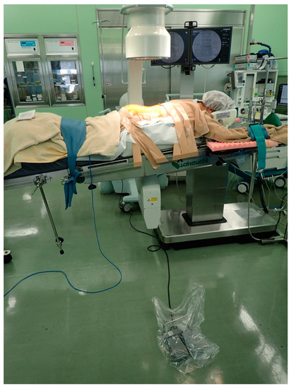


Figure 1. Operating room setup (the foot switch is at least 1 m away from the operating table).
Figure 2. (a) Insertion of K-wires at outer edge of pedicles; (b) confirmation of K-wire placement under fluoroscopy.
Figure 3. (a) A tubular retractor placed over the outside of the facet joint along the K-wire; (b) exposure of the lateral margin of the facet joint.
Figure 4. (a) A self-drilling pin; (b) a self-drilling pin inserted into the pedicle.
Figure 5. (a) Posteroanterior radiograph showing direction of self-drilling pins; (b) Pedicle screw insertion into the opposite side of the laminectomy via the tubular retractor.
Figure 6. (a,b) Insertion of the pedicle screw under the microscope.
Figure 7. (a,b) Computed tomography scan of the screw deviation to the medial pedicle wall in grade D breach.
Figure 8. Lateral radiograph of ossification of the posterior longitudinal ligament of the lumbar spine.
Table 1. Patient demographic and treatment information.
Table 1. Patient demographic and treatment information.
CharacteristicValue (%)Number of patients24Mean patient age in years (range)64.8 (24–88)Men (%)9 (37.5)Women (%)15 (62.5)Body Mass Index (kg/m2) (range)25.5 (17.3–32.9)Primary diagnosis Spondylolisthesis13 (51)Degenerative disc disease5 (22)Disc herniation6 (25)Lumbar level L1/21L3/45L4/514L5/S14Table 2. Surgery and radiation data for 24 patients who underwent MIS TLIF.
Table 2. Surgery and radiation data for 24 patients who underwent MIS TLIF.
FactorMean Value (Range)Operating time (minutes)201.8 (145–246)Fluoroscopic time/case (seconds)26.8 (8–56)Radiation dose of DAP (mGy∗m2)0.0706 (0.018–0.133)Radiation dose of AK (mGy)6.0 (1.071–21.74)Radiation dose of ESD (mGy)11.31 (2.199–44.64)Extra time required for this procedure (minutes)39 (16–69)Table 3. Statistical examination of surgery and radiation data according to patient characteristics.
Table 3. Statistical examination of surgery and radiation data according to patient characteristics.
Age<65 Years Old (n = 12)≥65 Years Old (n = 12) Fluoroscopic time (seconds)26.5 (8–56)24.5 (15–49)p = 0.582Radiation dose of DAP (mGy∗m2)0.061 (0.018–0.128)0.058 (0.034–0.26)p = 0.931Radiation dose of ESD (mGy)9.884 (3.002–21.928)9.823 (2.199–44.637)p = 0.908Extra time required for this procedure (minutes)40 (27–71)33 (16–64)p = 0.111GenderMen (n = 9)Women (n = 15) Fluoroscopic time (seconds)24 (8–49)27 (15–56)p = 0.881Radiation dose of DAP (mGy∗m2)0.057 (0.018–0.102)0.061 (0.032–0.26)p = 0.811Radiation dose of ESD (mGy)9.747 (3.002–19.341)0.447 (2.199–44.637)p = 0.743Extra time required for this procedure (minutes)33 (27–48)39 (16–71)p = 0.367BMI<25 kg/m2 (n = 12)≥25 kg/m2 (n = 12) Fluoroscopic time (seconds)24 (15–49)26 (8–56)p = 0.862Radiation dose of DAP (mGy∗m2)0.0495 (0.032–0.102)0.066 (0.018–0.26)p = 0.119Radiation dose of ESD (mGy)9.232 (5.433–19.341)10.59 (2.199–44.637)p = 0.299Extra time required for this procedure (minutes)37 (16–69)33 (18–71)p = 0.977Primary diagnosisSpondylolisthesisTable 4. Pedicle screw breach rate (total and per vertebral level).
Table 4. Pedicle screw breach rate (total and per vertebral level).
Vertebral LevelScrews Per Vertebral LevelNumber of BreachesDirection of Breach (n)Grade of Breach (n)Breach Rate (%)L-120 0L-220 0L-3100 0L-4383Medial (1), Lateral (2)C (2), D (1)3.2L-5360 S-180 0Total963 3.2
留言 (0)